

The DOI matches its Crossref record; 51 of the 51 references it cites were found in a registry or an index.
Amyloid beta, tau, synaptic, neurodegeneration, and glial biomarkers in the preclinical stage of the Alzheimer's continuum
Open accessAbstract
Introduction: The biological pathways involved in the preclinical stage of the Alzheimer's continuum are not well understood. Methods: We used NeuroToolKit and Elecsys ® immunoassays to measure cerebrospinal fluid (CSF) amyloid‐β (Aβ)42, Aβ40, phosphorylated tau (p‐tau), total tau (t‐tau), neurofilament light (NfL), neurogranin, sTREM2, YKL40, GFAP, IL6, S100, and α‐synuclein in cognitively unimpaired participants of the ALFA+ study, many within the Alzheimer's continuum . Results: CSF t‐tau, p‐tau, and neurogranin increase throughout aging only in Aβ‐positive individuals, whereas NfL and glial biomarkers increase with aging regardless of Aβ status. We modelled biomarker changes as a function of CSF Aβ42/40, p‐tau and p‐tau/Aβ42 as proxies of disease progression. The first change observed in the Alzheimer's continuum was a decrease in the CSF Aβ42/40 ratio. This is followed by a steep increase in CSF p‐tau; t‐tau; neurogranin; and, to a lesser extent, in NfL and glial biomarkers. Discussion: Multiple biological pathways are altered and could be targeted very early in the Alzheimer's continuum .
Overview
The claims and statistics of this paper haven’t been checked yet.
Run the check — free, no account needed. It reads the open-access text and checks the sources it cites.
Integrity signals
Registry
Cites
Statistics
Figures
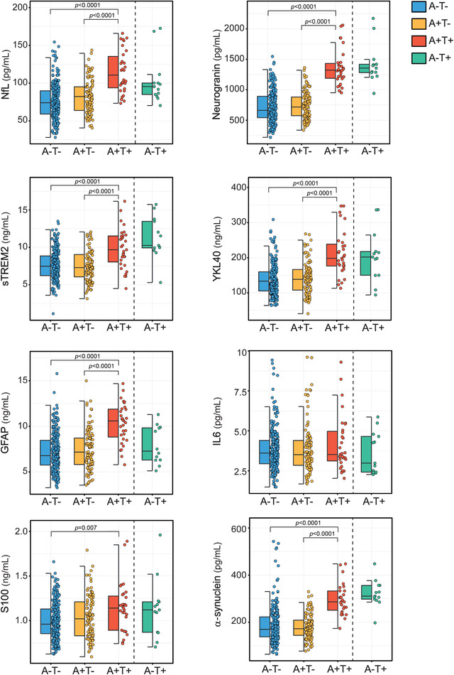
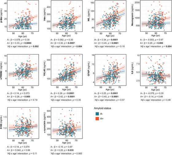
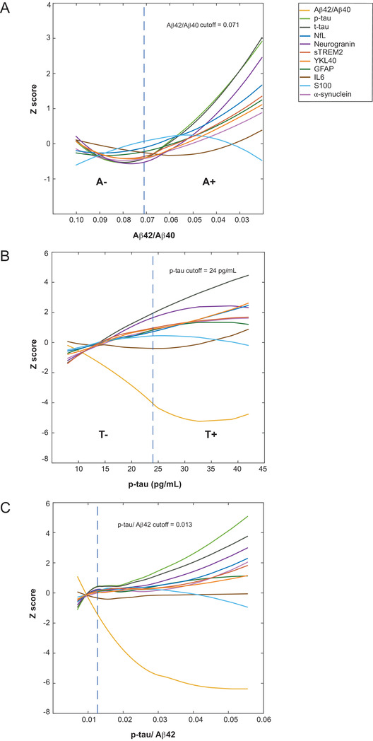
Verification
Claims the paper makes
The claims and statistics of this paper haven’t been checked yet.
Run the check — free, no account needed. It reads the open-access text and checks the sources it cites.
Claims other papers make about this one
Each is checked against this paper’s own text: does it say what the citing paper says it does?
The source backs part of the claim, not all of it — or backs it for a different population, condition or comparison.
In the citing paper
“The CSF sample collection and processing followed standard procedures25 and have been described previously15.”
From Plasma p-tau231 and p-tau217 as state markers of amyloid-β pathology in preclinical Alzheimer’s disease (2022) · cites this paper as [15]
Experimental — accuracy not yet measured
Self-check
Percentages, sums and reported differences re-computed from the paper’s own numbers.
The self-check needs the paper’s full text.
Citations
Cites
Controlling the False Discovery Rate: A Practical and Powerful Approach to Multiple Testing
Benjamini, Y., & Hochberg, Y. (1995) · Journal of the Royal Statistical Society Series B: Statistical Methodology · doi:10.1111/j.2517-6161.1995.tb02031.x · Cited by 109,482 · cited 1×
“The Elecsys β-Amyloid (1-42) CSF immunoassay in use is not a commercially available IVD assay.”
MethodRobust Locally Weighted Regression and Smoothing Scatterplots
Cleveland, W. S. (1979) · Journal of the American Statistical Association · doi:10.1080/01621459.1979.10481038 · Cited by 11,594 · cited 1×
“The Elecsys β-Amyloid (1-42) CSF immunoassay in use is not a commercially available IVD assay.”
MethodNIA‐AA Research Framework: Toward a biological definition of Alzheimer's disease
Jack, C. R., Bennett, D. A., Blennow, K., Carrillo, M. C., Dunn, B., Haeberlein, S. B., Holtzman, D. M., Jagust, W., Jessen, F., Karlawish, J., et al. (2018) · Alzheimer's & Dementia · doi:10.1016/j.jalz.2018.02.018 · Cited by 10,537
Self-citationNeuroinflammation in Alzheimer's disease
Heneka, M. T., Carson, M. J., Khoury, J. E., Landreth, G. E., Brosseron, F., Feinstein, D. L., Jacobs, A. H., Wyss-Coray, T., Vitorica, J., Ransohoff, R. M., et al. (2015) · The Lancet Neurology · doi:10.1016/s1474-4422(15)70016-5 · Cited by 6,226 · cited 1×
“The Elecsys β-Amyloid (1-42) CSF immunoassay in use is not a commercially available IVD assay.”
MethodClinical and biomarker changes in dominantly inherited Alzheimer's disease
Bateman, R. J., Xiong, C., Benzinger, T. L. S., Fagan, A. M., Goate, A., Fox, N. C., Marcus, D. S., Cairns, N. J., Xie, X., Blazey, T. M., et al. (2012) · New England Journal of Medicine · doi:10.1056/nejmoa1202753 · Cited by 3,814 · cited 5×
“(2) Aβ- and tau-pathology cutoffs derived herein might not be applied to clinical cohorts because ALFA+ is a very specific cohort aimed at studying preclinical AD and with a high percentage of APOEε4 carriers and Aβ-positive individuals.”
BackgroundMethodInfluentialSynapse loss in frontal cortex biopsies in Alzheimer's disease: Correlation with cognitive severity
DeKosky, S. T., & Scheff, S. W. (1990) · Annals of Neurology · doi:10.1002/ana.410270502 · Cited by 2,376 · cited 1×
“The Elecsys β-Amyloid (1-42) CSF immunoassay in use is not a commercially available IVD assay.”
MethodOlsson, B., Lautner, R., Andreasson, U., Öhrfelt, A., Portelius, E., Bjerke, M., Hölttä, M., Rosén, C., Olsson, C., Strobel, G., et al. (2016) · The Lancet Neurology · doi:10.1016/s1474-4422(16)00070-3 · Cited by 2,049
Self-citationA/T/N: An unbiased descriptive classification scheme for Alzheimer disease biomarkers
Jack, C. R., Bennett, D. A., Blennow, K., Carrillo, M. C., Feldman, H. H., Frisoni, G. B., Hampel, H., Jagust, W. J., Johnson, K. A., Knopman, D. S., et al. (2016) · Neurology · doi:10.1212/wnl.0000000000002923 · Cited by 1,864
Self-citationBiomarker Modeling of Alzheimer’s Disease
Jack, C. R., & Holtzman, D. M. (2013) · Neuron · doi:10.1016/j.neuron.2013.12.003 · Cited by 1,002 · cited 5×
“(3) Tau-related and synaptic dysfunction biomarkers increase across age only in Aβ-positive individuals, whereas axonal damage and glial biomarkers increase during aging in both Aβ-positive and -negative individuals.”
BackgroundMethodInfluentialTDP‐43 immunoreactivity in hippocampal sclerosis and Alzheimer's disease
Amador‐Ortiz, C., Lin, W., Ahmed, Z., Personett, D., Davies, P., Duara, R., Graff‐Radford, N. R., Hutton, M. L., & Dickson, D. W. (2007) · Annals of Neurology · doi:10.1002/ana.21154 · Cited by 916 · cited 1×
“The Elecsys β-Amyloid (1-42) CSF immunoassay in use is not a commercially available IVD assay.”
Method
Cited by
Sorted by citations
Plasma p-tau231: a new biomarker for incipient Alzheimer’s disease pathology.
Ashton, N. J., Pascoal, T. A., Karikari, T. K., Benedet, A. L., Lantero-Rodriguez, J., Brinkmalm, G., Snellman, A., Schöll, M., Troakes, C., Hye, A., et al. (2021) · Acta Neuropathologica · doi:10.1007/s00401-021-02275-6 · Cited by 719
Self-citationBenedet, A. L., Milà-Alomà, M., Vrillon, A., Ashton, N. J., Pascoal, T. A., Lussier, F., Karikari, T. K., Hourregue, C., Cognat, E., Dumurgier, J., et al. (2021) · JAMA Neurology · doi:10.1001/jamaneurol.2021.3671 · Cited by 638
Self-citationSuárez‐Calvet, M., Karikari, T. K., Ashton, N. J., Lantero Rodríguez, J., Milà‐Alomà, M., Gispert, J. D., Salvadó, G., Minguillon, C., Fauria, K., Shekari, M., et al. (2020) · EMBO Molecular Medicine · doi:10.15252/emmm.202012921 · Cited by 401
Self-citationMilà-Alomà, M., Ashton, N. J., Shekari, M., Salvadó, G., Ortiz-Romero, P., Montoliu-Gaya, L., Benedet, A. L., Karikari, T. K., Lantero-Rodriguez, J., Vanmechelen, E., et al. (2022) · Nature Medicine · doi:10.1038/s41591-022-01925-w · Cited by 353
Self-citationLiss, J. L., Seleri Assunção, S., Cummings, J., Atri, A., Geldmacher, D. S., Candela, S. F., Devanand, D. P., Fillit, H. M., Susman, J., Mintzer, J., et al. (2021) · Journal of Internal Medicine · doi:10.1111/joim.13244 · Cited by 312
Metadata
- Authors
- Milà‐Alomà, M., Salvadó, G., Gispert, J. D., Vilor‐Tejedor, N., Grau‐Rivera, O., Sala‐Vila, A., Sánchez‐Benavides, G., Arenaza‐Urquijo, E. M., Crous‐Bou, M., González‐de‐Echávarri, J. M., Minguillon, C., Fauria, K., Simon, M., Kollmorgen, G., Zetterberg, H., Blennow, K., Suárez‐Calvet, M., Molinuevo, J. L., & study, F. T. A.
- Affiliations
- Universitat Pompeu Fabra (ES)
- Pasqual Maragall Foundation (ES)
- Barcelonaβeta Brain Research Center (BBRC) Pasqual Maragall Foundation Barcelona Spain
- Harvard University (US)
- F. Hoffmann-La Roche AG (CH)
- Roche Diagnostics GmbH Penzberg Germany
- Sahlgrenska University Hospital (SE)
- Corresponding author
- Suárez‐Calvet, Marc
- ORCID
- Venue
- Alzheimer's & Dementia
- Publisher
- Wiley
- Published
- 2020-06-23
- Volume
- 16
- Issue
- 10
- Pages
- 1358-1371
- ISSN
- 1552-5260 (ISSN-L)
- 1552-5279
- PMC
- PMC7586814
- OpenAlex
- W3037038790
- Cited by
- 241
- References
- 51
- Citations by year
- 2026: 30 · 2025: 45 · 2024: 53 · 2023: 34 · 2022: 41 · 2021: 33 · 2020: 5
- FWCI
- 17.00
- Type
- journal article
- Language
- en
- Open access
- hybrid
- doi.org
- Licence
- Locations
- Alzheimer s & Dementia (publishedVersion, cc-by-nc-nd)
- Europe PMC (PubMed Central) (publishedVersion, other-oa)
- PubMed Central (publishedVersion, cc-by-nc-nd)
- PubMed (publishedVersion)
- Repositori digital de la UPF (Universitat Pompeu Fabra) (publishedVersion, cc-by-nc-nd)
- EUR Research Repository (Erasmus University Rotterdam) (acceptedVersion, other-oa)
- EUR Research Repository (Erasmus University Rotterdam) (submittedVersion, other-oa)
- RePub (Erasmus University Rotterdam) (submittedVersion, other-oa)
- UCL Discovery (University College London) (submittedVersion)
- Funders
- “la Caixa” Foundation (ID 100010434)
- Topics
- Life Sciences › Neuroscience › Cellular and Molecular Neuroscience › Alzheimer's disease research and treatments
- Health Sciences › Medicine › Psychiatry and Mental health › Dementia and Cognitive Impairment Research
- Life Sciences › Neuroscience › Neurology › Neuroinflammation and Neurodegeneration Mechanisms
- Keywords
- preclinical Alzheimer's disease
- MeSH terms
- Aged, Alzheimer Disease, Brain, Female, Humans, Longitudinal Studies, Male, Middle Aged, Nerve Degeneration, Neuroglia, Synapses, Biomarkers, Amyloid beta-Peptides, tau Proteins, Prodromal Symptoms
About these checks
Registry, retraction, Cites and statistics results are computed, not judged, with one exception: a printed reference that no index lists is read once by a model before it counts as a citation error. Claims are extracted by a model and kept only when their quote appears verbatim in the text; each cited source is then checked, with a model’s help, for whether it says what the claim relies on. Those verdicts can be wrong — read the claim’s quote and the cited source before relying on one.
Statistics and arithmetic are recomputed, not judged.
Confidence bands are shown only for fields calibrated on our annotated test set. None is calibrated yet, so no band is shown.
What counts as a claim ›How we measure confidence ›How we check this ›
Are you an author, or do you see a problem with this page? Request a review
