

The DOI matches its Crossref record; 26 of the 26 references it cites were found in a registry or an index.
PSMA PET total tumor volume predicts outcome of patients with advanced prostate cancer receiving [177Lu]Lu-PSMA-617 radioligand therapy in a bicentric analysis
Open accessAbstract
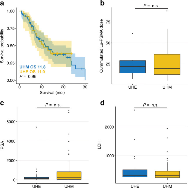
Abstract Introduction [ 177 Lu]Lu-PSMA-617 (Lu-PSMA) radioligand therapy is an emerging treatment option for patients with end-stage prostate cancer. However, response to Lu-PSMA therapy is only achieved in approximately half of patients. It is clinically important to identify patients at risk of poor outcome. Therefore, the aim of this study was to evaluate pretherapeutic PSMA PET derived total tumor volume and related metrics as prognosticators of overall survival in patients receiving Lu-PSMA therapy. Methods A total number of 110 patients form the Departments of Nuclear Medicine Münster and Essen were included in this retrospective analysis. Baseline PSMA PET-CT was available for all patients. Employing a previously published approach, all tumor lesions were semi-automatically delineated in PSMA PET-CT acquisitions. Total lesion number, total tumor volume (PSMA-TV), total lesion uptake (PSMA-TLU = PSMA-TV * SUV mean ), and total lesion quotient (PSMA-TLQ = PSMA-TV / SUV mean) were quantified for each patient. Log2 transformation was used for regressions. Results Lesion number, PSMA-TV, and PSMA-TLQ were prognosticators of overall survival (HR = 1.255, p = 0.009; HR = 1.299, p = 0.005; HR = 1.326, p = 0.002). In a stepwise backward Cox regression including lesion number, PSMA-TV, PSA, LDH, and PSMA-TLQ, only the latter two remained independent and statistically significant negative prognosticators of overall survival (HR = 1.632, p = 0.011; HR = 1.239, p = 0.024). PSMA-TLQ and LDH were significant negative prognosticators in multivariate Cox regression in contrast to PSA value. Conclusion PSMA-TV was a statistically significant negative prognosticator of overall survival in patients receiving Lu-PSMA therapy. PSMA-TLQ was an independent and superior prognosticator of overall survival compared with PSMA-TV.
Overview
The claims and statistics of this paper haven’t been checked yet.
Run the check — free, no account needed. It reads the open-access text and checks the sources it cites.
Integrity signals
Registry
Cites
Statistics
Figures
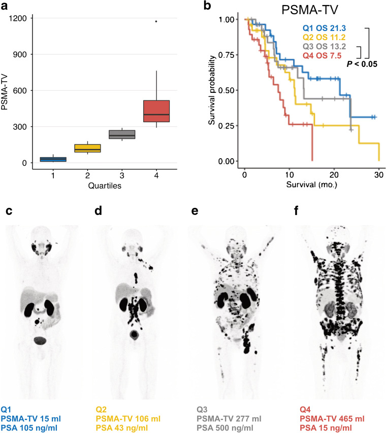
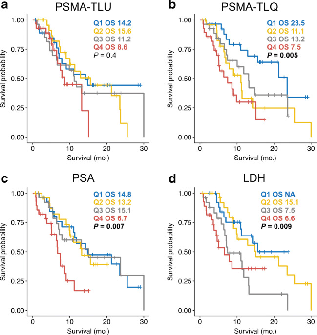
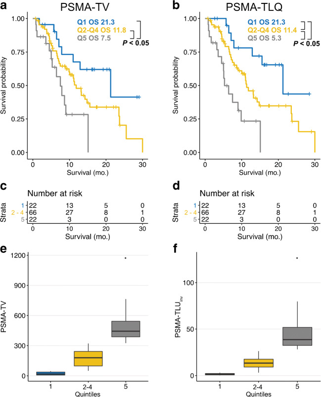
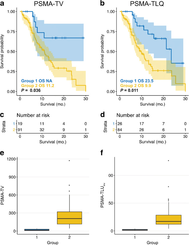
Verification
Claims the paper makes
The claims and statistics of this paper haven’t been checked yet.
Run the check — free, no account needed. It reads the open-access text and checks the sources it cites.
Claims other papers make about this one
Each is checked against this paper’s own text: does it say what the citing paper says it does?
The cited source says what the claim attributes to it.
In the citing paper
“Initial results also show a prognostic value of PSMA-PET tumor volume in prostate cancer prior to radioligand therapy with [177Lu]Lu-PSMA-617 [204].”
From Influences on PET Quantification and Interpretation (2022) · cites this paper as [204]
Self-check
Percentages, sums and reported differences re-computed from the paper’s own numbers.
The self-check needs the paper’s full text.
Citations
Cites
Chemohormonal Therapy in Metastatic Hormone-Sensitive Prostate Cancer
Sweeney, C. J., Chen, Y., Carducci, M. A., Liu, G., Jarrard, D. F., Eisenberger, M. A., Wong, Y., Hahn, N. M., Kohli, M., Cooney, M. M., et al. (2015) · New England Journal of Medicine · doi:10.1056/nejmoa1503747 · Cited by 2,958
Scher, H. I., Morris, M. J., Stadler, W. M., Higano, C. S., Basch, E., Fizazi, K. S., Antonarakis, E. S., Beer, T. M., Carducci, M. A., Chi, K. N., et al. (2016) · Journal of Clinical Oncology · doi:10.1200/jco.2015.64.2702 · Cited by 1,707
Hofman, M. S., Violet, J. A., Hicks, R. J., Ferdinandus, J., Thang, S. P., Akhurst, T. J., Iravani, A., Kong, G., Kumar, A. S. R., Murphy, D. G., et al. (2018) · The Lancet Oncology · doi:10.1016/s1470-2045(18)30198-0 · Cited by 1,184
Kyriakopoulos, C. E., Chen, Y., Carducci, M. A., Liu, G., Jarrard, D. F., Hahn, N. M., Shevrin, D. H., Dreicer, R., Hussain, M., Eisenberger, M. A., et al. (2018) · Journal of Clinical Oncology · doi:10.1200/jco.2017.75.3657 · Cited by 1,108
Rahbar, K., Ahmadzadehfar, H., Kratochwil, C., Haberkorn, U., Schäfers, M. A., Essler, M., Baum, R. P., Kulkarni, H., Schmidt, M. C., Drzezga, A. E., et al. (2016) · Journal of Nuclear Medicine · doi:10.2967/jnumed.116.183194 · Cited by 861
Self-citationAggarwal, R., Huang, J., Alumkal, J. J., Zhang, L., Feng, F. Y., Thomas, G. V., Weinstein, A. S., Friedl, V., Zhang, C., Witte, O. N., et al. (2018) · Journal of Clinical Oncology · doi:10.1200/jco.2017.77.6880 · Cited by 810
On the exact distribution of maximally selected rank statistics
Hothorn, T., & Lausen, B. (2003) · Computational Statistics & Data Analysis · doi:10.1016/s0167-9473(02)00225-6 · Cited by 694
Sartor, O., & de Bono, J. S. (2018) · New England Journal of Medicine · doi:10.1056/nejmra1701695 · Cited by 546
Violet, J., Jackson, P., Ferdinandus, J., Sandhu, S., Akhurst, T., Iravani, A., Kong, G., Kumar, A. R., Thang, S. P., Eu, P., et al. (2019) · Journal of Nuclear Medicine · doi:10.2967/jnumed.118.219352 · Cited by 504 · cited 2×
“In line with the concept of theranostics, high SUVmean is associated with higher tumor doses of Lu-PSMA therapy and should therefore be linked to a favorable outcome [25].”
BackgroundHalabi, S., Kelly, W. K., Ma, H., Zhou, H., Solomon, N. C., Fizazi, K., Tangen, C. M., Rosenthal, M., Petrylak, D. P., Hussain, M., et al. (2016) · Journal of Clinical Oncology · doi:10.1200/jco.2015.65.7270 · Cited by 461
Cited by
Sorted by citations
Seifert, R., Emmett, L., Rowe, S. P., Herrmann, K., Hadaschik, B. A., Calais, J., Giesel, F. L., Reiter, R. E., Maurer, T., Heck, M. M., et al. (2023) · European Urology · doi:10.1016/j.eururo.2023.02.002 · Cited by 333
Self-citationSayar, E., Patel, R. A., Coleman, I. M., Roudier, M. P., Zhang, A., Mustafi, P., Low, J., Hanratty, B. P., Ang, L. S., Bhatia, V., et al. (2023) · JCI Insight · doi:10.1172/jci.insight.162907 · Cited by 95
Johnsson, K., Brynolfsson, J., Sahlstedt, H., Nickols, N. G., Rettig, M. B., Probst, S., Morris, M. J., Bjartell, A. S., Eiber, M., & Anand, A. (2021) · European Journal of Nuclear Medicine and Molecular Imaging · doi:10.1007/s00259-021-05497-8 · Cited by 64
Rosar, F., Krause, J., Bartholomä, M. D., Maus, S., Stemler, T., Hierlmeier, I., Linxweiler, J., Ezziddin, S., & Khreish, F. (2021) · Pharmaceutics · doi:10.3390/pharmaceutics13050722 · Cited by 61
Pathmanandavel, S., Crumbaker, M., Yam, A. O. W., Nguyen, A., Rofe, C., Hovey, E. J., Gedye, C. A., Kwan, E. M., Hauser, C., Azad, A., et al. (2021) · Journal of Nuclear Medicine · doi:10.2967/jnumed.121.262552 · Cited by 60
Applications of Artificial Intelligence in PSMA PET/CT for Prostate Cancer Imaging
Belal, S. L., Frantz, S., Minarik, D., Enqvist, O., Wikström, E., Edenbrandt, L., & Trägårdh, E. (2023) · Seminars in Nuclear Medicine · doi:10.1053/j.semnuclmed.2023.06.001 · Cited by 63
Corpetti, M., Müller, C., Beltran, H., Bono, J. S. D., & Theurillat, J.-P. P. (2023) · European Urology · doi:10.1016/j.eururo.2023.11.018 · Cited by 55
Yazdani, E., Geramifar, P., Ziarati, N. K., Sadeghi, M., Amini, P., & Rahmim, A. (2024) · Diagnostics · doi:10.3390/diagnostics14020181 · Cited by 55
Influences on PET Quantification and Interpretation
Rogasch, J. M. M., Hofheinz, F., van Heek, L., Voltin, C.-A., Boellaard, R., & Kobe, C. (2022) · Diagnostics · doi:10.3390/diagnostics12020451 · Cited by 57
Total tumor volume reduction and low PSMA expression in patients receiving Lu-PSMA therapy
Seifert, R., Kessel, K., Schlack, K., Weckesser, M., Kersting, D., Seitzer, K. E., Weber, M., Bögemann, M., & Rahbar, K. (2021) · Theranostics · doi:10.7150/thno.60222 · Cited by 52
Self-citationA Prognostic Risk Score for Prostate Cancer Based on PSMA PET–derived Organ-specific Tumor Volumes
Seifert, R., Rasul, S., Seitzer, K. E., Eveslage, M., Nikoukar, L. R., Kessel, K., Schäfers, M. A., Yu, J., Haug, A. R., Hacker, M., et al. (2023) · Radiology · doi:10.1148/radiol.222010 · Cited by 48
Self-citationAssessing Response to 177Lu-PSMA Radioligand Therapy Using Modified PSMA PET Progression Criteria
Michalski, K., Klein, C., Brüggemann, T., Meyer, P. T., Jilg, C. A., & Ruf, J. (2021) · Journal of Nuclear Medicine · doi:10.2967/jnumed.120.260836 · Cited by 48
Capobianco, N., Sibille, L., Chantadisai, M., Gafita, A., Langbein, T., Platsch, G., Solari, E. L., Shah, V. P., Spottiswoode, B. S., Eiber, M., et al. (2021) · European Journal of Nuclear Medicine and Molecular Imaging · doi:10.1007/s00259-021-05473-2 · Cited by 49
Kendrick, J., Francis, R. J., Hassan, G. M., Rowshanfarzad, P., Ong, J. S. L., & Ebert, M. A. (2022) · European Journal of Nuclear Medicine and Molecular Imaging · doi:10.1007/s00259-022-05927-1 · Cited by 49
Clinical Use of PET/MR in Oncology: An Update
Seifert, R., Kersting, D., Rischpler, C., Opitz, M. K., Kirchner, J., Pabst, K. M., Mavroeidi, I. A., Laschinsky, C., Grueneisen, J. S., Schaarschmidt, B. M., et al. (2021) · Seminars in Nuclear Medicine · doi:10.1053/j.semnuclmed.2021.11.012 · Cited by 48
Self-citationSeifert, R., Telli, T., Hadaschik, B. A., Fendler, W. P., Kuo, P. H., & Herrmann, K. (2022) · Journal of Nuclear Medicine · doi:10.2967/jnumed.122.264741 · Cited by 49
Self-citationHartrampf, P. E., Hüttmann, T., Seitz, A. K., Kübler, H. R., Serfling, S. E., Schlötelburg, W., Michalski, K., Rowe, S. P., Pomper, M. G., Buck, A. K., et al. (2023) · European Journal of Nuclear Medicine and Molecular Imaging · doi:10.1007/s00259-023-06281-6 · Cited by 43
Predictors and Real-World Use of Prostate-Specific Radioligand Therapy: PSMA and Beyond
Gafita, A., Marcus, C. V., Kostos, L. K., Schuster, D. M., Calais, J., & Hofman, M. S. (2022) · American Society of Clinical Oncology Educational Book · doi:10.1200/edbk_350946 · Cited by 47
PSMA Theranostics: Science and Practice
Mokoala, K. M. G., Lawal, I. O., Lengana, T., Kgatle, M., Giesel, F. L., Vorster, M., & Sathekge, M. M. (2021) · Cancers · doi:10.3390/cancers13153904 · Cited by 44
von Eyben, F. E., Bauman, G. S., von Eyben, R., Rahbar, K., Soydal, Ç., Haug, A. R., Virgolini, I. J., Kulkarni, H., Baum, R. P., & Paganelli, G. (2020) · International Journal of Molecular Sciences · doi:10.3390/ijms21239054 · Cited by 40
Self-citationEmmett, L., Papa, N., Subramaniam, S., Crumbaker, M., Nguyen, A. D. K., Joshua, A. M., Sandhu, S., Weickhardt, A., Lee, S.-T., Ng, S., et al. (2025) · The Lancet Oncology · doi:10.1016/s1470-2045(25)00339-0 · Cited by 39
Ahmadzadehfar, H., Matern, R., Baum, R. P., Seifert, R., Kessel, K., Bögemann, M., Kratochwil, C., Rathke, H. G., Ilhan, H., Svirydenka, H., et al. (2021) · European Journal of Nuclear Medicine and Molecular Imaging · doi:10.1007/s00259-021-05383-3 · Cited by 38
Self-citationLuining, W. I., Cysouw, M. C. F., Meijer, D., Hendrikse, N. H., Boellaard, R., Vis, A. N., & Oprea‐Lager, D. E. (2022) · Cancers · doi:10.3390/cancers14051169 · Cited by 38
Widjaja, L., Werner, R. A., Roß, T. L., Bengel, F. M., & Derlin, T. (2021) · Cancers · doi:10.3390/cancers13122938 · Cited by 36
Roll, W., Schindler, P., Masthoff, M., Seifert, R., Schlack, K., Bögemann, M., Stegger, L., Weckesser, M., & Rahbar, K. (2021) · Cancers · doi:10.3390/cancers13153849 · Cited by 36
Self-citationSeifert, R., Alberts, I., Afshar‐Oromieh, A., & Rahbar, K. (2021) · PET Clinics · doi:10.1016/j.cpet.2021.03.004 · Cited by 37
Self-citationHerrmann, K., Walz, J. C., MacLennan, S., Briganti, A., Cornford, P. A., Czernin, J. G., Eiber, M. J., Fanti, S., Fendler, W. P., Fizazi, K. S., et al. (2025) · European Urology · doi:10.1016/j.eururo.2025.08.005 · Cited by 36
Self-citationTrägårdh, E., Enqvist, O., Ulén, J., Hvittfeldt, E., Garpered, S., Belal, S. L., Bjartell, A. S., & Edenbrandt, L. (2022) · European Journal of Nuclear Medicine and Molecular Imaging · doi:10.1007/s00259-022-05806-9 · Cited by 34
Prostate-Specific Membrane Antigen: Gateway to Management of Advanced Prostate Cancer
Unterrainer, L. M., Calais, J., & Bander, N. H. (2024) · Annual Review of Medicine · doi:10.1146/annurev-med-081522-031439 · Cited by 32
Mittlmeier, L. M., Brendel, M., Beyer, L., Albert, N. L., Todica, A. S., Zacherl, M. J., Wenter, V., Herlemann, A., Kretschmer, A., Ledderose, S., et al. (2021) · Frontiers in Oncology · doi:10.3389/fonc.2021.663631 · Cited by 31
Wang, G., Zang, J., Jiang, Y., Liu, Q., Sui, H., Wang, R., Fan, X., Zhang, J., Zhu, Z., & Chen, X. S. (2022) · Journal of Nuclear Medicine · doi:10.2967/jnumed.122.264857 · Cited by 33
PSMA-targeted therapy for non-prostate cancers
Wang, J. H., & Kiess, A. P. (2023) · Frontiers in Oncology · doi:10.3389/fonc.2023.1220586 · Cited by 33
Sun, Y., Chen, G., He, J., Huang, Z., Li, S., Yang, Y., Zhong, L.-Y., Ji, S.-F., Huang, Y., Chen, X., et al. (2020) · Bioengineered · doi:10.1080/21655979.2020.1867405 · Cited by 29
Heidegger, I. M., Kesch, C. V., Kretschmer, A., Tsaur, I., Ceci, F., Valério, M., Tilki, D., Marra, G., Preißer, F., Fankhauser, C. D., et al. (2022) · Therapeutic Advances in Medical Oncology · doi:10.1177/17588359221081922 · Cited by 32
Karpinski, M. J., Rahbar, K., Bögemann, M., Nikoukar, L. R., Schäfers, M. A., Hoberück, S., Miederer, M., Hölscher, T., Rasul, S., Miszczyk, M., et al. (2025) · European Urology · doi:10.1016/j.eururo.2025.04.017 · Cited by 29
Self-citationProstate Cancer Theranostics With 177Lu-PSMA
Ahmadzadehfar, H., Seifert, R., Afshar‐Oromieh, A., Kratochwil, C., & Rahbar, K. (2024) · Seminars in Nuclear Medicine · doi:10.1053/j.semnuclmed.2024.02.007 · Cited by 29
Self-citationSeifert, K. E., Seifert, R., Kessel, K., Roll, W., Schlack, K., Boegemann, M., & Rahbar, K. (2021) · Cancers · doi:10.3390/cancers13153715 · Cited by 25
Self-citationUnterrainer, L. M., Beyer, L., Zacherl, M. J., Gildehaus, F. J., Todica, A. S., Kunte, S. C., Holzgreve, A., Sheikh, G. T., Herlemann, A., Casuscelli, J., et al. (2022) · Biomedicines · doi:10.3390/biomedicines10050946 · Cited by 25
Wang, Q., & Zhang, J. (2022) · European Journal of Medicinal Chemistry · doi:10.1016/j.ejmech.2022.114690 · Cited by 25
Prognostic Value of PSMA PET/CT in Prostate Cancer
Lawal, I. O., Ndlovu, H., Kgatle, M., Mokoala, K. M. G., & Sathekge, M. M. (2023) · Seminars in Nuclear Medicine · doi:10.1053/j.semnuclmed.2023.07.003 · Cited by 26
Chen, R., Ng, Y. L., Yang, X., Zhu, Y., Li, L., Zhao, H., Zhou, Y., Huang, G., & Liu, J. (2023) · European Journal of Nuclear Medicine and Molecular Imaging · doi:10.1007/s00259-023-06456-1 · Cited by 25
Hong, S., Leonio, M. I., Ferri, V., Duan, H., Aparici, C. M., Davidzon, G., Franc, B. L., Moradi, F., Shah, J. C., Bergstrom, C. P., et al. (2024) · European Journal of Nuclear Medicine and Molecular Imaging · doi:10.1007/s00259-024-06718-6 · Cited by 26
Demirci, R. A., Ghodsi, A., Gulati, R., Behnia, F., Nelson, P. S., Cheng, H. H., Yezefski, T., Haffner, M. C., Hawley, J. E., Montgomery, B., et al. (2024) · Journal of Nuclear Medicine · doi:10.2967/jnumed.124.268167 · Cited by 25
New PSMA-Targeting Ligands: Transformation from Diagnosis (Ga-68) to Radionuclide Therapy (Lu-177)
Zha, Z., Choi, S., Li, L., Zhao, R., Plöessl, K., Yao, X., Alexoff, D., Zhu, L., & Kung, H. F. (2022) · Journal of Medicinal Chemistry · doi:10.1021/acs.jmedchem.2c00852 · Cited by 22
Giunta, E. F., Brighi, N., Gurioli, G., Matteucci, F., Paganelli, G., & Giorgi, U. D. (2024) · Cancer Treatment Reviews · doi:10.1016/j.ctrv.2024.102699 · Cited by 23
Collins, K., & Liang Cheng, C. (2022) · Human Pathology · doi:10.1016/j.humpath.2022.06.004 · Cited by 21
Lang, D., Ritzberger, L., Rambousek, V., Horner, A., Wass, R. E., Akbari, K., Kaiser, B., Kronbichler, J., Lamprecht, B., & Gabriel, M. (2021) · Cancers · doi:10.3390/cancers13236096 · Cited by 20
Şimşek, D. H., Kuyumcu, S., Karadogan, S., Oflas, M., Işık, E. G., Özkan, Z. G., Paksoy, N., Ekmekçioğlu, Ö., Ekenel, M., & Şanlı, Y. (2021) · Annals of Nuclear Medicine · doi:10.1007/s12149-021-01610-x · Cited by 20
Şimşek, D. H., Kuyumcu, S., Karadogan, S., Özkan, Z. G., Işık, E. G., Başaran, M., Şanlı, Ö., & Şanlı, Y. (2022) · Clinical Nuclear Medicine · doi:10.1097/rlu.0000000000004222 · Cited by 19
Peterson, J. R., Cole, J. A., Pfeiffer, J. R., Norris, G. H., Zhang, Y., Lopez-Ramos, D., Pandey, T., Biancalana, M., Esslinger, H. R., Antony, A. K., et al. (2023) · Breast Cancer Research · doi:10.1186/s13058-023-01654-z · Cited by 18
Akhavanallaf, A., Joshi, S., Mohan, A., Worden, F. P., Krauss, J. C., Zaidi, H., Frey, K. A., Suresh, K., Dewaraja, Y. K., & Wong, K. K. (2024) · Theranostics · doi:10.7150/thno.98053 · Cited by 18
PSMA‐Hornet: Fully‐automated, multi‐target segmentation of healthy organs in PSMA PET/CT images
Klyuzhin, I. S., Chaussé, G., Bloise, I., Harsini, S., Ferres, J. L., Uribe, C., & Rahmim, A. (2023) · Medical Physics · doi:10.1002/mp.16658 · Cited by 18
Lutetium Lu 177 vipivotide tetraxetan for metastatic castration-resistant prostate cancer
Shah, H., Ravi, P. K., Sonpavde, G., & Jacene, H. A. (2022) · Expert Review of Anticancer Therapy · doi:10.1080/14737140.2022.2139679 · Cited by 17
Standardized PSMA-PET Imaging of Advanced Prostate Cancer
Seifert, R., Gafita, A., Telli, T., Voter, A. F., Herrmann, K., Pomper, M. G., Hadaschik, B. A., Rowe, S. P., & Fendler, W. P. (2023) · Seminars in Nuclear Medicine · doi:10.1053/j.semnuclmed.2023.07.005 · Cited by 16
Self-citationBurgard, C., Hein, C., Blickle, A., Bartholomä, M. D., Maus, S., Petto, S., Schaefer-Schuler, A., Ezziddin, S., & Rosar, F. (2023) · European Journal of Nuclear Medicine and Molecular Imaging · doi:10.1007/s00259-023-06476-x · Cited by 17
Zang, S., Meng, Q., Li, X., Guo, T., Zhang, L., Zhao, Z., Yu, F., Zhang, P., Wu, W., Ni, Y., et al. (2025) · European Journal of Nuclear Medicine and Molecular Imaging · doi:10.1007/s00259-025-07628-x · Cited by 18
Ng, T. S. C., Gao, X., Salari, K., Zlatev, D. V., Heidari, P., & Kamran, S. C. (2021) · Frontiers in Oncology · doi:10.3389/fonc.2021.722277 · Cited by 14
Güzel, Y., Kömek, H., Can, C., Kaplan, İ., Akdeniz, N., Kepenek, F., & Gündoğan, C. (2023) · Annals of Nuclear Medicine · doi:10.1007/s12149-023-01854-9 · Cited by 14
Saffar, H. A., Chen, D. C., Delgado, C., Ingvar, J., Hofman, M. S., Lawrentschuk, N., Perera, M., Murphy, D. G., & Eapen, R. S. (2024) · Cancers · doi:10.3390/cancers16050939 · Cited by 15
Kluge, K., Einspieler, H., Haberl, D., Spielvogel, C. P., Amereller, D., Egger, G., Kramer, G., Grubmüller, B., Shariat, S. F., Hacker, M., et al. (2024) · European Journal of Nuclear Medicine and Molecular Imaging · doi:10.1007/s00259-024-06698-7 · Cited by 13
Caracciolo, M., Castello, A., Castellani, M., Bartolomei, M., & Lopci, E. (2024) · Biomedicines · doi:10.3390/biomedicines12102355 · Cited by 13
Hu, J., Seifert, R., Karkampouna, S., Gomes, C. V., Xue, S., Afshar‐Oromieh, A., Rominger, A., & Shi, K. (2024) · European Journal of Nuclear Medicine and Molecular Imaging · doi:10.1007/s00259-024-07010-3 · Cited by 14
Self-citationMurthy, V., Ludwig, V. B., Gafita, A., Hope, T. A., & Calais, J. (2025) · Journal of Nuclear Medicine · doi:10.2967/jnumed.124.268818 · Cited by 13
Repeatability of 68Ga-PSMA-HBED-CC PET/CT-derived total molecular tumor volume
Seifert, R., Sandach, P., Kersting, D., Fendler, W. P., Hadaschik, B. A., Herrmann, K., Sunderland, J. J., & Pollard, J. H. (2021) · Journal of Nuclear Medicine · doi:10.2967/jnumed.121.262528 · Cited by 13
Self-citationAndela, S. B., Amthauer, H., Furth, C., Rogasch, J. M. M., Beck, M., Mehrhof, F. B., Ghadjar, P., van den Hoff, J., Klatte, T., Tahbaz, R., et al. (2024) · Radiation Oncology · doi:10.1186/s13014-024-02483-w · Cited by 12
To Scan or Not to Scan: An Unnecessary Dilemma for PSMA Radioligand Therapy
Srinivas, S., & Iagaru, A. H. (2021) · Journal of Nuclear Medicine · doi:10.2967/jnumed.121.263035 · Cited by 12
Recent advances in the molecular targeted drugs for prostate cancer
Gao, P., Li, T., Zhang, K., & Luo, G. (2023) · International Urology and Nephrology · doi:10.1007/s11255-023-03487-3 · Cited by 13
Shagera, Q. A., Karfis, I., Sideris, S., Guiot, T., Woff, E., Chanzá, N. M., Roumeguère, T., Gil, T., Flamen, P., & Artigas, C. (2023) · Clinical Nuclear Medicine · doi:10.1097/rlu.0000000000004763 · Cited by 11
Chua, W. M., Lam, W. W.-C., Tong, A. K. T., Sultana, R., Kua, S. M. Y., Kanesvaran, R., Wong, A. S. C., Tay, K. J., Cheng, T. J. L., Ng, D. C. E., et al. (2023) · Asia-Pacific Journal of Clinical Oncology · doi:10.1111/ajco.13944 · Cited by 10
Is There a Role of Artificial Intelligence in Preclinical Imaging?
Küper, A. T., Blanc‐Durand, P., Gafita, A., Kersting, D., Fendler, W. P., Seibold, C., Moraitis, A., Lückerath, K., James, M. L., & Seifert, R. (2023) · Seminars in Nuclear Medicine · doi:10.1053/j.semnuclmed.2023.03.003 · Cited by 10
Self-citationTrägårdh, E., Ulén, J., Enqvist, O., Larsson, M., Valind, K., Minarik, D., & Edenbrandt, L. (2025) · EJNMMI Physics · doi:10.1186/s40658-025-00786-9 · Cited by 10
Telli, T. A., Özgüven, S., Alan, Ö., Filizoğlu, N., Öztürk, M. A., Busery, N. S., Işık, S., Arıkan, R., Demircan, N. C., Başoğlu, T., et al. (2022) · Annals of Nuclear Medicine · doi:10.1007/s12149-022-01785-x · Cited by 10
Utility of PET to Appropriately Select Patients for PSMA-Targeted Theranostics
Eshghi, A., Covington, M. F., Eshghi, N., & Kuo, P. H. (2022) · Clinical Nuclear Medicine · doi:10.1097/rlu.0000000000004196 · Cited by 10
Advances in PSMA Alpha Theragnostics
Vorster, M., & Sathekge, M. M. (2024) · Seminars in Nuclear Medicine · doi:10.1053/j.semnuclmed.2024.03.004 · Cited by 9
Treatment response assessment in mCRPC: is PSMA-PET/CT going to take the lead?
Franco, M. D., Mei, R., Garcia, C., & Fanti, S. (2024) · Therapeutic Advances in Medical Oncology · doi:10.1177/17588359241258367 · Cited by 9
Staging of prostate Cancer with ultra-fast PSMA-PET scans enhanced by AI
Kersting, D., Borys, K., Küper, A. T., Kim, M., Haubold, J., Goerttler, T., Umutlu, L., Costa, P. F., Kleesiek, J., Rischpler, C., et al. (2025) · European Journal of Nuclear Medicine and Molecular Imaging · doi:10.1007/s00259-024-07060-7 · Cited by 11
Self-citationSantoro-Fernandes, V., Schott, B., Weisman, A. J., Lokre, O., Cho, S. Y., Perlman, S. B., Perk, T., & Jeraj, R. (2025) · Journal of Nuclear Medicine · doi:10.2967/jnumed.124.267809 · Cited by 9
177Lu-PSMA-RLT of metastatic castration-resistant prostate cancer: limitations and improvements
Cao, J., Chen, Y., Hu, M. L., & Zhang, W. (2021) · Annals of Nuclear Medicine · doi:10.1007/s12149-021-01649-w · Cited by 9
Schneider, C., Täger, P., Hammes, J., Fischer, T. H., Drzezga, A. E., Pfister, D. J. K. P., Heidenreich, A., & Schmidt, M. C. (2021) · Nuklearmedizin - NuclearMedicine · doi:10.1055/a-1670-9500 · Cited by 9
PSMA-Targeted Radiopharmaceuticals for Prostate Cancer Diagnosis and Therapy
Oldan, J., Almaguel, F., Voter, A. F., Durán, A., Gafita, A., Pomper, M., Hope, T., & Rowe, S. P. (2024) · Cancer Journal · doi:10.1097/ppo.0000000000000718 · Cited by 5
Kemppainen, J., Kangasmäki, A., Malaspina, S., Pape, B., Jalomäki, J., Kairemo, K., Kononen, J., & Joensuu, T. K. (2022) · Cancers · doi:10.3390/cancers14246155 · Cited by 8
Siripongsatian, D., Jantarato, A., Promteangtrong, C., Kunawudhi, A., Kiatkittikul, P., Boonkawin, N., Yaset, S., Somboon, S., & Chotipanich, C. (2024) · Indian journal of radiology and imaging - new series/Indian journal of radiology and imaging/Indian Journal of Radiology & Imaging · doi:10.1055/s-0044-1779634 · Cited by 7
Alkahtani, T. O. (2025) · BMC Medical Imaging · doi:10.1186/s12880-025-01571-x · Cited by 7
Kluge, K., Lotz, V., Einspieler, H., Haberl, D., Spielvogel, C. P., Amereller, D., Kramer, G., Grubmüller, B., Shariat, S. F., Haug, A. R., et al. (2025) · Clinical Epigenetics · doi:10.1186/s13148-025-01811-5 · Cited by 7
Widjaja, L., Hornfeck, J., Siegmund, S. C., Gildehaus, F. J., Schmidt-Hegemann, N.-S., Wenter, V., Sheikh, G. T., Klimek, K., Casuscelli, J., Stief, C. G., et al. (2025) · European Journal of Nuclear Medicine and Molecular Imaging · doi:10.1007/s00259-025-07632-1 · Cited by 8
Molin, K., Barry, N., Gill, S., Hassan, G. M., Francis, R. J., Ong, J. S. L., Ebert, M. A., & Kendrick, J. (2025) · Physical and Engineering Sciences in Medicine · doi:10.1007/s13246-024-01516-8 · Cited by 6
Koh, N., Wei, M.-C., Lam, W., Tong, A., Kanesvaran, R., Wong, A., Chen, K., & Thang, S. (2025) · Asia-Pacific Journal of Clinical Oncology · doi:10.1111/ajco.14174 · Cited by 5
Zamanian, A., Rousseau, É., Buteau, F.-A., Arsenault, F., Beaulieu, A., April, G., Juneau, D., Plouznikoff, N., Turcotte, É. E., Allard, C., et al. (2025) · Cancer Imaging · doi:10.1186/s40644-025-00910-z · Cited by 6
Kiani, I., Siri, M. A., Babaei, M., Aliasghari, M. M., Mohammadzadeh, S., Mohammadi, S., & Salavati, A. (2026) · Journal of Nuclear Medicine · doi:10.2967/jnumed.125.270872 · Cited by 8
Theranostics in oncology: What radiologists want to know
Ferdinandus, J., Fendler, W. P., Morigi, J. J., & Fanti, S. (2021) · European Journal of Radiology · doi:10.1016/j.ejrad.2021.109875 · Cited by 6
Self-citationKarimzadeh, A., Hansen, K., Hasa, E., Haller, B., Heck, M. M., Tauber, R., D’Alessandria, C., Weber, W., Eiber, M., & Rauscher, I. (2025) · European Journal of Nuclear Medicine and Molecular Imaging · doi:10.1007/s00259-024-07003-2 · Cited by 5
Murthy, V., Kimura, K., Theus, L., Nguyen, A., Grogan, T., Lokre, O., Perk, T., Rettig, M. B., Drakaki, A., Shen, J., et al. (2025) · Journal of Nuclear Medicine · doi:10.2967/jnumed.125.270916 · Cited by 5
Ghodsi, A., Owens, L., Demirci, R. A., Gafita, A., Cheng, H. H., Hawley, J. E., Raychaudhuri, R., Khan, H. M., Sunkara, R., Hsieh, A. C., et al. (2026) · Radiology · doi:10.1148/radiol.250649 · Cited by 5
Chandekar, K. R., Satapathy, S., Singh, H., Kumar, R., Kumar, S., Kakkar, N., Mittal, B. R., & Singh, S. K. (2024) · Nuclear Medicine Communications · doi:10.1097/mnm.0000000000001874 · Cited by 5
Roll, W., Plagwitz, L., Ventura, D., Masthoff, M., Backhaus, C., Varghese, J., Rahbar, K., & Schindler, P. (2025) · European Journal of Nuclear Medicine and Molecular Imaging · doi:10.1007/s00259-025-07416-7 · Cited by 5
Self-citationFuoco, V., Argiroffi, G., Mazzaglia, S., Lorenzoni, A., Guadalupi, V., Franza, A., Scalorbi, F., Aliberti, G., Chiesa, C., Procopio, G., et al. (2021) · Tumori Journal · doi:10.1177/03008916211037732 · Cited by 5
Yechiel, Y., Orr, Y., Gurevich, K., Gill, R., & Keidar, Z. (2023) · Cancers · doi:10.3390/cancers15041020 · Cited by 6
Duran, A. O., İnanç, M., Bozkurt, O., Özaslan, E., & Özkan, M. (2023) · Clinical Lung Cancer · doi:10.1016/j.cllc.2023.02.009 · Cited by 5
Siebinga, H., Hendrikx, J. J. M. A., Huitema, A. D. R., & der Veen, B. J. D. W. (2023) · EJNMMI Research · doi:10.1186/s13550-023-01008-y · Cited by 4
Diffuse Bone Marrow Involvement of Multiple Myeloma on [18F]PSMA-1007 PET/CT
Michalski, K., Jilg, C. A., Engelhardt, M. M., Meyer, P. T., & Ruf, J. (2022) · Clinical Nuclear Medicine · doi:10.1097/rlu.0000000000004286 · Cited by 4
Metadata
- Authors
- Seifert, R., Kessel, K., Schlack, K., Weber, M., Herrmann, K., Spanke, M., Fendler, W. P., Hadaschik, B., Kleesiek, J., Schäfers, M., Weckesser, M., Boegemann, M., & Rahbar, K.
- Affiliations
- University Hospital Münster (DE)
- Essen University Hospital (DE)
- German Cancer Research Center (DE)
- Corresponding author
- Rahbar, Kambiz
- ORCID
- Venue
- European Journal of Nuclear Medicine and Molecular Imaging
- Published
- 2020-09-24
- Volume
- 48
- Issue
- 4
- Pages
- 1200-1210
- ISSN
- 1619-7070 (ISSN-L)
- 1619-7089
- PMC
- PMC8041668
- OpenAlex
- W3088497176
- Cited by
- 160
- References
- 26
- Citations by year
- 2026: 31 · 2025: 31 · 2024: 25 · 2023: 26 · 2022: 20 · 2021: 24 · 2020: 2
- FWCI
- 8.77
- Type
- journal article
- Language
- en
- Open access
- hybrid
- doi.org
- Locations
- European Journal of Nuclear Medicine and Molecular Imaging (publishedVersion, cc-by)
- PubMed Central (publishedVersion, cc-by)
- PubMed (publishedVersion)
- Informationszentrum Lebenswissenschaften (ZB MED) (submittedVersion, cc-by)
- Universitätsbibliographie, Universität Duisburg-Essen (submittedVersion)
- Funders
- Westfälische Wilhelms-Universität Münster
- Topics
- Health Sciences › Medicine › Radiology, Nuclear Medicine and Imaging › Radiopharmaceutical Chemistry and Applications
- Health Sciences › Medicine › Radiology, Nuclear Medicine and Imaging › Medical Imaging Techniques and Applications
- Health Sciences › Medicine › Urology › Prostate Cancer Diagnosis and Treatment
- Keywords
- 177Lu-PSMA-617, radioligand therapy, advanced prostate cancer, PSMA PET/CT, total tumor volume, prognostic factors, overall survival, PSMA-617, PSMA-11, tumor volume measurement
- MeSH terms
- Positron Emission Tomography Computed Tomography, Dipeptides, Heterocyclic Compounds, 1-Ring, Humans, Male, Retrospective Studies, Treatment Outcome, Prostate-Specific Antigen, Tumor Burden, Prostatic Neoplasms, Castration-Resistant
About these checks
Registry, retraction, Cites and statistics results are computed, not judged, with one exception: a printed reference that no index lists is read once by a model before it counts as a citation error. Claims are extracted by a model and kept only when their quote appears verbatim in the text; each cited source is then checked, with a model’s help, for whether it says what the claim relies on. Those verdicts can be wrong — read the claim’s quote and the cited source before relying on one.
Statistics and arithmetic are recomputed, not judged.
Confidence bands are shown only for fields calibrated on our annotated test set. None is calibrated yet, so no band is shown.
What counts as a claim ›How we measure confidence ›How we check this ›
Are you an author, or do you see a problem with this page? Request a review
