

The DOI matches its Crossref record; 3 of the 3 references it cites were found in a registry or an index.
Pre-clinical safety and efficacy evaluation of Helicobacter Pylori neutrophil-activating protein (NAP)-armed CAR-T cells targeting B cell lymphomas
Open accessPublished version of a preprint
Abstract
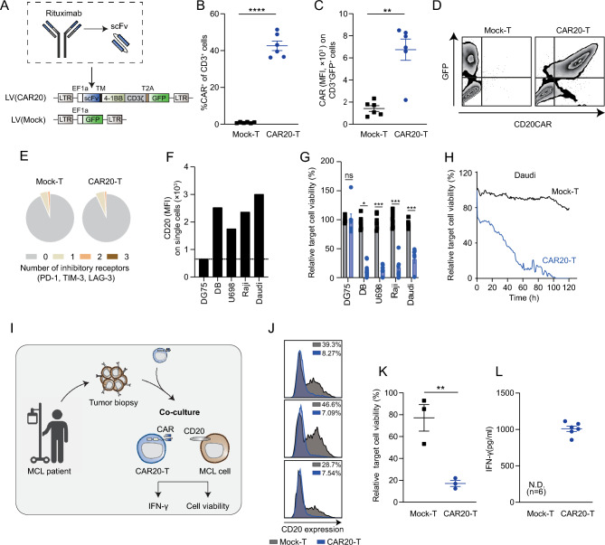
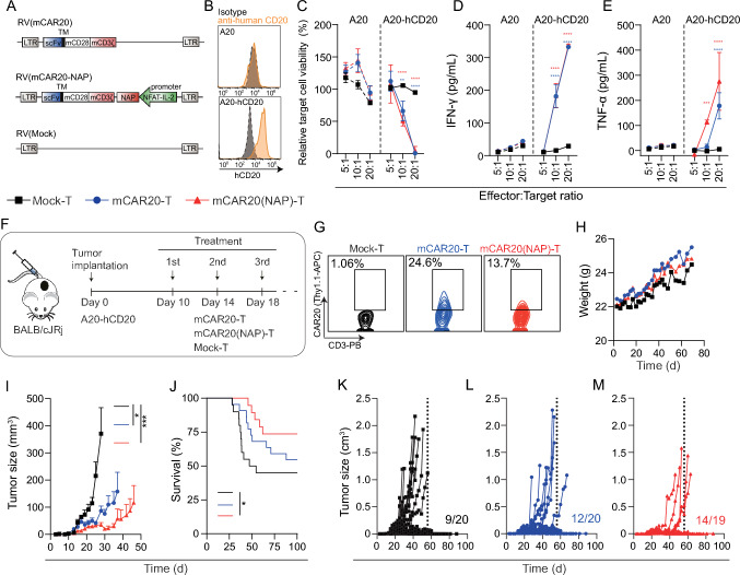
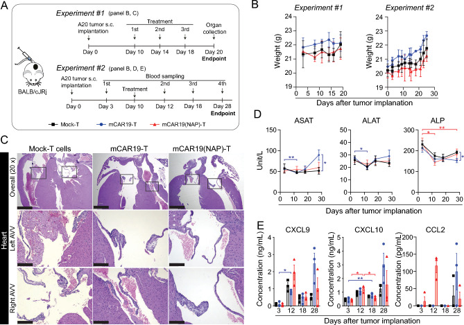
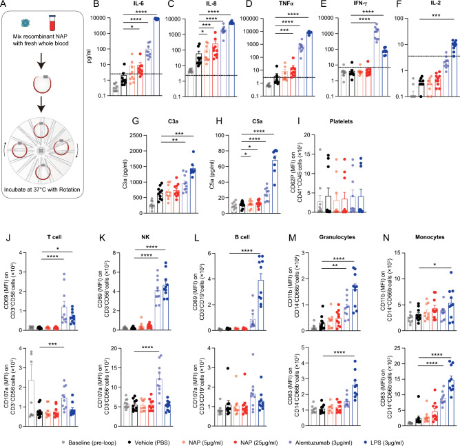
CD19 CAR-T cell therapy shows striking results in treating B cell malignancies. However, approximately two-thirds of the lymphoma patients eventually relapse, with about one-third displaying CD19-negative tumors at relapse. Our previous study showed that CAR-T cells armed with the Helicobacter pylori neutrophil-activating protein (NAP), CAR(NAP)-T cells, can trigger a bystander immune response and eliminate CAR-target-antigen-negative tumor cells. Here, we report the development of CD20-targeted CAR-T cells (CAR20-T cells), with the targeting moiety from rituximab, and the safety and efficacy of NAP-armed CAR-T cells. CAR20-T cells displayed efficient and specific cytotoxic potential against multiple human B cell lymphoma cell lines in vitro. In addition, primary mantle cell lymphoma cells, isolated from a patient who relapsed after rituximab treatment, can also be eliminated by CAR20-T cells. CAR20(NAP)-T cells delayed tumor growth and prolonged survival of mice with lymphoma. No obvious histopathological alteration in major organs were observed in mice treated with CAR(NAP)-T cells. Further, no excessive cytokine release or immune cell activation was observed when human blood from healthy volunteers was exposed to recombinant NAP protein in an ex vivo blood loop assay, suggesting a safe therapeutic profile for NAP. Taken together, these results warrant the clinical investigation of CAR20(NAP)-T cells.
Overview
The claims and statistics of this paper haven’t been checked yet.
Run the check — free, no account needed. It reads the open-access text and checks the sources it cites.
Integrity signals
Registry
Cites
Statistics
Figures
Verification
Claims the paper makes
The claims and statistics of this paper haven’t been checked yet.
Run the check — free, no account needed. It reads the open-access text and checks the sources it cites.
Self-check
Percentages, sums and reported differences re-computed from the paper’s own numbers.
The self-check needs the paper’s full text.
Citations
Cites
Cohen, J. A., Coles, A. J., Arnold, D. L., Confavreux, C., Fox, E. J., Hartung, H.-P., Havrdova, E., Selmaj, K. W., Weiner, H. L., Fisher, E., et al. (2012) · The Lancet · doi:10.1016/s0140-6736(12)61769-3 · Cited by 1,205
Milone, M. C., Fish, J. D., Carpenito, C., Carroll, R. G., Binder, G. K., Teachey, D., Samanta, M., Lakhal, M., Gloss, B., Danet-Desnoyers, G., et al. (2009) · Molecular Therapy · doi:10.1038/mt.2009.83 · Cited by 1,197
Milo, R. (2013) · BioEssays · doi:10.1002/bies.201300066 · Cited by 614
Research works printed but not linked: 52
- Maude SL, Laetsch TW, Buechner J et al (2018) Tisagenlecleucel in children and young adults with B-cell lymphoblastic leukemia. N Engl J Med 378:439–448. https://doi.org/10.1056/NEJMoa1709866 · DOI · Paper page
- Shah BD, Ghobadi A, Oluwole OO et al (2021) KTE-X19 for relapsed or refractory adult B-cell acute lymphoblastic leukaemia: phase 2 results of the single-arm, open-label, multicentre ZUMA-3 study. Lancet 398:491–502. https://doi.org/10.1016/S0140-6736(21)01222-8 · DOI
- Locke FL, Miklos DB, Jacobson CA et al (2022) Axicabtagene ciloleucel as second-line therapy for large B-cell lymphoma. N Engl J Med 386:640–654. https://doi.org/10.1056/NEJMoa2116133 · DOI · Paper page
- Neelapu SS, Dickinson M, Munoz J et al (2022) Axicabtagene ciloleucel as first-line therapy in high-risk large B-cell lymphoma: the phase 2 ZUMA-12 trial. Nat Med 28:735–742. https://doi.org/10.1038/s41591-022-01731-4 · DOI
- Abramson JS, Palomba ML, Gordon LI et al (2020) Lisocabtagene maraleucel for patients with relapsed or refractory large B-cell lymphomas (TRANSCEND NHL 001): a multicentre seamless design study. Lancet 396:839–852. https://doi.org/10.1016/S0140-6736(20)31366-0 · DOI · Paper page
- Wang M, Munoz J, Goy A et al (2020) KTE-X19 CAR t-cell therapy in relapsed or refractory mantle-cell lymphoma. N Engl J Med 382:1331–1342. https://doi.org/10.1056/NEJMoa1914347 · DOI · Paper page
- Neelapu SS, Locke FL, Bartlett NL et al (2017) Axicabtagene ciloleucel CAR T-cell therapy in refractory large B-cell lymphoma. N Engl J Med 377:2531–2544. https://doi.org/10.1056/NEJMoa1707447 · DOI · Paper page
- Ghilardi G, Lee YG, Porazzi P et al (2022) Overcoming CD19-negative relapses in patients with B-cell lymphomas treated with tisagenlecleucel. Blood 140:7371–7373. https://doi.org/10.1182/blood-2022-167601 · DOI
- Shalabi H, Kraft IL, Wang HW et al (2018) Sequential loss of tumor surface antigens following chimeric antigen receptor T-cell therapies in diffuse large B-cell lymphoma. Haematologica 103:e215–e218. https://doi.org/10.3324/haematol.2017.183459 · DOI
- Plaks V, Rossi JM, Chou J et al (2021) CD19 target evasion as a mechanism of relapse in large B-cell lymphoma treated with axicabtagene ciloleucel. Blood 138:1081–1085. https://doi.org/10.1182/blood.2021010930 · DOI · Paper page
- Saleh K, Khalife N, Arbab A et al (2024) Updates on chimeric antigen receptor T-cells in large B-cell lymphoma. Biomedicines. https://doi.org/10.3390/biomedicines12122810 · DOI
- Till BG, Jensen MC, Wang J et al (2008) Adoptive immunotherapy for indolent non-Hodgkin lymphoma and mantle cell lymphoma using genetically modified autologous CD20-specific T cells. Blood 112:2261–2271. https://doi.org/10.1182/blood-2007-12-128843 · DOI
- Till BG, Jensen MC, Wang J et al (2012) CD20-specific adoptive immunotherapy for lymphoma using a chimeric antigen receptor with both CD28 and 4–1BB domains: pilot clinical trial results. Blood 119:3940–3950. https://doi.org/10.1182/blood-2011-10-387969 · DOI
- McLaughlin P, Grillo-Lopez AJ, Link BK et al (1998) Rituximab chimeric anti-CD20 monoclonal antibody therapy for relapsed indolent lymphoma: half of patients respond to a four-dose treatment program. J Clin Oncol 16:2825–2833. https://doi.org/10.1200/JCO.1998.16.8.2825 · DOI
- Davis TA, White CA, Grillo-Lopez AJ et al (1999) Single-agent monoclonal antibody efficacy in bulky non-Hodgkin’s lymphoma: results of a phase II trial of rituximab. J Clin Oncol 17:1851–1857. https://doi.org/10.1200/JCO.1999.17.6.1851 · DOI
- Kluin-Nelemans HC, Hoster E, Hermine O et al (2012) Treatment of older patients with mantle-cell lymphoma. N Engl J Med 367:520–531. https://doi.org/10.1056/NEJMoa1200920 · DOI
- Habermann TM, Weller EA, Morrison VA et al (2006) Rituximab-CHOP versus CHOP alone or with maintenance rituximab in older patients with diffuse large B-cell lymphoma. J Clin Oncol 24:3121–3127. https://doi.org/10.1200/JCO.2005.05.1003 · DOI
- Coiffier B, Lepage E, Briere J et al (2002) CHOP chemotherapy plus rituximab compared with CHOP alone in elderly patients with diffuse large-B-cell lymphoma. N Engl J Med 346:235–242. https://doi.org/10.1056/NEJMoa011795 · DOI · Paper page
- Hiddemann W, Kneba M, Dreyling M et al (2005) Frontline therapy with rituximab added to the combination of cyclophosphamide, doxorubicin, vincristine, and prednisone (CHOP) significantly improves the outcome for patients with advanced-stage follicular lymphoma compared with therapy with CHOP alone: results of a prospective randomized study of the German Low-Grade Lymphoma Study Group. Blood 106:3725–3732. https://doi.org/10.1182/blood-2005-01-0016 · DOI
- Johnson NA, Leach S, Woolcock B et al (2009) CD20 mutations involving the rituximab epitope are rare in diffuse large B-cell lymphomas and are not a significant cause of R-CHOP failure. Haematologica 94:423–427. https://doi.org/10.3324/haematol.2008.001024 · DOI
- Chen X, Chen LC, Khericha M et al (2023) Rational protein design yields a CD20 CAR with superior antitumor efficacy compared with CD19 CAR. Cancer Immunol Res 11:150–163. https://doi.org/10.1158/2326-6066.CIR-22-0504 · DOI
- Amedei A, Cappon A, Codolo G et al (2006) The neutrophil-activating protein of Helicobacter pylori promotes Th1 immune responses. J Clin Invest 116:1092–1101. https://doi.org/10.1172/JCI27177 · DOI
- Codolo G, Fassan M, Munari F, Volpe A, Bassi P, Rugge M, Pagano F, D’Elios MM, de Bernard M (2012) HP-NAP inhibits the growth of bladder cancer in mice by activating a cytotoxic Th1 response. Cancer Immunol Immunother 61:31–40. https://doi.org/10.1007/s00262-011-1087-2 · DOI
- Ramachandran M, Yu D, Wanders A, Essand M, Eriksson F (2013) An infection-enhanced oncolytic adenovirus secreting H. pylori neutrophil-activating protein with therapeutic effects on neuroendocrine tumors. Mol Ther 21:2008–2018. https://doi.org/10.1038/mt.2013.153 · DOI
- Satin B, Del Giudice G, Della Bianca V et al (2000) The neutrophil-activating protein (HP-NAP) of Helicobacter pylori is a protective antigen and a major virulence factor. J Exp Med 191:1467–1476. https://doi.org/10.1084/jem.191.9.1467 · DOI
- Ramachandran M, Jin C, Yu D, Eriksson F, Essand M (2014) Vector-encoded Helicobacter pylori neutrophil-activating protein promotes maturation of dendritic cells with Th1 polarization and improved migration. J Immunol 193:2287–2296. https://doi.org/10.4049/jimmunol.1400339 · DOI
- Jin C, Ma J, Ramachandran M, Yu D, Essand M (2022) CAR T cells expressing a bacterial virulence factor trigger potent bystander antitumour responses in solid cancers. Nat Biomed Eng 6:830–841. https://doi.org/10.1038/s41551-022-00875-5 · DOI
- Long AH, Haso WM, Shern JF et al (2015) 4–1BB costimulation ameliorates T cell exhaustion induced by tonic signaling of chimeric antigen receptors. Nat Med 21:581–590. https://doi.org/10.1038/nm.3838 · DOI · Paper page
- Saren T, Saronio G, Marti Torrell P et al (2023) Complementarity-determining region clustering may cause CAR-T cell dysfunction. Nat Commun 14:4732. https://doi.org/10.1038/s41467-023-40303-z · DOI
- Fletcher EAK, Eltahir M, Lindqvist F, Rieth J, Tornqvist G, Leja-Jarblad J, Mangsbo SM (2018) Extracorporeal human whole blood in motion, as a tool to predict first-infusion reactions and mechanism-of-action of immunotherapeutics. Int Immunopharmacol 54:1–11. https://doi.org/10.1016/j.intimp.2017.10.021 · DOI
- Thomas K, Eisele J, Rodriguez-Leal FA, Hainke U, Ziemssen T (2016) Acute effects of alemtuzumab infusion in patients with active relapsing-remitting MS. Neurol Neuroimmunol Neuroinflamm 3:e228. https://doi.org/10.1212/NXI.0000000000000228 · DOI
- Caon C, Namey M, Meyer C, Mayer L, Oyuela P, Margolin DH, Rizzo M (2015) Prevention and management of infusion-associated reactions in the comparison of Alemtuzumab and Rebif((R)) efficacy in multiple sclerosis (CARE-MS) program. Int J MS Care 17:191–198. https://doi.org/10.7224/1537-2073.2014-030 · DOI
- Tucureanu MM, Rebleanu D, Constantinescu CA, Deleanu M, Voicu G, Butoi E, Calin M, Manduteanu I (2018) Lipopolysaccharide-induced inflammation in monocytes/macrophages is blocked by liposomal delivery of G(i)-protein inhibitor. Int J Nanomed 13:63–76. https://doi.org/10.2147/IJN.S150918 · DOI
- Schuster SJ, Bishop MR, Tam CS et al (2019) Tisagenlecleucel in adult relapsed or refractory diffuse large B-cell lymphoma. N Engl J Med 380:45–56. https://doi.org/10.1056/NEJMoa1804980 · DOI · Paper page
- Wang Y, Zhang WY, Han QW et al (2014) Effective response and delayed toxicities of refractory advanced diffuse large B-cell lymphoma treated by CD20-directed chimeric antigen receptor-modified T cells. Clin Immunol 155:160–175. https://doi.org/10.1016/j.clim.2014.10.002 · DOI
- Zhang WY, Wang Y, Guo YL et al (2016) Treatment of CD20-directed chimeric antigen receptor-modified T cells in patients with relapsed or refractory B-cell non-Hodgkin lymphoma: an early phase IIa trial report. Signal Transduct Target Ther 1:16002. https://doi.org/10.1038/sigtrans.2016.2 · DOI
- Li P, Liu W, Zhou L et al (2024) C-CAR066, a novel fully human anti-CD20 CAR-T therapy for relapsed or refractory large B-cell lymphoma after failure of anti-CD19 CAR-T therapy: a phase I clinical study. Am J Hematol 99:2306–2312. https://doi.org/10.1002/ajh.27488 · DOI
- Zah E, Lin MY, Silva-Benedict A, Jensen MC, Chen YY (2016) T cells expressing CD19/CD20 bispecific chimeric antigen receptors prevent antigen escape by malignant B cells. Cancer Immunol Res 4:498–508. https://doi.org/10.1158/2326-6066.CIR-15-0231 · DOI
- Fousek K, Watanabe J, Joseph SK et al (2021) CAR T-cells that target acute B-lineage leukemia irrespective of CD19 expression. Leukemia 35:75–89. https://doi.org/10.1038/s41375-020-0792-2 · DOI
- Zhang Y, Wang Y, Liu Y et al (2022) Long-term activity of tandem CD19/CD20 CAR therapy in refractory/relapsed B-cell lymphoma: a single-arm, phase 1–2 trial. Leukemia 36:189–196. https://doi.org/10.1038/s41375-021-01345-8 · DOI
- Tong C, Zhang Y, Liu Y et al (2020) Optimized tandem CD19/CD20 CAR-engineered T cells in refractory/relapsed B-cell lymphoma. Blood 136:1632–1644. https://doi.org/10.1182/blood.2020005278 · DOI
- Kochenderfer JN, Dudley ME, Carpenter RO et al (2013) Donor-derived CD19-targeted T cells cause regression of malignancy persisting after allogeneic hematopoietic stem cell transplantation. Blood 122:4129–4139. https://doi.org/10.1182/blood-2013-08-519413 · DOI
- Eltahir M, Fletcher E, Dynesius L et al (2021) Profiling of donor-specific immune effector signatures in response to rituximab in a human whole blood loop assay using blood from CLL patients. Int Immunopharmacol 90:107226. https://doi.org/10.1016/j.intimp.2020.107226 · DOI
- Lee DW, Santomasso BD, Locke FL et al (2019) ASTCT consensus grading for cytokine release syndrome and neurologic toxicity associated with immune effector cells. Biol Blood Marrow Transplant 25:625–638. https://doi.org/10.1016/j.bbmt.2018.12.758 · DOI · Paper page
- Ben-Bassat H, Goldblum N, Mitrani S et al (1977) Establishment in continuous culture of a new type of lymphocyte from a ]=“Burkitt like]=“ malignant lymphoma (line D.G.-75). Int J Cancer 19:27–33. https://doi.org/10.1002/ijc.2910190105 · DOI
- Nilsson K, Sundstrom C (1974) Establishment and characteristics of two unique cell lines from patients with lymphosarcoma. Int J Cancer 13:808–823. https://doi.org/10.1002/ijc.2910130609 · DOI
- Jin C, Fotaki G, Ramachandran M, Nilsson B, Essand M, Yu D (2016) Safe engineering of CAR T cells for adoptive cell therapy of cancer using long-term episomal gene transfer. EMBO Mol Med 8:702–711. https://doi.org/10.15252/emmm.201505869 · DOI
- Hillerdal V, Nilsson B, Carlsson B, Eriksson F, Essand M (2012) T cells engineered with a T cell receptor against the prostate antigen TARP specifically kill HLA-A2+ prostate and breast cancer cells. Proc Natl Acad Sci U S A 109:15877–15881. https://doi.org/10.1073/pnas.1209042109 · DOI
- Kochenderfer JN, Yu Z, Frasheri D, Restifo NP, Rosenberg SA (2010) Adoptive transfer of syngeneic T cells transduced with a chimeric antigen receptor that recognizes murine CD19 can eradicate lymphoma and normal B cells. Blood 116:3875–3886. https://doi.org/10.1182/blood-2010-01-265041 · DOI
- Refaeli Y, Van Parijs L, Alexander SI, Abbas AK (2002) Interferon gamma is required for activation-induced death of T lymphocytes. J Exp Med 196:999–1005. https://doi.org/10.1084/jem.20020666 · DOI
- Jin C, Yu D, Hillerdal V, Wallgren A, Karlsson-Parra A, Essand M (2014) Allogeneic lymphocyte-licensed DCs expand T cells with improved antitumor activity and resistance to oxidative stress and immunosuppressive factors. Mol Ther Methods Clin Dev 1:14001. https://doi.org/10.1038/mtm.2014.1 · DOI
- Kuse R, Schuster S, Schubbe H, Dix S, Hausmann K (1985) Blood lymphocyte volumes and diameters in patients with chronic lymphocytic leukemia and normal controls. Blut 50:243–248. https://doi.org/10.1007/BF00320301 · DOI
Metadata
- Authors
- Ma, J., Sarén, T., Jin, C., Kim, H. S., Contreras Pineda, P. D., de Bernard, M., Amini, R.-M., Rondahl, V., Enblad, G., Yu, D., & Essand, M.
- Affiliations
- Uppsala University (SE)
- University of Padua (IT)
- Swedish University of Agricultural Sciences (SE)
- Corresponding author
- Yu, Di
- ORCID
- Venue
- Cancer Immunology, Immunotherapy
- Published
- 2025-07-12
- Volume
- 74
- Issue
- 8
- Pages
- 262-262
- ISSN
- 0340-7004 (ISSN-L)
- 1432-0851
- PMC
- PMC12255594
- OpenAlex
- W4412362323
- Cited by
- 1
- References
- 3
- Citations by year
- 2026: 1
- FWCI
- 0.41
- Type
- journal article
- Language
- en
- Open access
- gold
- doi.org
- Locations
- Cancer Immunology Immunotherapy (publishedVersion, cc-by)
- Research Square (acceptedVersion, cc-by)
- Europe PMC (PubMed Central) (publishedVersion, cc-by)
- PubMed Central (publishedVersion, cc-by)
- PubMed (publishedVersion)
- Epsilon Open Archive (Sveriges lantbruksuniversitet biblioteket (Swedish University of Agricultural Sciences)) (acceptedVersion, cc-by)
- Uppsala University Publications (Uppsala University) (submittedVersion, cc-by)
- DOAJ (DOAJ: Directory of Open Access Journals) (submittedVersion, cc-by-sa)
- Padua Research Archive (University of Padova) (submittedVersion, cc-by)
- Funders
- Erik, Karin och Gösta Selanders stiftelse (2023)
- Cancerfonden (22-2229Pj, 19-0184Pj)
- Barncancerfonden (PR2022-0105, PR2020-0167)
- VINNOVA (2021-04488)
- Vetenskapsrådet (2019-01326)
- Uppsala University
- Topics
- Health Sciences › Medicine › Oncology › CAR-T cell therapy research
- Life Sciences › Immunology and Microbiology › Immunology › Immunotherapy and Immune Responses
- Life Sciences › Immunology and Microbiology › Immunology › T-cell and B-cell Immunology
- Keywords
- chimeric antigen receptor T cells, B-cell lymphoma, mantle cell lymphoma, relapsed lymphoma, rituximab, cytokine release syndrome, tumor growth inhibition
- MeSH terms
- Rituximab, Receptors, Chimeric Antigen, Animals, Bacterial Proteins, Female, Humans, T-Lymphocytes, Immunotherapy, Adoptive, Lymphoma, B-Cell, Helicobacter pylori, Xenograft Model Antitumor Assays, Cell Line, Tumor, Mice
About these checks
Registry, retraction, Cites and statistics results are computed, not judged, with one exception: a printed reference that no index lists is read once by a model before it counts as a citation error. Claims are extracted by a model and kept only when their quote appears verbatim in the text; each cited source is then checked, with a model’s help, for whether it says what the claim relies on. Those verdicts can be wrong — read the claim’s quote and the cited source before relying on one.
Statistics and arithmetic are recomputed, not judged.
Confidence bands are shown only for fields calibrated on our annotated test set. None is calibrated yet, so no band is shown.
What counts as a claim ›How we measure confidence ›How we check this ›
Are you an author, or do you see a problem with this page? Request a review
