

The DOI matches its Crossref record.
Endoscopic gastric atrophy is strongly associated with gastric cancer development after Helicobacter pylori eradication
Open accessAbstract
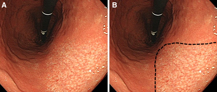
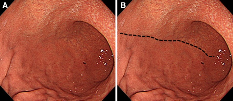
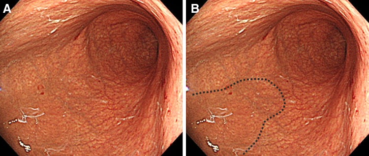
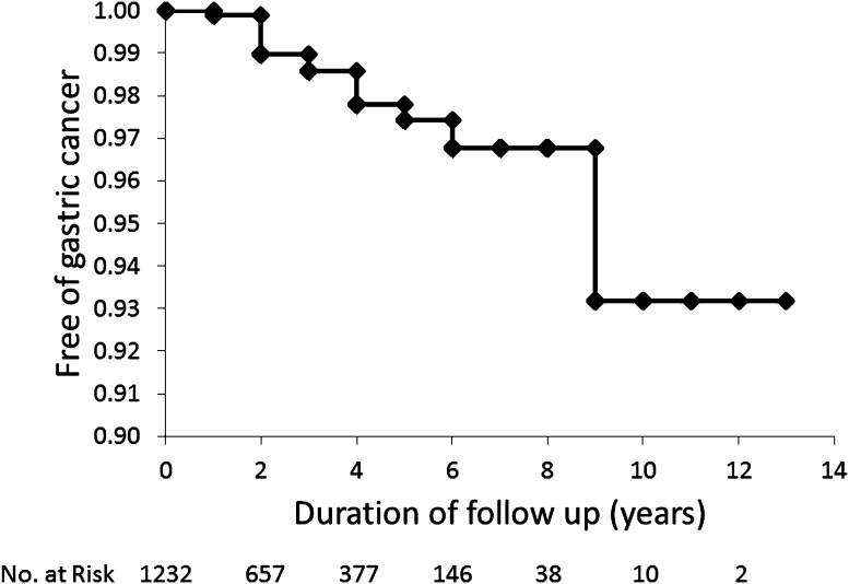
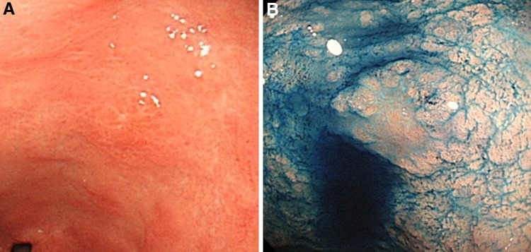
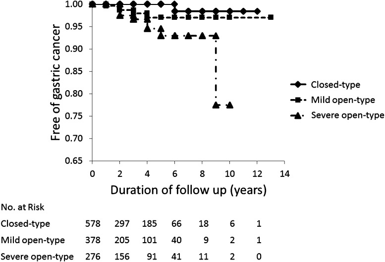
BACKGROUND: Risk factors for gastric cancer during continuous infection with Helicobacter pylori have been well documented; however, little has been reported on the risk factors for primary gastric cancer after H. pylori eradication. We conducted a retrospective, endoscopy-based, long-term, large-cohort study to clarify the risk factors for gastric cancer following H. pylori eradication. METHODS: Patients who achieved successful H. pylori eradication and periodically underwent esophagogastroduodenoscopy surveillance thereafter at Toyoshima Endoscopy Clinic were enrolled. The primary endpoint was the development of gastric cancer. Statistical analysis was performed using the Kaplan-Meier method and Cox's proportional hazards models. RESULTS: Gastric cancer developed in 15 of 1232 patients. The cumulative incidence rates were 1.0 % at 2 years, 2.6 % at 5 years, and 6.8 % at 10 years. Histology showed that all gastric cancers (17 lesions) in the 15 patients were of the intestinal type, within the mucosal layer, and <20 mm in diameter. Based on univariate analysis, older age and higher endoscopic grade of gastric atrophy were significantly associated with gastric cancer development after eradication of H. pylori, and gastric ulcers were marginally associated. Multivariate analysis identified higher grade of gastric atrophy (hazard ratio 1.77; 95 % confidence interval 1.12-2.78; P = 0.01) as the only independently associated parameter. CONCLUSIONS: Endoscopic gastric atrophy is a major risk factor for gastric cancer development after H. pylori eradication. Further long-term studies are required to determine whether H. pylori eradication leads to regression of H. pylori-related gastritis and reduces the risk of gastric cancer.
Overview
The claims and statistics of this paper haven’t been checked yet.
Run the check — free, no account needed. It reads the open-access text and checks the sources it cites.
Integrity signals
Registry
Cites
Statistics
Figures
Verification
Claims the paper makes
The claims and statistics of this paper haven’t been checked yet.
Run the check — free, no account needed. It reads the open-access text and checks the sources it cites.
Claims other papers make about this one
Each is checked against this paper’s own text: does it say what the citing paper says it does?
The source backs part of the claim, not all of it — or backs it for a different population, condition or comparison.
In the citing paper
“severe or extensive atrophy (02–03 types) has significantly higher cumulative risk of gastric cancer compared with mild atrophy (C1–C2 types)47–49”
From AGA Clinical Practice Update on the Diagnosis and Management of Atrophic Gastritis: Expert Review (2021) · cites this paper as [49]
Experimental — accuracy not yet measured
Self-check
Percentages, sums and reported differences re-computed from the paper’s own numbers.
The self-check needs the paper’s full text.
Citations
Cites
No references could be resolved yet.
Research works printed but not linked: 34
- International Agency for Research on Cancer. Helicobacter pylori. Biological agents, volume 100 B. A review of human carcinogens, pp 385–435. [Cited 2016 12/6]. http://monographs.iarc.fr/ENG/Monographs/vol100B/mono100B.pdf
- Plummer M, Franceschi S, Vignat J, Forman D, de Martel C (2015) Global burden of gastric cancer attributable to Helicobacter pylori. Int J Cancer 136:487–490 · DOI
- Ma JL, Zhang L, Brown LM, Li JY, Shen L, Pan KF, Liu WD, Hu Y, Han ZX, Crystal-Mansour S, Pee D, Blot WJ, Fraumeni JF Jr, You WC, Gail MH (2012) Fifteen-year effects of Helicobacter pylori, garlic, and vitamin treatments on gastric cancer incidence and mortality. J Natl Cancer Inst 104:488–492 · DOI
- Ford AC, Forman D, Hunt RH, Yuan Y, Moayyedi P (2014) Helicobacter pylori eradication therapy to prevent gastric cancer in healthy asymptomatic infected individuals: systematic review and meta-analysis of randomised controlled trials. BMJ 348:g3174 · DOI
- Asaka M, Kato M, Sakamoto N (2014) Roadmap to eliminate gastric cancer with Helicobacter pylori eradication and consecutive surveillance in Japan. J Gastroenterol 49:1–8 · DOI
- Uemura N, Okamoto S, Yamamoto S, Matsumura N, Yamaguchi S, Yamakido M, Taniyama K, Sasaki N, Schlemper RJ (2001) Helicobacter pylori infection and the development of gastric cancer. N Engl J Med 345:784–789 · DOI
- Wong BC, Lam SK, Wong WM, Chen JS, Zheng TT, Feng RE, Lai KC, Hu WH, Yuen ST, Leung SY, Fong DY, Ho J, Ching CK, Chen JS (2004) Helicobacter pylori eradication to prevent gastric cancer in a high-risk region of China: a randomized controlled trial. JAMA 291:187–194 · DOI
- Yamaji Y, Watabe H, Yoshida H, Kawabe T, Wada R, Mitsushima T, Omata M (2009) High-risk population for gastric cancer development based on serum pepsinogen status and lifestyle factors. Helicobacter 14:81–86 · DOI
- Tanikawa C, Urabe Y, Matsuo K, Kubo M, Takahashi A, Ito H, Tajima K, Kamatani N, Nakamura Y, Matsuda K (2012) A genome-wide association study identifies two susceptibility loci for duodenal ulcer in the Japanese population. Nat Genet 44:430–434 · DOI
- Dinis-Ribeiro M, Areia M, de Vries AC, Marcos-Pinto R, Monteiro-Soares M, O’Connor A, Pereira C, Pimentel-Nunes P, Correia R, Ensari A, Dumonceau JM, Machado JC, Macedo G, Malfertheiner P, Matysiak-Budnik T, Megraud F, Miki K, O’Morain C, Peek RM, Ponchon T, Ristimaki A, Rembacken B, Carneiro F, Kuipers EJ (2012) Management of precancerous conditions and lesions in the stomach (MAPS): guideline from the European Society of Gastrointestinal Endoscopy (ESGE), European Helicobacter Study Group (EHSG), European Society of Pathology (ESP), and the Sociedade Portuguesa de Endoscopia Digestiva (SPED). Endoscopy 44:74–94 · DOI
- Hanaoka N, Uedo N, Shiotani A, Inoue T, Takeuchi Y, Higashino K, Ishihara R, Iishi H, Haruma K, Tatsuta M (2010) Autofluorescence imaging for predicting development of metachronous gastric cancer after Helicobacter pylori eradication. J Gastroenterol Hepatol 25:1844–1849 · DOI
- Maehata Y, Nakamura S, Fujisawa K, Esaki M, Moriyama T, Asano K, Fuyuno Y, Yamaguchi K, Egashira I, Kim H, Kanda M, Hirahashi M, Matsumoto T (2012) Long-term effect of Helicobacter pylori eradication on the development of metachronous gastric cancer after endoscopic resection of early gastric cancer. Gastrointest Endosc 75:39–46 · DOI
- Watari J, Moriichi K, Tanabe H, Kashima S, Nomura Y, Fujiya M, Tomita T, Oshima T, Fukui H, Miwa H, Das KM, Kohgo Y (2012) Biomarkers predicting development of metachronous gastric cancer after endoscopic resection: an analysis of molecular pathology of Helicobacter pylori eradication. Int J Cancer 130:2349–2358 · DOI
- Sugimoto T, Yamaji Y, Sakitani K, Isomura Y, Yoshida S, Yamada A, Hirata Y, Ogura K, Okamoto M, Koike K (2015) Neutrophil infiltration and the distribution of intestinal metaplasia is associated with metachronous gastric cancer following endoscopic submucosal dissection. Can J Gastroenterol Hepatol 29:321–325 · DOI
- Kamada T, Hata J, Sugiu K, Kusunoki H, Ito M, Tanaka S, Inoue K, Kawamura Y, Chayama K, Haruma K (2005) Clinical features of gastric cancer discovered after successful eradication of Helicobacter pylori: results from a 9-year prospective follow-up study in Japan. Aliment Pharmacol Ther 21:1121–1126 · DOI
- Take S, Mizuno M, Ishiki K, Nagahara Y, Yoshida T, Yokota K, Oguma K (2007) Baseline gastric mucosal atrophy is a risk factor associated with the development of gastric cancer after Helicobacter pylori eradication therapy in patients with peptic ulcer diseases. J Gastroenterol 42(Suppl 17):21–27 · DOI
- Kodama M, Murakami K, Okimoto T, Abe H, Sato R, Ogawa R, Mizukami K, Shiota S, Nakagawa Y, Soma W, Arita T, Fujioka T (2013) Histological characteristics of gastric mucosa prior to Helicobacter pylori eradication may predict gastric cancer. Scand J Gastroenterol 48:1249–1256 · DOI
- Schlemper R, Riddell R, Kato Y, Borchard F, Cooper H, Dawsey S, Dixon M, Fenoglio-Preiser C, Flejou J, Geboes K, Hattori T, Hirota T, Itabashi M, Iwafuchi M, Iwashita A, Kim Y, Kirchner T, Klimpfinger M, Koike M, Lauwers G, Lewin K, Oberhuber G, Offner F, Price A, Rubio C, Shimizu M, Shimoda T, Sipponen P, Solcia E, Stolte M, Watanabe H, Yamabe H (2000) The Vienna classification of gastrointestinal epithelial neoplasia. Gut 47:251–255 · DOI
- Lauren P (1965) The two histological main types of gastric carcinoma: diffuse and so-called intestinal-type carcinoma. An attempt at a histo-clinical classification. Acta Pathol Microbiol Scand 64:31–49 · DOI
- Kimura K, Takemoto T (1969) An endoscopic recognition of the atrophic border and its significance in chronic gastritis. Endoscopy 3:87–97 · DOI
- Satoh K, Kimura K, Taniguchi Y, Yoshida Y, Kihira K, Takimoto T, Kawata H, Saifuku K, Ido K, Takemoto T, Ota Y, Tada M, Karita M, Sakaki N, Hoshihara Y (1996) Distribution of inflammation and atrophy in the stomach of Helicobacter pylori-positive and -negative patients with chronic gastritis. Am J Gastroenterol 91:963–969
- Ohkusa T, Fujiki K, Takashimizu I, Kumagai J, Tanizawa T, Eishi Y, Yokoyama T, Watanabe M (2001) Improvement in atrophic gastritis and intestinal metaplasia in patients in whom Helicobacter pylori was eradicated. Ann Intern Med 134:380–386 · DOI
- Mera R, Fontham ET, Bravo LE, Bravo JC, Piazuelo MB, Camargo MC, Correa P (2005) Long term follow up of patients treated for Helicobacter pylori infection. Gut 54:1536–1540 · DOI
- Kodama M, Murakami K, Okimoto T, Sato R, Uchida M, Abe T, Shiota S, Nakagawa Y, Mizukami K, Fujioka T (2012) Ten-year prospective follow-up of histological changes at five points on the gastric mucosa as recommended by the updated Sydney system after Helicobacter pylori eradication. J Gastroenterol 47:394–403 · DOI
- Iijima K, Sekine H, Koike T, Imatani A, Ohara S, Shimosegawa T (2004) Long-term effect of Helicobacter pylori eradication on the reversibility of acid secretion in profound hypochlorhydria. Aliment Pharmacol Ther 19:1181–1188 · DOI
- Shiotani A, Uedo N, Iishi H, Tatsuta M, Ishiguro S, Nakae Y, Kamada T, Haruma K, Merchant JL (2007) Re-expression of sonic hedgehog and reduction of CDX2 after Helicobacter pylori eradication prior to incomplete intestinal metaplasia. Int J Cancer 121:1182–1189 · DOI
- Perri F, Cotugno R, Piepoli A, Merla A, Quitadamo M, Gentile A, Pilotto A, Annese V, Andriulli A (2007) Aberrant DNA methylation in non-neoplastic gastric mucosa of H. pylori infected patients and effect of eradication. Am J Gastroenterol 102:1361–1371 · DOI
- Yanaoka K, Oka M, Ohata H, Yoshimura N, Deguchi H, Mukoubayashi C, Enomoto S, Inoue I, Iguchi M, Maekita T, Ueda K, Utsunomiya H, Tamai H, Fujishiro M, Iwane M, Takeshita T, Mohara O, Ichinose M (2009) Eradication of Helicobacter pylori prevents cancer development in subjects with mild gastric atrophy identified by serum pepsinogen levels. Int J Cancer 125:2697–2703 · DOI
- Correa P, Houghton J (2007) Carcinogenesis of Helicobacter pylori. Gastroenterology 133:659–672 · DOI
- Nardone G, Rocco A, Malfertheiner P (2004) Review article: Helicobacter pylori and molecular events in precancerous gastric lesions. Aliment Pharmacol Ther 20:261–270 · DOI
- Watanabe M, Kato J, Inoue I, Yoshimura N, Yoshida T, Mukoubayashi C, Deguchi H, Enomoto S, Ueda K, Maekita T, Iguchi M, Tamai H, Utsunomiya H, Yamamichi N, Fujishiro M, Iwane M, Tekeshita T, Mohara O, Ushijima T, Ichinose M (2012) Development of gastric cancer in nonatrophic stomach with highly active inflammation identified by serum levels of pepsinogen and Helicobacter pylori antibody together with endoscopic rugal hyperplastic gastritis. Int J Cancer 131:2632–2642 · DOI
- Kobayashi M, Hashimoto S, Nishikura K, Mizuno K, Takeuchi M, Sato Y, Ajioka Y, Aoyagi Y (2013) Magnifying narrow-band imaging of surface maturation in early differentiated-type gastric cancers after Helicobacter pylori eradication. J Gastroenterol 48:1332–1342 · DOI
- Yamamoto K, Kato M, Takahashi M, Haneda M, Shinada K, Nishida U, Yoshida T, Sonoda N, Ono S, Nakagawa M, Mori Y, Nakagawa S, Mabe K, Shimizu Y, Moriya J, Kubota K, Matsuno Y, Shimoda T, Watanabe H, Asaka M (2011) Clinicopathological analysis of early-stage gastric cancers detected after successful eradication of Helicobacter pylori. Helicobacter 16:210–216 · DOI
- Shichijo S, Hirata Y, Sakitani K, Yamamoto S, Serizawa T, Niikura R, Watabe H, Yoshida S, Yamada A, Yamaji Y, Ushiku T, Fukayama M, Koike K (2015) Distribution of intestinal metaplasia as a predictor of gastric cancer development. J Gastroenterol Hepatol 30:1260–1264 · DOI
Cited by
Sorted by citations
Banks, M., Graham, D., Jansen, M., Gotoda, T., Coda, S., di Pietro, M., Uedo, N., Bhandari, P., Pritchard, D. M., Kuipers, E. J., et al. (2019) · Gut · doi:10.1136/gutjnl-2018-318126 · Cited by 647
AGA Clinical Practice Update on the Diagnosis and Management of Atrophic Gastritis: Expert Review
Shah, S. C., Piazuelo, M. B., Kuipers, E. J., & Li, D. (2021) · Gastroenterology · doi:10.1053/j.gastro.2021.06.078 · Cited by 560
Metadata
- Authors
- Toyoshima, O., Yamaji, Y., Yoshida, S., Matsumoto, S., Yamashita, H., Kanazawa, T., & Hata, K.
- Affiliations
- The University of Tokyo (JP)
- Corresponding author
- Toyoshima, Osamu
- ORCID
- Venue
- Surgical Endoscopy
- Published
- 2016-09-07
- Volume
- 31
- Issue
- 5
- Pages
- 2140-2148
- ISSN
- 0930-2794 (ISSN-L)
- 1432-2218
- PMC
- PMC5411409
- OpenAlex
- W2518082296
- Cited by
- 59
- References
- 0
- Citations by year
- 2026: 3 · 2025: 4 · 2024: 3 · 2023: 7 · 2022: 9 · 2021: 7 · 2020: 14 · 2019: 6 · 2018: 6
- FWCI
- 4.43
- Type
- journal article
- Language
- en
- Open access
- hybrid
- doi.org
- Locations
- Surgical Endoscopy (publishedVersion, cc-by)
- Europe PMC (PubMed Central) (publishedVersion, cc-by)
- PubMed Central (publishedVersion, cc-by)
- PubMed (publishedVersion)
- Funders
- Japan Society for the Promotion of Science (15H04806)
- Topics
- Health Sciences › Medicine › Gastroenterology › Helicobacter pylori-related gastroenterology studies
- Health Sciences › Medicine › Oncology › Gastric Cancer Management and Outcomes
- Health Sciences › Medicine › Gastroenterology › Gastrointestinal Bleeding Diagnosis and Treatment
- Keywords
- gastric cancer, Helicobacter pylori eradication, atrophic gastritis, risk factors, intestinal-type gastric cancer, gastric ulcer
- MeSH terms
- Atrophy, Female, Follow-Up Studies, Humans, Japan, Male, Middle Aged, Retrospective Studies, Risk Factors, Stomach, Stomach Neoplasms, Endoscopy, Digestive System, Helicobacter pylori, Helicobacter Infections
About these checks
Registry, retraction, Cites and statistics results are computed, not judged, with one exception: a printed reference that no index lists is read once by a model before it counts as a citation error. Claims are extracted by a model and kept only when their quote appears verbatim in the text; each cited source is then checked, with a model’s help, for whether it says what the claim relies on. Those verdicts can be wrong — read the claim’s quote and the cited source before relying on one.
Statistics and arithmetic are recomputed, not judged.
Confidence bands are shown only for fields calibrated on our annotated test set. None is calibrated yet, so no band is shown.
What counts as a claim ›How we measure confidence ›How we check this ›
Are you an author, or do you see a problem with this page? Request a review
How to cite
Cite CoChat for the checks on this page, and the paper for its own findings.
- This page
- CoChat. (2026). Citation check: Endoscopic gastric atrophy is strongly associated with gastric cancer development after Helicobacter pylori eradication. https://app.cochat.ai/papers/10.1007/s00464-016-5211-4
- The paper
- Toyoshima, O., Yamaji, Y., Yoshida, S., Matsumoto, S., Yamashita, H., Kanazawa, T., & Hata, K. (2017). Endoscopic gastric atrophy is strongly associated with gastric cancer development after Helicobacter pylori eradication. Surgical Endoscopy. https://doi.org/10.1007/s00464-016-5211-4
