

The DOI matches its Crossref record; 2 of the 2 references it cites were found in a registry or an index.
Impact of umbilical cord mesenchymal stromal/stem cell secretome and cord blood serum in prostate cancer progression
Open accessAbstract
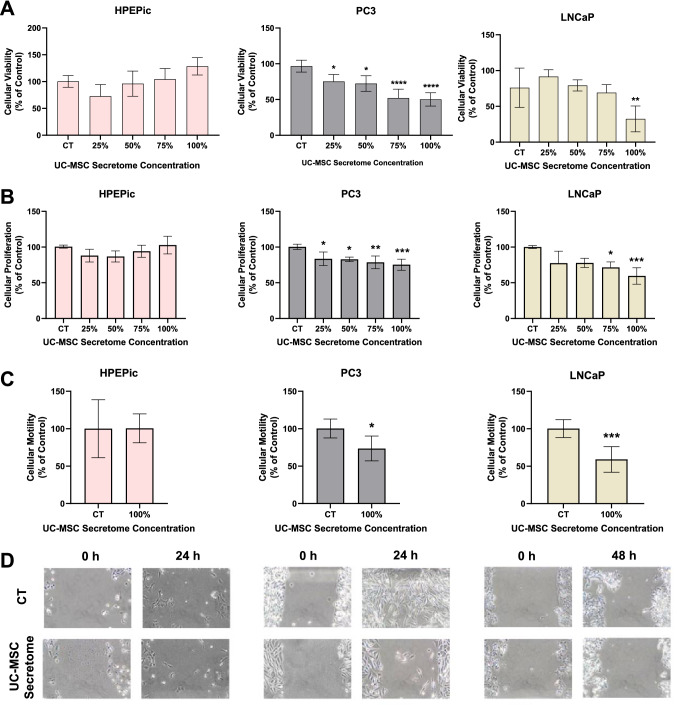
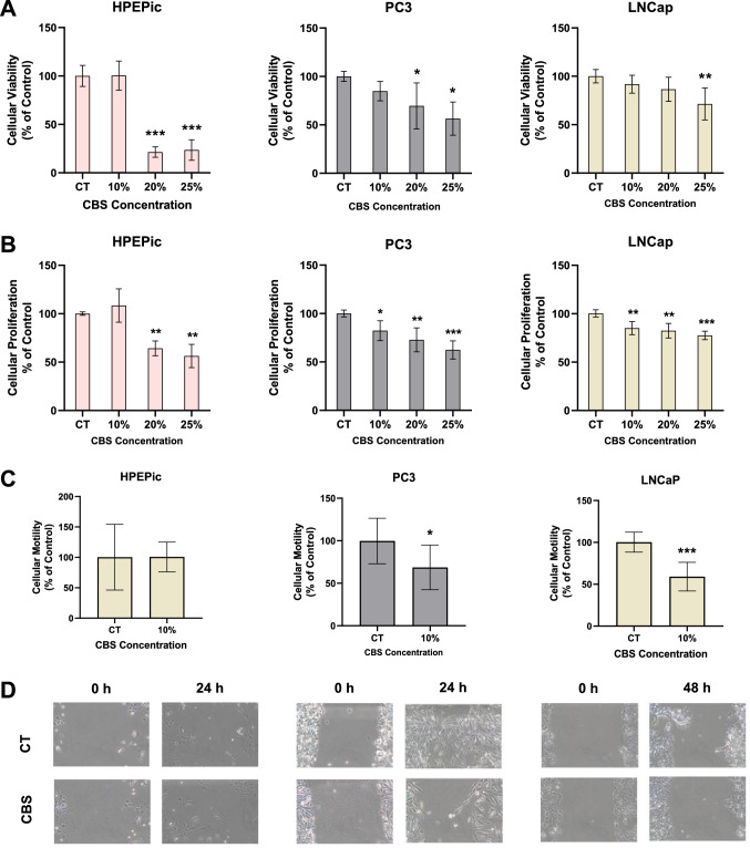
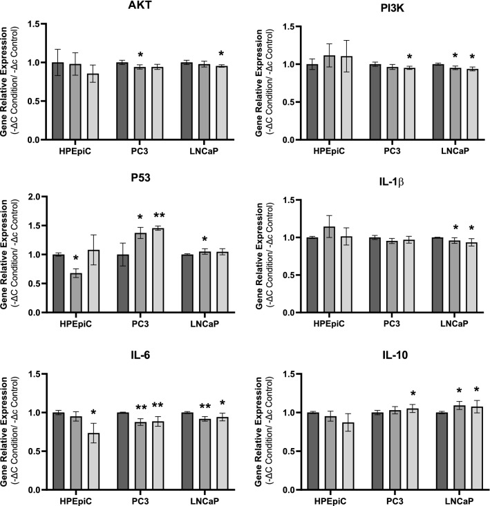
Prostate cancer (PCa) is the second most common malignancy in men, and the fifth leading cause of death worldwide. Mesenchymal stromal/stem cells (MSC) have been identified in PCa, although contradictory effects in malignant transformation and tumor progression have been described. Since umbilical cord (UC) MSC and cord blood serum (CBS) are rich in numerous growth and anti-inflammatory factors, UC-MSC secretome and CBS are able to modulate tumor cell proliferation and survival as well as immunity and angiogenesis. In the present study, we address this relationship and investigate the influence of UC-MSC secretome and CBS on two human PCa cell lines (PC3 and LNCaP) and a normal epithelial prostate cell line (HPEpiC). Our results disclosed that upon exposure to UC-MSC-conditioned medium or CBS, both PC3 and LNCaP cells exhibited reduced viability, proliferation, and motility while non-malignant epithelial prostate cells were unaffected. These findings were corroborated by expression analysis of AKT/PI3K signaling pathway, p53 and interleukin genes. UC-MSC and CBS factors decreased the expression of growth-stimulating AKT and PI3K effectors and simultaneously up-regulated the expression of tumor-suppressor p53. Moreover, a more anti-inflammatory expression profile was found in both malignant PCa cell lines. Altogether, these results shed light into possible mechanisms by which UC-MSC and CBS reduce PCa progression, further reinforcing their potential use as novel therapeutic agents in PCa. Graphical abstract
Overview
The claims and statistics of this paper haven’t been checked yet.
Run the check — free, no account needed. It reads the open-access text and checks the sources it cites.
Integrity signals
Registry
Cites
Statistics
Figures
Verification
Claims the paper makes
The claims and statistics of this paper haven’t been checked yet.
Run the check — free, no account needed. It reads the open-access text and checks the sources it cites.
Claims other papers make about this one
Each is checked against this paper’s own text: does it say what the citing paper says it does?
The source backs part of the claim, not all of it — or backs it for a different population, condition or comparison.
In the citing paper
“the disruption of the cancer-associated signaling pathways, such as PI3K/AKT [107], [113], via upregulation of PTEN, and of WNT/β-catenin signaling pathway by MSCs-secreted antagonist of the Wnt signaling DKK-1 [114] contributes to the inhibition of tumor cell migration, invasion, and survival [114], [115], [116], [117], [118].”
From Mesenchymal stem cell secretome for regenerative medicine: Where do we stand? (2025) · cites this paper as [107]
Experimental — accuracy not yet measured
Self-check
Percentages, sums and reported differences re-computed from the paper’s own numbers.
The self-check needs the paper’s full text.
Citations
Cites
Role of MSC in the Tumor Microenvironment
Hass, R. (2020) · Cancers · doi:10.3390/cancers12082107 · Cited by 116
Chen, L., Shi, Y., Zhu, X., Guo, W., Zhang, M., Che, Y., Tang, L., Yang, X., You, Q., & Liu, Z. (2019) · Oncology Reports · doi:10.3892/or.2019.7206 · Cited by 92
Research works printed but not linked: 51
- Rawla P. Epidemiology of prostate cancer. World J Oncol Elmer Press. 2016;10:107–25.
- Sharp VJ, Takacs EB, Powell CR. Prostatitis: diagnosis and treatment. Am Fam Physician. 2010;82(4):397–406.
- Bachmann C. Serenoa repens for benign prostatic hyperplasia. Schweiz Z für Ganzheitsmed/Swiss J Integr Med. 2015;27(4):202–6. · DOI
- Hulvat MC. Cancer incidence and trends. Surg Clin North Am. 2020;100(3):469–81. · DOI
- Bray F, et al. Global cancer statistics 2018: GLOBOCAN estimates of incidence and mortality worldwide for 36 cancers in 185 countries. CA Cancer J Clin. 2018;68(6):394–424. · DOI · Paper page
- Rahimi Tesiye M, Abrishami Kia Z, Rajabi-Maham H. Mesenchymal stem cells and prostate cancer: a concise review of therapeutic potentials and biological aspects. Stem Cell Res. 2022;63: 102864. · DOI
- Litwin MS, Tan HJ. The diagnosis and treatment of prostate cancer: a review. JAMA. 2017;317(24):2532–42. · DOI
- Abbasian Ardakani A, Rajaee J, Khoei S. Diagnosis of human prostate carcinoma cancer stem cells enriched from DU145 cell lines changes with microscopic texture analysis in radiation and hyperthermia treatment using run-length matrix. Int J Radiat Biol. 2017;93(11):1248–56. · DOI
- Driscoll J, Patel T. The mesenchymal stem cell secretome as an acellular regenerative therapy for liver disease. J Gastroenterol. 2019;54(9):763–73. · DOI
- Liau LL, et al. Characteristics and clinical applications of Wharton’s jelly-derived mesenchymal stromal cells. Curr Res Transl Med. 2020;68(1):5–16. · DOI
- Subramanian A, et al. Comparative characterization of cells from the various compartments of the human umbilical cord shows that the Wharton’s jelly compartment provides the best source of clinically utilizable mesenchymal stem cells. PLoS ONE. 2015;10(6): e0127992. · DOI
- Gomes A, et al. Human umbilical cord mesenchymal stem cells in type 2 diabetes mellitus: the emerging therapeutic approach. Cell Tissue Res. 2021;385(3):497–518. · DOI
- Shojaei S, et al. Effect of mesenchymal stem cells-derived exosomes on tumor microenvironment: tumor progression versus tumor suppression. J Cell Physiol. 2019;234(4):3394–409. · DOI
- Sandonà M, et al. Mesenchymal stromal cells and their secretome: new therapeutic perspectives for skeletal muscle regeneration. Front Bioeng Biotechnol. 2021;9: 652970. · DOI
- Noiseux N, et al. Mesenchymal stem cells overexpressing Akt dramatically repair infarcted myocardium and improve cardiac function despite infrequent cellular fusion or differentiation. Mol Ther. 2006;14(6):840–50. · DOI
- Caplan AI, Dennis JE. Mesenchymal stem cells as trophic mediators. J Cell Biochem. 2006;98(5):1076–84. · DOI · Paper page
- Yeo RWY, et al. Exosome: a novel and safer therapeutic refinement of mesenchymal stem cell. J Circulating Biomark. 2013. https://doi.org/10.5772/57460. · DOI
- Bian D, et al. The application of mesenchymal stromal cells (MSCs) and their derivative exosome in skin wound healing: a comprehensive review. Stem Cell Res Ther. 2022;13(1):24. · DOI
- Salgado AJ, et al. Adipose tissue derived stem cells secretome: soluble factors and their roles in regenerative medicine. Curr Stem Cell Res Ther. 2010;5(2):103–10. · DOI
- Liu CB, et al. Human umbilical cord-derived mesenchymal stromal cells improve left ventricular function, perfusion, and remodeling in a porcine model of chronic myocardial ischemia. Stem Cells Transl Med. 2016;5(8):1004–13. · DOI
- Si Y, et al. Infusion of mesenchymal stem cells ameliorates hyperglycemia in type 2 diabetic rats: identification of a novel role in improving insulin sensitivity. Diabetes. 2012;61(6):1616–25. · DOI
- Soria B, et al. Human mesenchymal stem cells prevent neurological complications of radiotherapy. Front Cell Neurosci. 2019;13:204. · DOI
- Song JY, et al. Umbilical cord-derived mesenchymal stem cell extracts ameliorate atopic dermatitis in mice by reducing the T cell responses. Sci Rep. 2019;9(1):6623. · DOI
- Eleuteri S, Fierabracci A. Insights into the secretome of mesenchymal stem cells and its potential applications. Int J Mol Sci. 2019;20(18):4597. · DOI
- Park CW, et al. Cytokine secretion profiling of human mesenchymal stem cells by antibody array. Int J Stem Cells. 2009;2(1):59–68. · DOI
- Zhang LN, et al. Fusion with mesenchymal stem cells differentially affects tumorigenic and metastatic abilities of lung cancer cells. J Cell Physiol. 2019;234(4):3570–82. · DOI
- Melzer C, von der Ohe J, Hass R. In vivo cell fusion between mesenchymal stroma/stem-like cells and breast cancer cells. Cancers. 2019;11(2):185. · DOI
- Hmadcha A, et al. Therapeutic potential of mesenchymal stem cells for cancer therapy. Front Bioeng Biotechnol. 2020. https://doi.org/10.3389/fbioe.2020.00043. · DOI
- Lu L, et al. Bone marrow mesenchymal stem cells suppress growth and promote the apoptosis of glioma U251 cells through downregulation of the PI3K/AKT signaling pathway. Biomed Pharmacother. 2019;112: 108625. · DOI
- Ho IA, et al. Human bone marrow-derived mesenchymal stem cells suppress human glioma growth through inhibition of angiogenesis. Stem Cells. 2013;31(1):146–55. · DOI
- Lee C, et al. Exosomes mediate the cytoprotective action of mesenchymal stromal cells on hypoxia-induced pulmonary hypertension. Circulation. 2012;126(22):2601–11. · DOI
- Pakravan K, et al. MicroRNA-100 shuttled by mesenchymal stem cell-derived exosomes suppresses in vitro angiogenesis through modulating the mTOR/HIF-1α/VEGF signaling axis in breast cancer cells. Cell Oncol (Dordr). 2017;40(5):457–70. · DOI
- Belderbos ME, et al. Plasma-mediated immune suppression: a neonatal perspective. Pediatr Allergy Immunol. 2013;24(2):102–13. · DOI
- Cox ST, et al. Functional characterisation and analysis of the soluble NKG2D ligand repertoire detected in umbilical cord blood plasma. Front Immunol. 2018;9:1282. · DOI
- Samarkanova D, et al. Cord blood platelet rich plasma derivatives for clinical applications in non-transfusion medicine. Front Immunol. 2020;11:942. · DOI
- Solves P, et al. Qualitative and quantitative cell recovery in umbilical cord blood processed by two automated devices in routine cord blood banking: a comparative study. Blood Transfus. 2013;11(3):405–11.
- Cervantes-Villagrana RD, et al. Tumor-induced neurogenesis and immune evasion as targets of innovative anti-cancer therapies. Signal Transduct Target Ther. 2020;5(1):99. · DOI
- Park S, et al. PI3K pathway in prostate cancer: all resistant roads lead to PI3K. Biochim Biophys Acta Rev Cancer. 2018;1870(2):198–206. · DOI
- Whiteside TL. Exosome and mesenchymal stem cell cross-talk in the tumor microenvironment. Semin Immunol. 2018;35:69–79. · DOI
- Dabrowski FA, et al. Comparison of the paracrine activity of mesenchymal stem cells derived from human umbilical cord, amniotic membrane and adipose tissue. J Obstet Gynaecol Res. 2017;43(11):1758–68. · DOI
- Sun X, et al. Human umbilical cord-derived mesenchymal stem cells ameliorate insulin resistance by suppressing NLRP3 inflammasome-mediated inflammation in type 2 diabetes rats. Stem Cell Res Ther. 2017;8(1):241. · DOI
- Yao J, et al. Extracellular vesicles derived from human umbilical cord mesenchymal stem cells alleviate rat hepatic ischemia-reperfusion injury by suppressing oxidative stress and neutrophil inflammatory response. Faseb j. 2019;33(2):1695–710. · DOI
- Versura P, et al. Efficacy of standardized and quality-controlled cord blood serum eye drop therapy in the healing of severe corneal epithelial damage in dry eye. Cornea. 2013;32(4):412–8. · DOI
- Riquelme I, et al. The gene expression status of the PI3K/AKT/mTOR pathway in gastric cancer tissues and cell lines. Pathol Oncol Res. 2016;22(4):797–805. · DOI
- Zhou J, et al. Genetic and bioinformatic analyses of the expression and function of PI3K regulatory subunit PIK3R3 in an Asian patient gastric cancer library. BMC Med Genom. 2012;5:34. https://doi.org/10.1186/1755-8794-5-34. · DOI
- Creighton CJ, et al. Proteomic and transcriptomic profiling reveals a link between the PI3K pathway and lower estrogen-receptor (ER) levels and activity in ER+ breast cancer. Breast Cancer Res. 2010;12(3):R40. · DOI
- Dasu MR, Devaraj S, Jialal I. High glucose induces IL-1beta expression in human monocytes: mechanistic insights. Am J Physiol Endocrinol Metab. 2007;293(1):E337–46. · DOI
- Chen MF, et al. Role of interleukin 1 beta in esophageal squamous cell carcinoma. J Mol Med (Berl). 2012;90(1):89–100. · DOI
- Kai H, et al. Involvement of proinflammatory cytokines IL-1beta and IL-6 in progression of human gastric carcinoma. Anticancer Res. 2005;25(2a):709–13.
- Deans DA, et al. Elevated tumour interleukin-1beta is associated with systemic inflammation: a marker of reduced survival in gastro-oesophageal cancer. Br J Cancer. 2006;95(11):1568–75. · DOI
- Jiang XP, et al. Down-regulation of expression of interleukin-6 and its receptor results in growth inhibition of MCF-7 breast cancer cells. Anticancer Res. 2011;31(9):2899–906.
Cited by
Sorted by citations
Mesenchymal stem cell secretome for regenerative medicine: Where do we stand?
Trigo, C. M., Rodrigues, J. S., Camões, S. P., Solá, S., & Miranda, J. P. (2025) · Journal of Advanced Research · doi:10.1016/j.jare.2024.05.004 · Cited by 252
“Indeed, many authors have been reported the effect of the secretome of MSCs in tumor cell proliferation and invasion in vitro and in vivo [104–107].”
Metadata
- Authors
- Sousa, A., Coelho, P., Leite, F., Teixeira, C., Rocha, A. C., Santos, I., Baylina, P., Fernandes, R., Soares, R., Costa, R., & Gomes, A.
- Affiliations
- Universidade do Porto (PT)
- Corresponding author
- Gomes, Andreia
- ORCID
- Venue
- Human Cell
- Published
- 2023-02-19
- Volume
- 36
- Issue
- 3
- Pages
- 1160-1172
- ISSN
- 0914-7470 (ISSN-L)
- 1749-0774
- PMC
- PMC10110723
- OpenAlex
- W4321366474
- Cited by
- 15
- References
- 2
- Citations by year
- 2026: 2 · 2025: 4 · 2024: 7 · 2023: 2
- FWCI
- 2.60
- Type
- journal article
- Language
- en
- Open access
- hybrid
- doi.org
- Locations
- Human Cell (publishedVersion, cc-by)
- PubMed Central (publishedVersion, cc-by)
- PubMed (publishedVersion)
- Portuguese National Funding Agency for Science, Research and Technology (RCAAP Project by FCT) (submittedVersion)
- Funders
- Bebé Vida - Ciências Para A Vida, S.A
- Fundação para a Ciência e a Tecnologia (UID/BIM/0493/2020, 2021.06521.BD)
- Instituto Politécnico do Porto (IPP4COVID/BI/02/2020)
- Porto Research, Technology & Innovation Center (PORTIC/LabMi/BI/2021/03)
- Universidade do Porto
- Topics
- Life Sciences › Biochemistry, Genetics and Molecular Biology › Cell Biology › Mesenchymal stem cell research
- Life Sciences › Biochemistry, Genetics and Molecular Biology › Cancer Research › Prostate Cancer Treatment and Research
- Health Sciences › Medicine › Oncology › Cancer Cells and Metastasis
- Keywords
- prostate cancer, human umbilical cord mesenchymal stem cells, MSC secretome, cord blood serum, tumor progression, PI3K/Akt signaling, p53, interleukin genes, anti-inflammatory factors, cell proliferation, cell motility, PC-3 cells, LNCaP cells, cancer stem cells
- MeSH terms
- Secretome, Humans, Male, Prostate, Prostatic Neoplasms, Stem Cells, Umbilical Cord, Tumor Suppressor Protein p53, Phosphatidylinositol 3-Kinases, Serum, Proto-Oncogene Proteins c-akt
About these checks
Registry, retraction, Cites and statistics results are computed, not judged, with one exception: a printed reference that no index lists is read once by a model before it counts as a citation error. Claims are extracted by a model and kept only when their quote appears verbatim in the text; each cited source is then checked, with a model’s help, for whether it says what the claim relies on. Those verdicts can be wrong — read the claim’s quote and the cited source before relying on one.
Statistics and arithmetic are recomputed, not judged.
Confidence bands are shown only for fields calibrated on our annotated test set. None is calibrated yet, so no band is shown.
What counts as a claim ›How we measure confidence ›How we check this ›
Are you an author, or do you see a problem with this page? Request a review
