Cited by 72

The DOI matches its Crossref record.

Injectable thermo-responsive nano-hydrogel loading triptolide for the anti-breast cancer enhancement via localized treatment based on “two strikes” effects

Luo, Yaoyao, Li, Jingjing, Hu, Yichen, Gao, Fei, Pak-Heng Leung, George, Geng, Funeng, Fu, Chaomei, Zhang, Jinming·2020·Acta Pharmaceutica Sinica B

Overview

The claims and statistics of this paper haven’t been checked yet.

Run the check — free, no account needed. It reads the open-access text and checks the sources it cites.

A few free checks a day · papers already verified open instantly · results are public

Integrity signals

Registry

Crossref · 10.1016/j.apsb.2020.05.011

Cites

Statistics

Figures

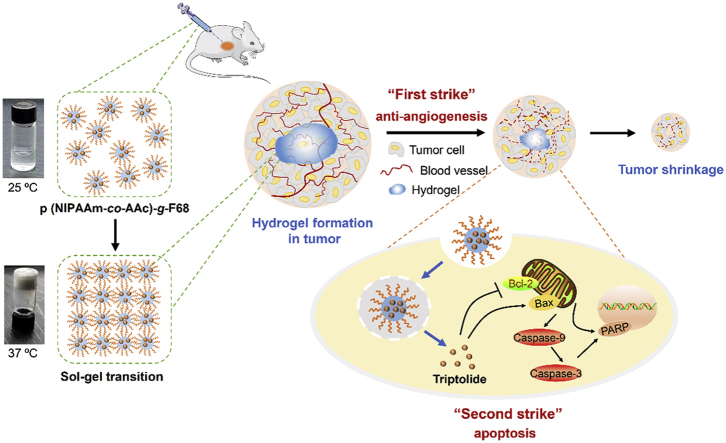
Graphical abstract Graphical abstract A themoresponsive nano-hydrogel was fabricated for the sustained release of triptolide (TPL) in the localized treatment of breast tumor. TPL@nano-gel exhibited the enhanced pro-apoptosis effects against breast tumor by the regulation of endogenous mitochondria pathway in vitro and in vivo.
Graphical abstract Graphical abstract A themoresponsive nano-hydrogel was fabricated for the sustained release of triptolide (TPL) in the localized treatment of breast tumor. TPL@nano-gel exhibited the enhanced pro-apoptosis effects against breast tumor by the regulation of endogenous mitochondria pathway in vitro and in vivo.CC BY-NC-ND · PubMed Central
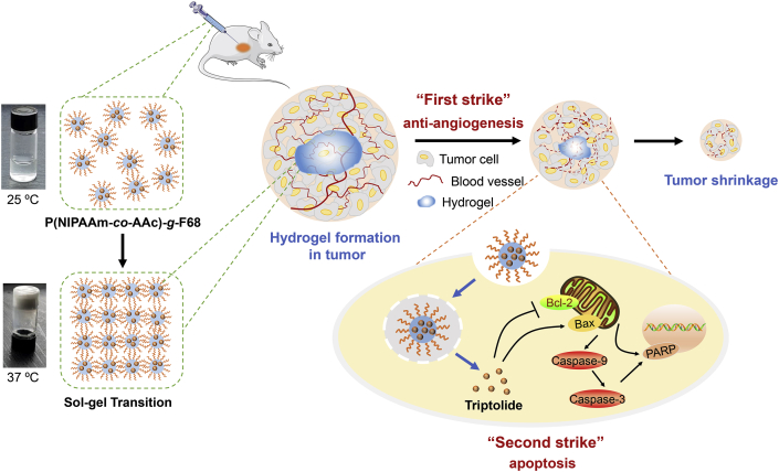
Scheme 1 Schematic illustration of injectable thermosensitive nano-hydrogel loading triptolide (TPL) composed by the self-assembly of p (NIPAAm-co-AAc)-g-F68 copolymer for localized tumor therapy, mediated by two strike approaches: anti-angiogenesis and cancer cell apoptosis.
Scheme 1 Schematic illustration of injectable thermosensitive nano-hydrogel loading triptolide (TPL) composed by the self-assembly of p (NIPAAm-co-AAc)-g-F68 copolymer for localized tumor therapy, mediated by two strike approaches: anti-angiogenesis and cancer cell apoptosis.CC BY-NC-ND · PubMed Central
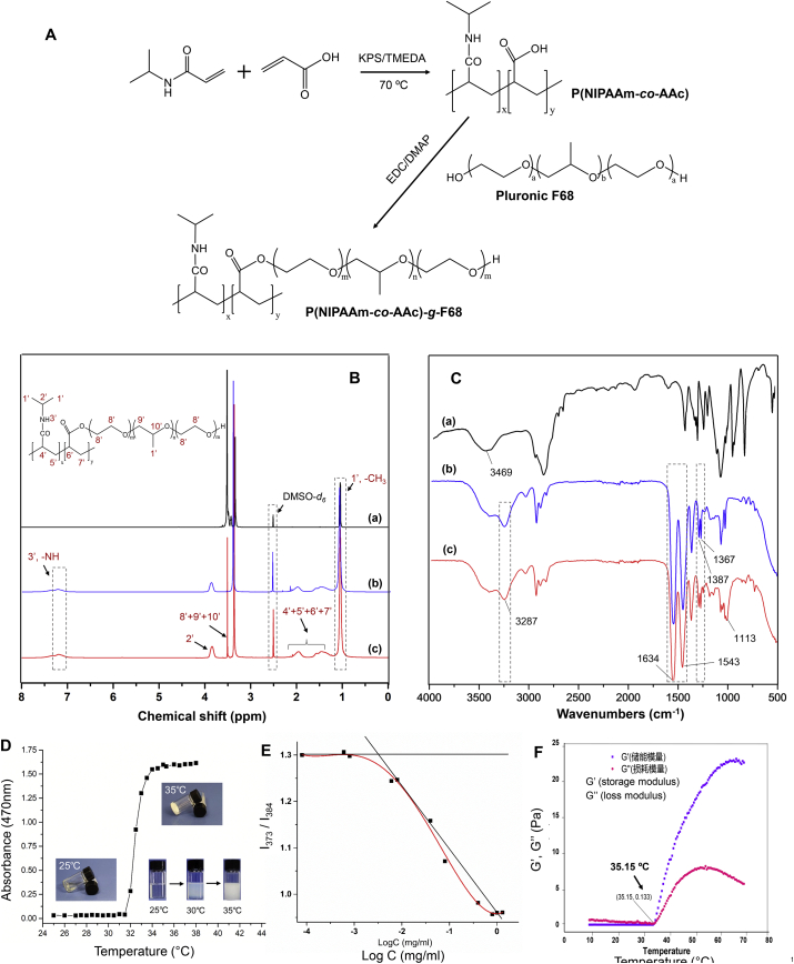
Figure 1 (A) Synthesis scheme of the p (NIPAAm-co-AAc)-g-F68 copolymer. 1H NMR spectra (B) and FT-IR spectra (C) of F68 (a), p (NIPAAm-co-AAc) (b), and p (NIPAAm-co-AAc)-g-F68 (c). (D)Temperature induced absorbance change of hydrogel by UV–Vis spectrophotometry to indicate phase transition (E) Plots of I373/I384 intensity ratio versus the logarithm of p (NIPAAm-co-AAc)-g-F68 copolymer concentration. (F) Rheological properties of the p (NIPAAm-co-AAc)-g-F68 hydrogel.
Figure 1 (A) Synthesis scheme of the p (NIPAAm-co-AAc)-g-F68 copolymer. 1H NMR spectra (B) and FT-IR spectra (C) of F68 (a), p (NIPAAm-co-AAc) (b), and p (NIPAAm-co-AAc)-g-F68 (c). (D)Temperature induced absorbance change of hydrogel by UV–Vis spectrophotometry to indicate phase transition (E) Plots of I373/I384 intensity ratio versus the logarithm of p (NIPAAm-co-AAc)-g-F68 copolymer concentration. (F) Rheological properties of the p (NIPAAm-co-AAc)-g-F68 hydrogel.CC BY-NC-ND · PubMed Central
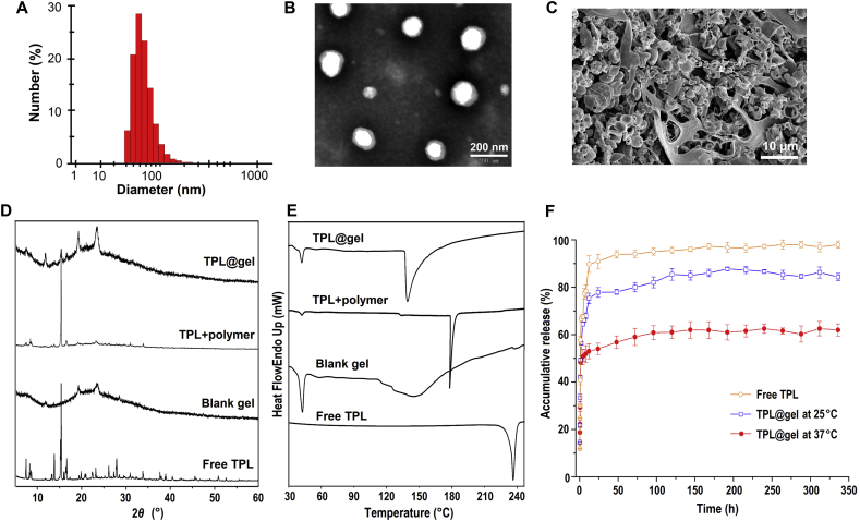
Figure 2 Characterization of TPL-loaded nano-micelles composed by p (NIPAAm-co-AAc)-g-F68 copolymer. (A) Particle distribution of nanomicelles by DLS (B) TEM image of TPL-loaded micelle solution. (C) SEM image of lyophilized hydrogel formed by sol–gel transition of micelle solution. XRD (D) and DSC (E) analysis of TPL-loaded hydrogel. (F) Cumulative release of TPL from hydrogels at 25 and 37 °C, respectively (mean ± SD, n = 3).
Figure 2 Characterization of TPL-loaded nano-micelles composed by p (NIPAAm-co-AAc)-g-F68 copolymer. (A) Particle distribution of nanomicelles by DLS (B) TEM image of TPL-loaded micelle solution. (C) SEM image of lyophilized hydrogel formed by sol–gel transition of micelle solution. XRD (D) and DSC (E) analysis of TPL-loaded hydrogel. (F) Cumulative release of TPL from hydrogels at 25 and 37 °C, respectively (mean ± SD, n = 3).CC BY-NC-ND · PubMed Central
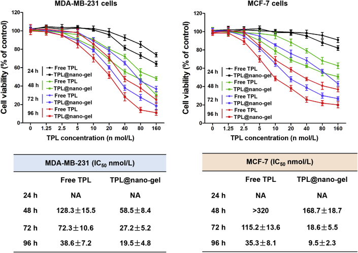
Figure 3 Cytotoxicity of TPL@nano-gel against MDA-MB-231 and MCF-7 human breast cancer cells, compared with free TPL. MDA-MB-231 and MCF-7 cells were treated with various concentrations of free TPL and TPL@nano-gel (0–160 nmol/L) for 24, 48, 72 and 96 h, respectively. Cell viability was assessed by MTT assay. IC50 values of cell viability in MDA-MB-231 and MCF-7 cells with free TPL and TPL@nano-gel treatment were calculated and analyzed by software GraphPad Prism 5.0 (mean ± SD, n = 3).
Figure 3 Cytotoxicity of TPL@nano-gel against MDA-MB-231 and MCF-7 human breast cancer cells, compared with free TPL. MDA-MB-231 and MCF-7 cells were treated with various concentrations of free TPL and TPL@nano-gel (0–160 nmol/L) for 24, 48, 72 and 96 h, respectively. Cell viability was assessed by MTT assay. IC50 values of cell viability in MDA-MB-231 and MCF-7 cells with free TPL and TPL@nano-gel treatment were calculated and analyzed by software GraphPad Prism 5.0 (mean ± SD, n = 3).CC BY-NC-ND · PubMed Central
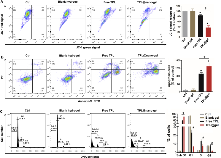
Figure 4 Effects of TPL@nano-gel on mitochondrial membrane potential (A), apoptosis (B) and cell cycle progression (C) of MDA-MB-231 cells using flow cytometry. MDA-MB-231 cells were treated with or without blank gel (160 μg/mL), or free TPL (20 nmol/L) and TPL@nano-gel (20 nmol/L) for 48 h (mean ± SD, n = 3). ∗P < 0.05 versus the control group. #P < 0.05 free TPL vs. TPL@nano-gel group.
Figure 4 Effects of TPL@nano-gel on mitochondrial membrane potential (A), apoptosis (B) and cell cycle progression (C) of MDA-MB-231 cells using flow cytometry. MDA-MB-231 cells were treated with or without blank gel (160 μg/mL), or free TPL (20 nmol/L) and TPL@nano-gel (20 nmol/L) for 48 h (mean ± SD, n = 3). ∗P < 0.05 versus the control group. #P < 0.05 free TPL vs. TPL@nano-gel group.CC BY-NC-ND · PubMed Central
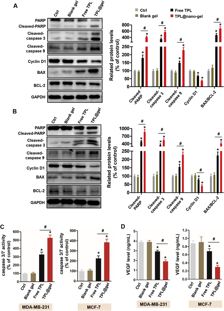
Figure 5 Effects of TPL@nano-gel on the expression levels of proteins related to apoptosis in MDA-MB-231 (A) and MCF-7 (B) breast cancer cells after 48 h treatment by Western blot analysis. Proteins were involved in the regulation of apoptosis including PARP, cleaved-PARP, cleaved-Caspase 3, cleaved-Caspase 9, Cyclin D1, Bax and Bcl-2. VEGF levels in the culture medium of MDA-MB-231 and MCF-7 cells after 48 h treatment of various agents were measured by a commercial VEGF ELISA kit (C). Caspase 3/7 activity in MDA-MB-231 and MCF-7 cells after 48 h treatment was measured by a commercial Caspase 3/7 activity kit (D). ∗P < 0.05 vs. the control group. #P < 0.05 TPL@gel versus free TPL group (mean ± SD, n = 3).
Figure 5 Effects of TPL@nano-gel on the expression levels of proteins related to apoptosis in MDA-MB-231 (A) and MCF-7 (B) breast cancer cells after 48 h treatment by Western blot analysis. Proteins were involved in the regulation of apoptosis including PARP, cleaved-PARP, cleaved-Caspase 3, cleaved-Caspase 9, Cyclin D1, Bax and Bcl-2. VEGF levels in the culture medium of MDA-MB-231 and MCF-7 cells after 48 h treatment of various agents were measured by a commercial VEGF ELISA kit (C). Caspase 3/7 activity in MDA-MB-231 and MCF-7 cells after 48 h treatment was measured by a commercial Caspase 3/7 activity kit (D). ∗P < 0.05 vs. the control group. #P < 0.05 TPL@gel versus free TPL group (mean ± SD, n = 3).CC BY-NC-ND · PubMed Central
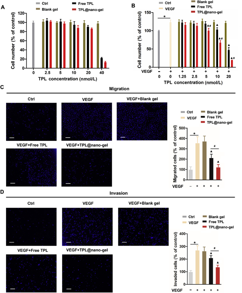
Figure 6 TPL@nano-gel inhibited VEGF-induced HUVEC cell proliferation, migration, and invasion. (A) HUVECs treated with blank gel, free TPL and TPL@nano-gel (0–40 nmol/L) for 48 h and then cell viability was measured by MTT assay. (B) HUVECs were pre-starved in low serum medium for 24 h, then treated without (control) or with VEGF (20 ng/mL) in the presence of blank gel, free TPL and TPL@nano-gel (0–20 nmol/L) for 48 h. Cell viability was measured by MTT assay. Endothelial cell transwell migration (C) and invasion (D) were studied after 24 h treatment of various agents with TPL at 10 nmol/L in the presence of VEGF (20 ng/mL). HUVECs received VEGF (20 ng/mL) treatment served as positive control. Scale bar is 200 μm, ∗P < 0.05, positive control with VEGF stimulation versus negative control group, ▲P < 0.05, TPL formulations versus positive control with VEGF stimulation, and #P < 0.05, TPL@nano-gel vs. free TPL group (mean ± SD, n = 3).
Figure 6 TPL@nano-gel inhibited VEGF-induced HUVEC cell proliferation, migration, and invasion. (A) HUVECs treated with blank gel, free TPL and TPL@nano-gel (0–40 nmol/L) for 48 h and then cell viability was measured by MTT assay. (B) HUVECs were pre-starved in low serum medium for 24 h, then treated without (control) or with VEGF (20 ng/mL) in the presence of blank gel, free TPL and TPL@nano-gel (0–20 nmol/L) for 48 h. Cell viability was measured by MTT assay. Endothelial cell transwell migration (C) and invasion (D) were studied after 24 h treatment of various agents with TPL at 10 nmol/L in the presence of VEGF (20 ng/mL). HUVECs received VEGF (20 ng/mL) treatment served as positive control. Scale bar is 200 μm, ∗P < 0.05, positive control with VEGF stimulation versus negative control group, ▲P < 0.05, TPL formulations versus positive control with VEGF stimulation, and #P < 0.05, TPL@nano-gel vs. free TPL group (mean ± SD, n = 3).CC BY-NC-ND · PubMed Central
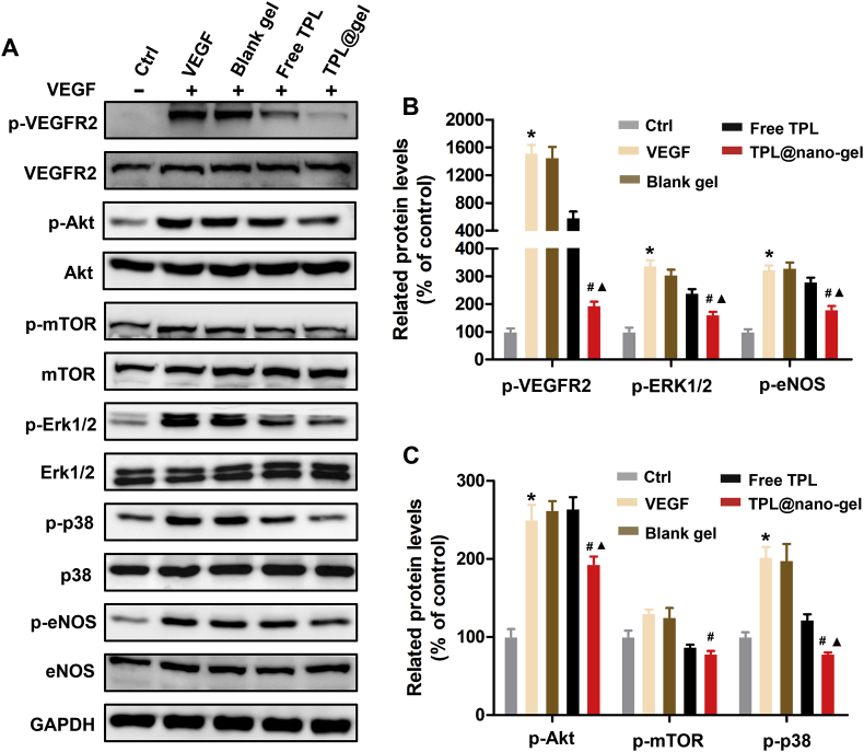
Figure 7 Effects of TPL@nano-gel on VEGFR-2 mediated angiogenesis signaling pathway. HUVECs were starved in low serum medium for 3 h and then treated with TPL formulations at 10 nmol/L for 24 h, followed by stimulation of VEGF (50 ng/mL) for 15 min. The expression levels of the major proteins involved in VEGFR2-mediated angiogenesis pathway such as VEGFR2, p-VEGFR2, AKT, p-AKT, mTOR, p-mTOR, ERK1/2, p-ERK1/2, P38, p-P38, eNOS and p-eNOS were detected by Western blot analysis (A). Quantitative analysis of the protein expression levels in HUVECs (B, C). ∗P < 0.05, positive control with VEGF stimulation vs. negative control group; ▲P < 0.05, TPL formulations vs. positive control with VEGF stimulation; #P < 0.05, TPL@nano-gel vs. free TPL group (mean ± SD, n = 3).
Figure 7 Effects of TPL@nano-gel on VEGFR-2 mediated angiogenesis signaling pathway. HUVECs were starved in low serum medium for 3 h and then treated with TPL formulations at 10 nmol/L for 24 h, followed by stimulation of VEGF (50 ng/mL) for 15 min. The expression levels of the major proteins involved in VEGFR2-mediated angiogenesis pathway such as VEGFR2, p-VEGFR2, AKT, p-AKT, mTOR, p-mTOR, ERK1/2, p-ERK1/2, P38, p-P38, eNOS and p-eNOS were detected by Western blot analysis (A). Quantitative analysis of the protein expression levels in HUVECs (B, C). ∗P < 0.05, positive control with VEGF stimulation vs. negative control group; ▲P < 0.05, TPL formulations vs. positive control with VEGF stimulation; #P < 0.05, TPL@nano-gel vs. free TPL group (mean ± SD, n = 3).CC BY-NC-ND · PubMed Central
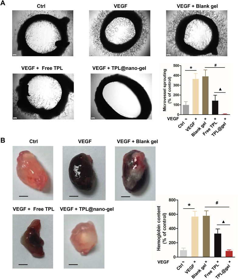
Figure 8 TPL@nano-gel inhibited angiogenic activity of aorta ring sprouting ex vivo and VEGF-induced angiogenesis matrigel plug in vivo. (A) Representative photographs of micro-vessel sprouting in rat aortic rings. Rat aortic rings were embedded in matrigel and treated with TPL formulations with 20 nmol/L of TPL in the presence of VEGF (100 ng/mL) for 10 days. Then the micro-vessel growth was recorded by an inverted microscope and the number of micro-vessel sprouting was quantified by ImageJ software. Aortic rings VEGF (100 ng/mL) served as positive control. Scale bar = 100 μm. Quantitative analysis of micro-vessel sprouting in each group. (B) Representative photographs of blood vessels formation in matrigel plugs. 6-Week-old BALB/c male mice were subcutaneously injected with 0.5 mL of growth factor-reduced matrigel containing VEGF (250 ng) and heparin (100 Unit) with TPL formulations with 20 nmol/L of TPL. After 14 days, the animals were sacrificed and the matrigel plugs were removed, photographed and weighted. Scale bar: 2.5 mm. To quantify the formation of functional blood vessels, the amount of hemoglobin was measured using the Drabkin reagent Kit (mean ± SD, n = 3), ∗P < 0.05, VEGF alone group vs. the control group, #P < 0.05, TPL@nano-gel vs. blank gel, and ▲P < 0.05, TPL@gel vs. free TPL group.
Figure 8 TPL@nano-gel inhibited angiogenic activity of aorta ring sprouting ex vivo and VEGF-induced angiogenesis matrigel plug in vivo. (A) Representative photographs of micro-vessel sprouting in rat aortic rings. Rat aortic rings were embedded in matrigel and treated with TPL formulations with 20 nmol/L of TPL in the presence of VEGF (100 ng/mL) for 10 days. Then the micro-vessel growth was recorded by an inverted microscope and the number of micro-vessel sprouting was quantified by ImageJ software. Aortic rings VEGF (100 ng/mL) served as positive control. Scale bar = 100 μm. Quantitative analysis of micro-vessel sprouting in each group. (B) Representative photographs of blood vessels formation in matrigel plugs. 6-Week-old BALB/c male mice were subcutaneously injected with 0.5 mL of growth factor-reduced matrigel containing VEGF (250 ng) and heparin (100 Unit) with TPL formulations with 20 nmol/L of TPL. After 14 days, the animals were sacrificed and the matrigel plugs were removed, photographed and weighted. Scale bar: 2.5 mm. To quantify the formation of functional blood vessels, the amount of hemoglobin was measured using the Drabkin reagent Kit (mean ± SD, n = 3), ∗P < 0.05, VEGF alone group vs. the control group, #P < 0.05, TPL@nano-gel vs. blank gel, and ▲P < 0.05, TPL@gel vs. free TPL group.CC BY-NC-ND · PubMed Central
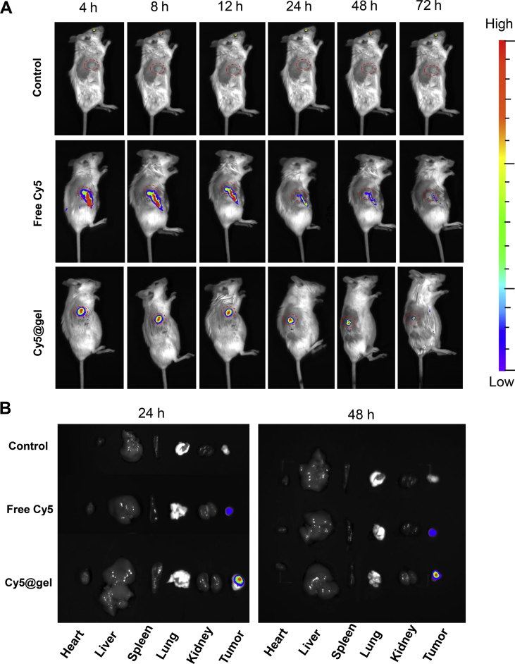
Figure 9 Retention of free Cy5 and Cy5-labled gel in tumor observed by IVIS spectrum. (A) Fluorescent images of the retention of Cy5 formulations in tumors after intra-tumoral injection at predetermined time intervals, i.e., 4, 8, 12, 24, 48 and 72 h after injection (B) Images of dissected organs of the mice treated with Cy5 formulations for 24 and 48 h post-injection.
Figure 9 Retention of free Cy5 and Cy5-labled gel in tumor observed by IVIS spectrum. (A) Fluorescent images of the retention of Cy5 formulations in tumors after intra-tumoral injection at predetermined time intervals, i.e., 4, 8, 12, 24, 48 and 72 h after injection (B) Images of dissected organs of the mice treated with Cy5 formulations for 24 and 48 h post-injection.CC BY-NC-ND · PubMed Central
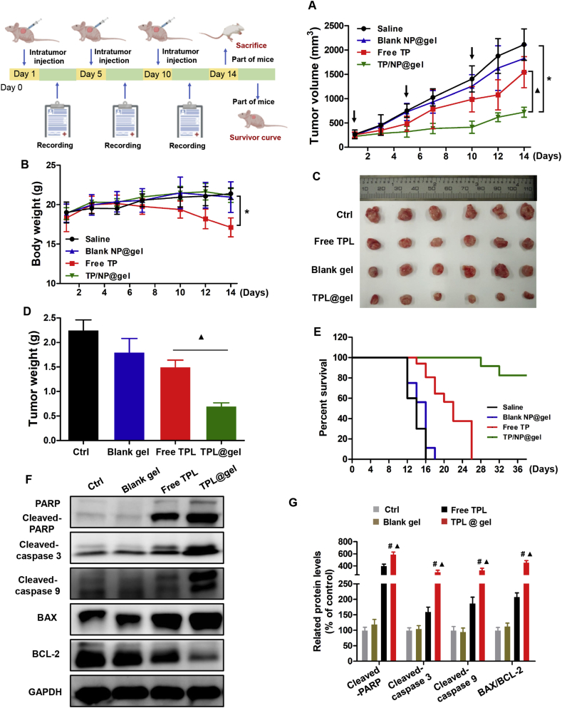
Figure 10 In vivo anticancer activity of TPL@nano-gel on 4T1 xenograft model. Tumor volume changes (A), body weight changes (B), Photographs of tumors (C), final mean tumor weights (D), Kaplan–Meier survival curve (E) of mice receiving the above-indicated treatments. Expressions of apoptosis-related proteins in tumor tissue by Western Blot analysis (F). Quantitative analysis of those apoptosis-related protein expressions (G) (mean ± SD, n = 3), ∗P < 0.05 versus the control group; #P < 0.05, TPL@nano-gel vs. blank gel; and ▲P < 0.05, TPL@nano-gel vs. free TPL group.
Figure 10 In vivo anticancer activity of TPL@nano-gel on 4T1 xenograft model. Tumor volume changes (A), body weight changes (B), Photographs of tumors (C), final mean tumor weights (D), Kaplan–Meier survival curve (E) of mice receiving the above-indicated treatments. Expressions of apoptosis-related proteins in tumor tissue by Western Blot analysis (F). Quantitative analysis of those apoptosis-related protein expressions (G) (mean ± SD, n = 3), ∗P < 0.05 versus the control group; #P < 0.05, TPL@nano-gel vs. blank gel; and ▲P < 0.05, TPL@nano-gel vs. free TPL group.CC BY-NC-ND · PubMed Central
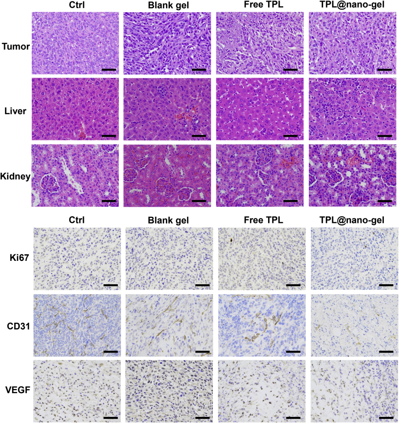
Figure 11 Representative histological microphotographs of tumor, liver, kidney of mice receiving the above-indicated treatments by H&E staining, and tumor tissues by immunohistochemical staining with primary antibodies against Ki-67, CD31, and VEGF. Scale bar = 100 μm.
Figure 11 Representative histological microphotographs of tumor, liver, kidney of mice receiving the above-indicated treatments by H&E staining, and tumor tissues by immunohistochemical staining with primary antibodies against Ki-67, CD31, and VEGF. Scale bar = 100 μm.CC BY-NC-ND · PubMed Central

Verification

Claims the paper makes

The claims and statistics of this paper haven’t been checked yet.

Run the check — free, no account needed. It reads the open-access text and checks the sources it cites.

A few free checks a day · papers already verified open instantly · results are public

Claims other papers make about this one

Each is checked against this paper’s own text: does it say what the citing paper says it does?

Self-check

Percentages, sums and reported differences re-computed from the paper’s own numbers.

The self-check needs the paper’s full text.

Citations

Cites

No references could be resolved yet.

Research works printed but not linked: 34

  1. Hou, 2019, Triptolide: medicinal chemistry, chemical biology and clinical progress, Eur J Med Chem, 176, 378, 10.1016/j.ejmech.2019.05.032 · DOI
  2. Meng, 2014, Targets and molecular mechanisms of triptolide in cancer therapy, Chin J Canc Res, 26, 622
  3. Liu, 2019, The effect of triptolide-loaded exosomes on the proliferation and apoptosis of human ovarian cancer SKOV3 cells, BioMed Res Int, 2019, 2595801
  4. Zhang, 2016, Nanoparticle‒hydrogel: a hybrid biomaterial system for localized drug delivery, ACS Appl Mater Interfaces, 44, 2049
  5. Deng, 2019, Injectable thermosensitive hydrogel systems based on functional PEG/PCL block polymer for local drug delivery, J Control Release, 297, 60, 10.1016/j.jconrel.2019.01.026 · DOI
  6. Norouzi, 2016, Injectable hydrogel-based drug delivery systems for local cancer therapy, Drug Discov Today, 21, 1835, 10.1016/j.drudis.2016.07.006 · DOI
  7. Yang, 2018, Nanogel-incorporated injectable hydrogel for synergistic therapy based on sequential local delivery of combretastatin-A4 phosphate (CA4P) and doxorubicin (DOX), ACS Appl Mater Interfaces, 10, 18560, 10.1021/acsami.8b04394 · DOI
  8. Gaowa, 2014, Combination of hybrid peptide with biodegradable gelatin hydrogel for controlled release and enhancement of anti-tumor activity in vivo, J Control Release, 176, 1, 10.1016/j.jconrel.2013.12.021 · DOI
  9. Lai, 2016, Design and fabrication of hydrogel-based nanoparticulate systems for in vivo drug delivery, J Control Release, 243, 269, 10.1016/j.jconrel.2016.10.013 · DOI
  10. Zhao, 2019, Injectable peptide hydrogel as intraperitoneal triptolide depot for the treatment of orthotopic hepatocellular carcinoma, Acta Pharm Sin B, 9, 1050, 10.1016/j.apsb.2019.06.001 · DOI
  11. Alexander, 2014, Polyethylene glycol (PEG)-poly(N-isopropylacrylamide) (PNIPAAm) based thermosensitive injectable hydrogels for biomedical applications, Eur J Pharm Biopharm, 88, 575, 10.1016/j.ejpb.2014.07.005 · DOI · Paper page
  12. Das, 2015, Stimulus-responsive,biodegradable, biocompatible, covalently cross-linked hydrogel based on dextrin and poly(N-isopropylacrylamide) for in vitro/in vivo controlled drug release, ACS Appl Mater Interfaces, 7, 14338, 10.1021/acsami.5b02975 · DOI
  13. Ma, 2015, Thermally responsive hydrogel blends: a general drug carrier model for controlled drug release, Angew Chem Int Ed Engl, 54, 7376, 10.1002/anie.201501705 · DOI
  14. Zhang, 2013, Dual stimuli-responsive N-phthaloylchitosan-graft-(poly(N-isopropylacrylamide)-block-poly(acrylic acid)) copolymer prepared via RAFT polymerization, Carbohydr Polym, 92, 662, 10.1016/j.carbpol.2012.09.003 · DOI
  15. Liu, 2017, Injectable thermoresponsive hydrogel formed by alginate-g-poly(N-isopropylacrylamide) that releases doxorubicin-encapsulated micelles as a smart drug delivery system, ACS Appl Mater Interfaces, 9, 35673, 10.1021/acsami.7b12849 · DOI
  16. Shen, 2015, Thermogelling polymer-platinum(IV) conjugates for long-term delivery of cisplatin, Biomacromolecules, 16, 105, 10.1021/bm501220a · DOI
  17. Wang, 2012, Adjustable degradation and drug release of a thermosensitive hydrogel based on a pendant cyclic ether modified poly(epsilon-caprolactone) and poly(ethylene glycol)co-polymer, Acta Biomater, 8, 3963, 10.1016/j.actbio.2012.07.021 · DOI
  18. Agarwal, 2013, Injectable implants for the sustained release of protein and peptide drugs, Drug Discov Today, 18, 337, 10.1016/j.drudis.2013.01.013 · DOI
  19. Wang, 2017, Chitosan cross-linked poly(acrylic acid) hydrogels: drug release control and mechanism, Colloids Surf B Biointerfaces, 152, 252, 10.1016/j.colsurfb.2017.01.008 · DOI
  20. Li, 2018, Heparin-poloxamer thermosensitive hydrogel loaded with bFGF and NGF enhances peripheral nerve regeneration in diabetic rats, Biomaterials, 168, 24, 10.1016/j.biomaterials.2018.03.044 · DOI
  21. Shieh, 2016, Temperature-, pH- and CO2-sensitive poly(N-isopropylacryl amide-co-acrylic acid) copolymers with high glass transition temperatures, Polymers, 8, 434, 10.3390/polym8120434 · DOI
  22. Wang, 2015, Preparation and characterization of CS-g-PNIPAAm microgels and application in a water vapour-permeable fabric, Carbohydr Polym, 127, 11, 10.1016/j.carbpol.2015.03.040 · DOI
  23. Li, 2017, A bioorthogonal nanosystem for imaging and in vivo tumor inhibition, Biomaterials, 138, 57, 10.1016/j.biomaterials.2017.05.036 · DOI
  24. Gao, 2019, Menthol-modified casein nanoparticles loading 10-hydroxycamptothecin for glioma targeting therapy, Acta Pharm Sin B, 9, 843, 10.1016/j.apsb.2019.01.006 · DOI
  25. Fathi, 2019, Dual thermo-and pH-sensitive injectable hydrogels of chitosan/(poly(N-isopropylacrylamide-co-itaconic acid)) for doxorubicin delivery in breast cancer, Int J Biol Macromol, 128, 957, 10.1016/j.ijbiomac.2019.01.122 · DOI · Paper page
  26. Song, 2016, Preparation of thermo-responsive graft copolymer by using a novel macro-RAFT agent and its application for drug delivery, Mater Sci Eng C Mater Biol Appl, 62, 45, 10.1016/j.msec.2016.01.026 · DOI
  27. Wei, 2015, A novel thermo-responsive hydrogel based on salecan and poly(N-isopropylacrylamide): synthesis and characterization, Colloids Surf B Biointerfaces, 125, 1, 10.1016/j.colsurfb.2014.10.057 · DOI
  28. Leal, 2013, Preparation and characterization of hydrogels based on homopolymeric fractions of sodium alginate and PNIPAAm, Carbohydr Polym, 92, 157, 10.1016/j.carbpol.2012.09.031 · DOI
  29. Gao, 2018, Dual pH and glucose sensitive gel gated mesoporous silica nanoparticles for drug delivery, Chin Chem Lett, 29, 107, 10.1016/j.cclet.2018.05.022 · DOI
  30. Wang, 2019, Synergistic antitumour effects of triptolide plus 10-hydroxycamptothecin onbladder cancer, Biomed Pharmacother, 115, 108899, 10.1016/j.biopha.2019.108899 · DOI
  31. Zhang, 2017, pH-sensitive polymeric nanoparticles for co-delivery of doxorubicin and curcumin to treat cancer via enhanced pro-apoptotic and anti-angiogenic activities, Acta Biomater, 58, 349, 10.1016/j.actbio.2017.04.029 · DOI
  32. Lien, 2019, A novel 2-aminobenzimidazole-based compound Jzu 17 exhibits anti-angiogenesis effects by targeting VEGFR-2 signalling, Br J Pharmacol, 176, 4034, 10.1111/bph.14813 · DOI
  33. Li, 2016, Sigma receptor-mediated targeted delivery of anti-angiogenic multifunctional nanodrugs for combination tumor therapy, J Control Release, 228, 107, 10.1016/j.jconrel.2016.02.044 · DOI
  34. Ruan, 2015, Matrix metalloproteinase-sensitive size-shrinkable nanoparticles for deep tumor penetration and pH triggered doxorubicin release, Biomaterials, 60, 100, 10.1016/j.biomaterials.2015.05.006 · DOI

Cited by

Sorted by citations

Metadata

Authors
Luo, Y., Li, J., Hu, Y., Gao, F., Pak-Heng Leung, G., Geng, F., Fu, C., & Zhang, J.
Venue
Acta Pharmaceutica Sinica B
Publisher
Elsevier BV
Published
2020-11
Volume
10
Issue
11
Pages
2227-2245
ISSN
2211-3835
Cited by
72
References
0
Language
en
Funders
  • National Natural Science Foundation of China (8170371, 81973662)
  • China Postdoctoral Science Foundation (2017M612930, 2019T120817)
  • China Academy of Space Technology (2018QNRC1-01)

About these checks

Registry, retraction, Cites and statistics results are computed, not judged, with one exception: a printed reference that no index lists is read once by a model before it counts as a citation error. Claims are extracted by a model and kept only when their quote appears verbatim in the text; each cited source is then checked, with a model’s help, for whether it says what the claim relies on. Those verdicts can be wrong — read the claim’s quote and the cited source before relying on one.

Statistics and arithmetic are recomputed, not judged.

Confidence bands are shown only for fields calibrated on our annotated test set. None is calibrated yet, so no band is shown.

What counts as a claim ›How we measure confidence ›How we check this ›

Are you an author, or do you see a problem with this page? Request a review