

The DOI matches its Crossref record.
Exosomal microRNA-1 and MYO15A as a target for therapy and diagnosis in renal cell carcinoma
Overview
The claims and statistics of this paper haven’t been checked yet.
Run the check — free, no account needed. It reads the open-access text and checks the sources it cites.
Integrity signals
Registry
Cites
Statistics
Figures
Verification
Claims the paper makes
The claims and statistics of this paper haven’t been checked yet.
Run the check — free, no account needed. It reads the open-access text and checks the sources it cites.
Claims other papers make about this one
Each is checked against this paper’s own text: does it say what the citing paper says it does?
In the citing paper
“This finding suggests that using exosomes to deliver miR-1 could be a promising therapeutic approach for RCC [249].”
From Emerging role of exosomes in cancer therapy: progress and challenges (2025) · cites this paper as [249]
In the citing paper
“For example, Yoshino et al. discovered that miRNA-1 (miR-1) could effectively inhibit RCC proliferation and infiltration.”
From Emerging role of exosomes in cancer therapy: progress and challenges (2025) · cites this paper as [249]
In the citing paper
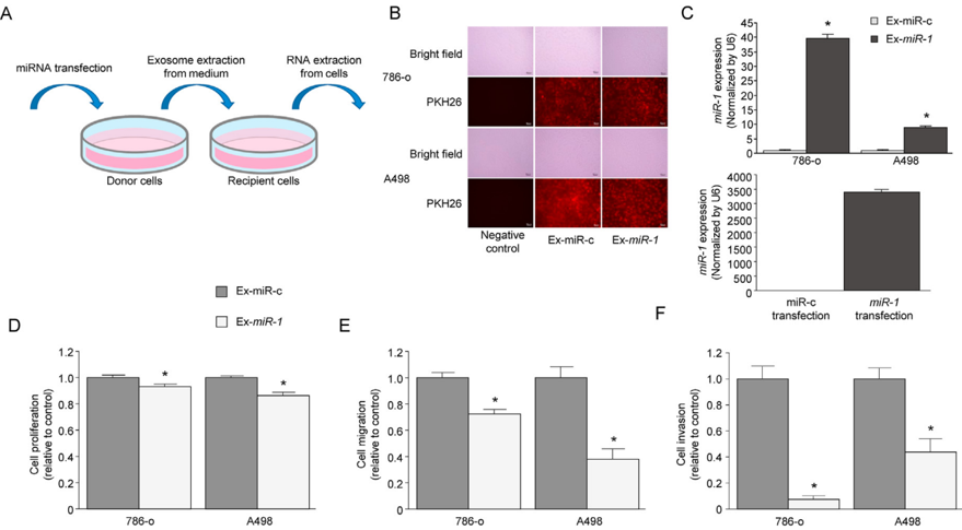
“Their study demonstrated that when RCC cells were handled with exosomes originated from miR-1-transfected cells, miR-1 expression increased significantly within these cells. miR-1 expression increased approximately 10 to 40 times compared to the control group. It significantly inhibited the growth, migration, and invasion of 786-o and A498 cells.”
From Emerging role of exosomes in cancer therapy: progress and challenges (2025) · cites this paper as [249]
In the citing paper
“| | Exosomes to deliver miR-1 | Suppressed RCC growth and invasion | [249] |”
From Emerging role of exosomes in cancer therapy: progress and challenges (2025) · cites this paper as [249]
Self-check
Percentages, sums and reported differences re-computed from the paper’s own numbers.
The self-check needs the paper’s full text.
Citations
Cites
No references could be resolved yet.
Research works printed but not linked: 20
- Rathmell, 2022, Management of metastatic clear cell renal cell carcinoma: ASCO guideline, J. Clin. Oncol., 10.1200/JCO.22.00868 · DOI
- Valadi, 2007, Exosome-mediated transfer of mRNAs and microRNAs is a novel mechanism of genetic exchange between cells, Nat. Cell Biol., 9, 654, 10.1038/ncb1596 · DOI · Paper page
- Tsuruda, 2020, Oncogenic effects of RAB27B through exosome independent function in renal cell carcinoma including sunitinib-resistant, PLoS One, 15, 10.1371/journal.pone.0232545 · DOI
- Kawakami, 2012, vol. 48, 827
- Yoshino, 2011, The tumour-suppressive function of miR-1 and miR-133a targeting TAGLN2 in bladder cancer, Br. J. Cancer, 104, 808, 10.1038/bjc.2011.23 · DOI
- Gerlinger, 2012, Intratumor heterogeneity and branched evolution revealed by multiregion sequencing, N. Engl. J. Med., 366, 883, 10.1056/NEJMoa1113205 · DOI · Paper page
- Aceto, 2014, Circulating tumor cell clusters are oligoclonal precursors of breast cancer metastasis, Cell, 158, 1110, 10.1016/j.cell.2014.07.013 · DOI
- Christensen, 2017, Liquid biopsy analysis of FGFR3 and PIK3CA hotspot mutations for disease surveillance in bladder cancer, Eur. Urol., 71, 961, 10.1016/j.eururo.2016.12.016 · DOI
- Alix-Panabières, 2016, Clinical applications of circulating tumor cells and circulating tumor DNA as liquid biopsy, Cancer Discov., 6, 479, 10.1158/2159-8290.CD-15-1483 · DOI · Paper page
- Piao, 2021, Role of exosomal miRNA in bladder cancer: a promising liquid biopsy biomarker, Int. J. Mol. Sci., 22, 10.3390/ijms22041713 · DOI
- Soares Martins, 2018, Exosome isolation from distinct biofluids using precipitation and column-based approaches, PLoS One, 13, 10.1371/journal.pone.0198820 · DOI
- Ayala-Mar, 2019, Recent advances and challenges in the recovery and purification of cellular exosomes, Electrophoresis, 40, 3036, 10.1002/elps.201800526 · DOI
- Sidhom, 2020, A review of exosomal isolation methods: is size exclusion chromatography the best option?, Int. J. Mol. Sci., 21, 10.3390/ijms21186466 · DOI
- Enderle, 2015, Characterization of RNA from exosomes and other extracellular vesicles isolated by a novel spin column-based method, PLoS One, 10, 10.1371/journal.pone.0136133 · DOI
- Chen, 2016, Effects of genetic correction on the differentiation of hair cell-like cells from iPSCs with MYO15A mutation, Cell Death Differ., 23, 1347, 10.1038/cdd.2016.16 · DOI
- Thind, 2016, Exosomal miRNAs as cancer biomarkers and therapeutic targets, J. Extracell. Vesicles, 5
- Mathieu, 2019, Specificities of secretion and uptake of exosomes and other extracellular vesicles for cell-to-cell communication, Nat. Cell Biol., 21, 9, 10.1038/s41556-018-0250-9 · DOI
- Wang, 2019, CD103-positive CSC exosome promotes EMT of clear cell renal cell carcinoma: role of remote MiR-19b-3p, Mol. Cancer, 18, 86, 10.1186/s12943-019-0997-z · DOI
- Liu, 2022, Tumor-associated macrophage-derived exosomes transmitting miR-193a-5p promote the progression of renal cell carcinoma via TIMP2-dependent vasculogenic mimicry, Cell Death Dis., 13, 382, 10.1038/s41419-022-04814-9 · DOI
- Lou, 2015, Exosomes derived from miR-122-modified adipose tissue-derived MSCs increase chemosensitivity of hepatocellular carcinoma, J. Hematol. Oncol., 8, 122, 10.1186/s13045-015-0220-7 · DOI · Paper page
Cited by
Sorted by citations
Emerging role of exosomes in cancer therapy: progress and challenges
Li, J., Wang, J., & Chen, Z. (2025) · Molecular Cancer · doi:10.1186/s12943-024-02215-4 · Cited by 297
Metadata
- Authors
- Yoshino, H., Tatarano, S., Tamai, M., Tsuruda, M., Iizasa, S., Arima, J., Kawakami, I., Fukumoto, W., Kawahara, I., Li, G., Sakaguchi, T., Inoguchi, S., Yamada, Y., & Enokida, H.
- Venue
- Biochemical and Biophysical Research Communications
- Publisher
- Elsevier BV
- Published
- 2022-11
- Volume
- 630
- Pages
- 71-76
- ISSN
- 0006-291X
- Cited by
- 24
- References
- 0
- Language
- en
- Licence
- Funders
- Kagoshima University
- Japan Society for the Promotion of Science (21K09404, 21K09430, 22K09452, 22K09507, 22K16792, 22K16820)
- Takeda Science Foundation
About these checks
Registry, retraction, Cites and statistics results are computed, not judged, with one exception: a printed reference that no index lists is read once by a model before it counts as a citation error. Claims are extracted by a model and kept only when their quote appears verbatim in the text; each cited source is then checked, with a model’s help, for whether it says what the claim relies on. Those verdicts can be wrong — read the claim’s quote and the cited source before relying on one.
Statistics and arithmetic are recomputed, not judged.
Confidence bands are shown only for fields calibrated on our annotated test set. None is calibrated yet, so no band is shown.
What counts as a claim ›How we measure confidence ›How we check this ›
Are you an author, or do you see a problem with this page? Request a review
How to cite
Cite CoChat for the checks on this page, and the paper for its own findings.
- This page
- CoChat. (2026). Citation check: Exosomal microRNA-1 and MYO15A as a target for therapy and diagnosis in renal cell carcinoma. https://app.cochat.ai/papers/10.1016/j.bbrc.2022.09.056
- The paper
- Yoshino, H., Tatarano, S., Tamai, M., Tsuruda, M., Iizasa, S., Arima, J., Kawakami, I., Fukumoto, W., Kawahara, I., Li, G., Sakaguchi, T., Inoguchi, S., Yamada, Y., & Enokida, H. (2022). Exosomal microRNA-1 and MYO15A as a target for therapy and diagnosis in renal cell carcinoma. Biochemical and Biophysical Research Communications. https://doi.org/10.1016/j.bbrc.2022.09.056
