

The DOI matches its Crossref record; 1 of the 1 references it cites were found in a registry or an index.
Missense mutations in the C-terminal portion of the B4GALNT2-encoded glycosyltransferase underlying the Sd(a−) phenotype
Overview
The claims and statistics of this paper haven’t been checked yet.
Run the check — free, no account needed. It reads the open-access text and checks the sources it cites.
Integrity signals
Registry
Cites
Statistics
Figures
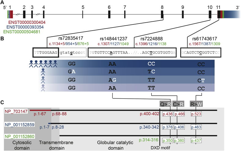
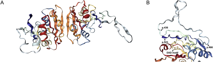
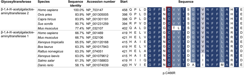
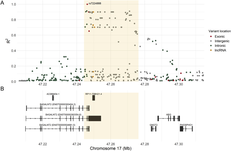
Verification
Claims the paper makes
The claims and statistics of this paper haven’t been checked yet.
Run the check — free, no account needed. It reads the open-access text and checks the sources it cites.
Claims other papers make about this one
Each is checked against this paper’s own text: does it say what the citing paper says it does?
The cited source says what the claim attributes to it.
In the citing paper
“Although the human B4GALNT2 gene is functional22, its expression levels vary and naturally occurring mutations have been identified that correlate with the Sd(a-) phenotype23.”
From Design and testing of a humanized porcine donor for xenotransplantation (2023) · cites this paper as [23]
Self-check
Percentages, sums and reported differences re-computed from the paper’s own numbers.
The self-check needs the paper’s full text.
Citations
Cites
Cunningham, F., Achuthan, P., Akanni, W., Allen, J., Amode, M. R., Armean, I. M., Bennett, R., Bhai, J., Billis, K., Boddu, S., et al. (2019) · Nucleic Acids Research · doi:10.1093/nar/gky1113 · Cited by 925
Research works printed but not linked: 40
- Renton, 1967, Anti-Sda, a new blood group antibody, Vox Sang., 13, 493
- Macvie, 1967, The reactions and inheritance of a new blood group antigen, Sda, Vox Sang., 13, 485
- Morton, 1970, The Sda blood group antigen in tissues and body fluids, Vox Sang., 19, 472
- Blanchard, 1985, Comparative study of glycophorin A derived O-glycans from human Cad, Sd(a+) and Sd(a-) erythrocytes, Biochem. J., 232, 813, 10.1042/bj2320813 · DOI
- Donald, 1983, A blood group Sda-active pentasaccharide isolated from Tamm-Horsfall urinary glycoprotein, Biochem. Biophys. Res. Commun., 115, 625, 10.1016/S0006-291X(83)80190-9 · DOI
- Dohi, 1996, Detection of N-acetylgalactosaminyltransferase mRNA which determines expression of Sda blood group carbohydrate structure in human gastrointestinal mucosa and cancer, Int. J. Cancer, 67, 626, 10.1002/(SICI)1097-0215(19960904)67:5<626::AID-IJC6>3.0.CO;2-W · DOI
- Lo Presti, 2003, Molecular cloning of the human β1,4 N-acetylgalactosaminyltransferase responsible for the biosynthesis of the Sda histo-blood group antigen: the sequence predicts a very long cytoplasmic domain, J. Biochem., 134, 675, 10.1093/jb/mvg192 · DOI
- Montiel, 2003, Molecular cloning, gene organization and expression of the human UDP-GalNAc:Neu5Acα2-3Galβ-R β1,4-N-acetylgalactosaminyltransferase responsible for the biosynthesis of the blood group Sda/Cad antigen: evidence for an unusual extended cytoplasmic domain, Biochem. J., 373, 369, 10.1042/bj20021892 · DOI · Paper page
- Jöud, 2018, Identification of human glycosyltransferase genes expressed in erythroid cells predicts potential carbohydrate blood group loci, Sci. Rep., 8, 6040, 10.1038/s41598-018-24445-5 · DOI
- Takeya, 1987, Identification and characterization of UDP-GalNAc: NeuAcα2−3Galβ1−4Glc(NAc) β1−4(GalNAc to Gal)N-acetylgalactosaminyltransferase in human blood plasma, J. Biochem., 101, 251, 10.1093/oxfordjournals.jbchem.a121898 · DOI
- Spitalnik, 1982, The serology of Sda effects of transfusion and pregnancy, Vox Sang., 42, 308
- Malagolini, 1989, Expression of UDP-GalNAc:NeuAcα2,3Galβ-R β1,4(GalNAc to Gal)N-acetylgalactosaminyltransferase involved in the synthesis of Sda antigen in human large intestine and colorectal carcinomas, Cancer Res., 49, 6466
- Serafini-Cessi, 2005, N-Glycans carried by Tamm-Horsfall glycoprotein have a crucial role in the defense against urinary tract diseases, Glycoconj. J., 22, 383, 10.1007/s10719-005-2142-z · DOI
- Cartron, 1983, Susceptibility to invasion by Plasmodium falciparum of some human erythrocytes carrying rare blood group antigens, Br. J. Haematol., 55, 639, 10.1111/j.1365-2141.1983.tb02846.x · DOI
- Heaton, 2017, A CRISPR activation screen identifies a pan-avian influenza virus inhibitory host factor, Cell Rep., 20, 1503, 10.1016/j.celrep.2017.07.060 · DOI
- Tanaka-Okamoto, 2018, Identification of internally sialylated carbohydrate tumor marker candidates, including Sda/CAD antigens, by focused glycomic analyses utilizing the substrate specificity of neuraminidase, Glycobiology, 28, 247, 10.1093/glycob/cwy010 · DOI
- Guo, 2018, Molecular cloning of the B4GALNT2 gene and its single nucleotide polymorphisms association with litter size in small tail han sheep, Animals, 8, 160, 10.3390/ani8100160 · DOI
- Byrne, 2018, B4GALNT2 and xenotransplantation: a newly appreciated xenogeneic antigen, Xenotransplantation, 25, 10.1111/xen.12394 · DOI
- Dall’Olio, 2014, The expanding roles of the Sda/Cad carbohydrate antigen and its cognate glycosyltransferase B4GALNT2, Biochim. Biophys. Acta, 1840, 443, 10.1016/j.bbagen.2013.09.036 · DOI
- Miller, 1988, A simple salting out procedure for extracting DNA from human nucleated cells, Nucleic Acids Res., 16, 1215, 10.1093/nar/16.3.1215 · DOI
- Hellberg, 2003, Additional molecular bases of the clinically important p blood group phenotype, Transfusion, 43, 899, 10.1046/j.1537-2995.2003.00425.x · DOI
- The 1000 Genomes Project Consortium, 2015, A global reference for human genetic variation, Nature, 526, 68, 10.1038/nature15393 · DOI · Paper page
- Möller, 2016, Erythrogene: a database for in-depth analysis of the extensive variation in 36 blood group systems in the 1000 Genomes Project, Blood Adv, 1, 240, 10.1182/bloodadvances.2016001867 · DOI
- Sudmant, 2015, An integrated map of structural variation in 2,504 human genomes, Nature, 526, 75, 10.1038/nature15394 · DOI · Paper page
- Adzhubei, 2010, A method and server for predicting damaging missense mutations, Nat. Methods, 7, 248, 10.1038/nmeth0410-248 · DOI · Paper page
- Vaser, 2016, SIFT missense predictions for genomes, Nat. Protoc., 11, 1, 10.1038/nprot.2015.123 · DOI · Paper page
- Zerbino, 2018, Ensembl 2018, Nucleic Acids Res., 46, D754, 10.1093/nar/gkx1098 · DOI · Paper page
- Lek, 2016, Analysis of protein-coding genetic variation in 60,706 humans, Nature, 536, 285, 10.1038/nature19057 · DOI · Paper page
- Ameur, 2017, SweGen: a whole-genome data resource of genetic variability in a cross-section of the Swedish population, Eur. J. Hum. Genet., 25, 1253, 10.1038/ejhg.2017.130 · DOI
- Berman, 2000, The Protein Data Bank, Nucleic Acids Res., 28, 235, 10.1093/nar/28.1.235 · DOI · Paper page
- Waterhouse, 2018, SWISS-MODEL: homology modelling of protein structures and complexes, Nucleic Acids Res., 46, W296, 10.1093/nar/gky427 · DOI
- Osawa, 2009, Crystal structure of chondroitin polymerase from Escherichia coli K4, Biochem. Biophys. Res. Commun., 378, 10, 10.1016/j.bbrc.2008.08.121 · DOI
- Rose, 2018, NGL viewer: web-based molecular graphics for large complexes, Bioinformatics, 34, 3755, 10.1093/bioinformatics/bty419 · DOI · Paper page
- Altschul, 1990, Basic local alignment search tool, J. Mol. Biol., 215, 403, 10.1016/S0022-2836(05)80360-2 · DOI · Paper page
- Machiela, 2015, LDlink: a web-based application for exploring population-specific haplotype structure and linking correlated alleles of possible functional variants, Bioinformatics, 31, 3555, 10.1093/bioinformatics/btv402 · DOI · Paper page
- Desmet, 2009, Human Splicing Finder: an online bioinformatics tool to predict splicing signals, Nucleic Acids Res., 37, e67, 10.1093/nar/gkp215 · DOI
- Barretina, 2012, The Cancer Cell Line Encyclopedia enables predictive modelling of anticancer drug sensitivity, Nature, 483, 603, 10.1038/nature11003 · DOI · Paper page
- Breton, 2006, Structures and mechanisms of glycosyltransferases, Glycobiology, 16, 29r, 10.1093/glycob/cwj016 · DOI
- Sringarm, 1974, Cad receptor in Thai blood donors, Vox Sang., 26, 462, 10.1111/j.1423-0410.1974.tb02721.x · DOI
- Sun, 2017, Down-regulation of long non-coding RNA RP11-708H21.4 is associated with poor prognosis for colorectal cancer and promotes tumorigenesis through regulating AKT/mTOR pathway, Oncotarget, 8, 27929, 10.18632/oncotarget.15846 · DOI
Cited by
Sorted by citations
Design and testing of a humanized porcine donor for xenotransplantation
Anand, R. P., Layer, J. V., Heja, D., Hirose, T., Lassiter, G., Firl, D. J., Paragas, V. B., Akkad, A., Chhangawala, S., Colvin, R. B., et al. (2023) · Nature · doi:10.1038/s41586-023-06594-4 · Cited by 302
Metadata
- Authors
- Stenfelt, L., Hellberg, Å., Möller, M., Thornton, N., Larson, G., & Olsson, M. L.
- Venue
- Biochemistry and Biophysics Reports
- Publisher
- Elsevier BV
- Published
- 2019-09
- Volume
- 19
- Pages
- 100659
- ISSN
- 2405-5808
- PMC
- PMC6646742
- Cited by
- 15
- References
- 1
- Language
- en
- Licence
- Funders
- Vetenskapsrådet
- Knut och Alice Wallenbergs Stiftelse
About these checks
Registry, retraction, Cites and statistics results are computed, not judged, with one exception: a printed reference that no index lists is read once by a model before it counts as a citation error. Claims are extracted by a model and kept only when their quote appears verbatim in the text; each cited source is then checked, with a model’s help, for whether it says what the claim relies on. Those verdicts can be wrong — read the claim’s quote and the cited source before relying on one.
Statistics and arithmetic are recomputed, not judged.
Confidence bands are shown only for fields calibrated on our annotated test set. None is calibrated yet, so no band is shown.
What counts as a claim ›How we measure confidence ›How we check this ›
Are you an author, or do you see a problem with this page? Request a review
