

The DOI matches its Crossref record; 56 of the 56 references it cites were found in a registry or an index.
Neuronal-Specific TUBB3 Is Not Required for Normal Neuronal Function but Is Essential for Timely Axon Regeneration
Abstract
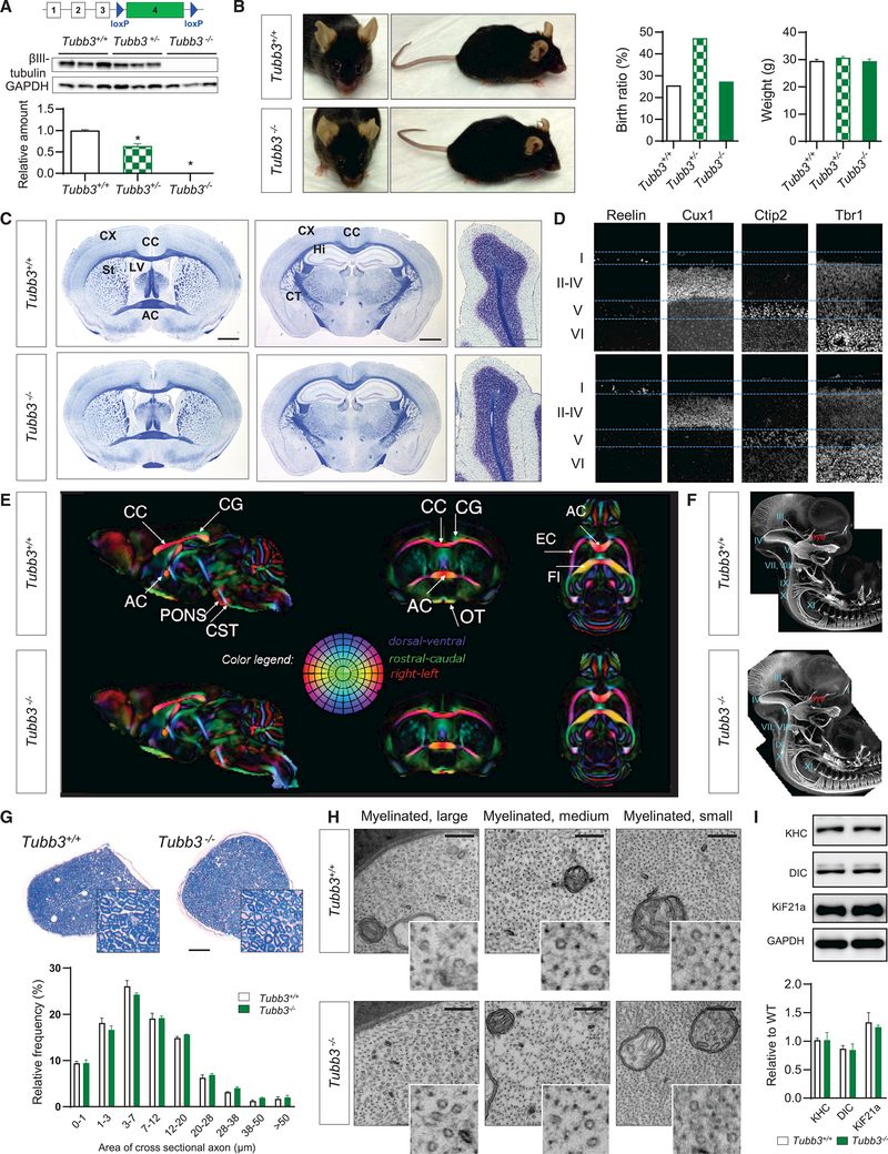
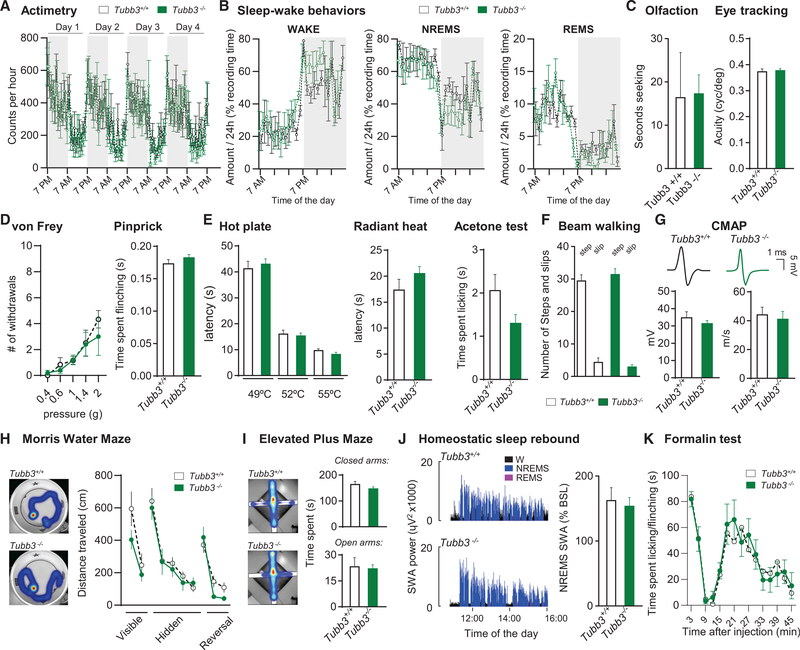
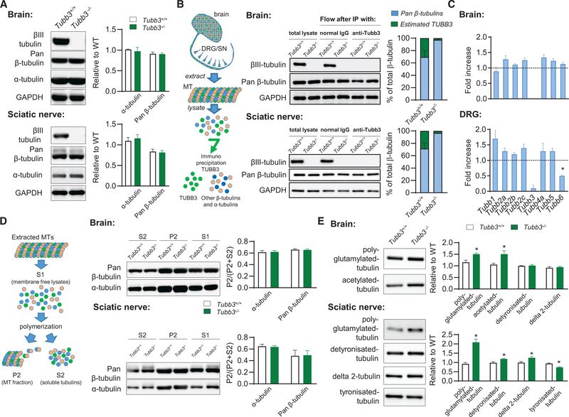
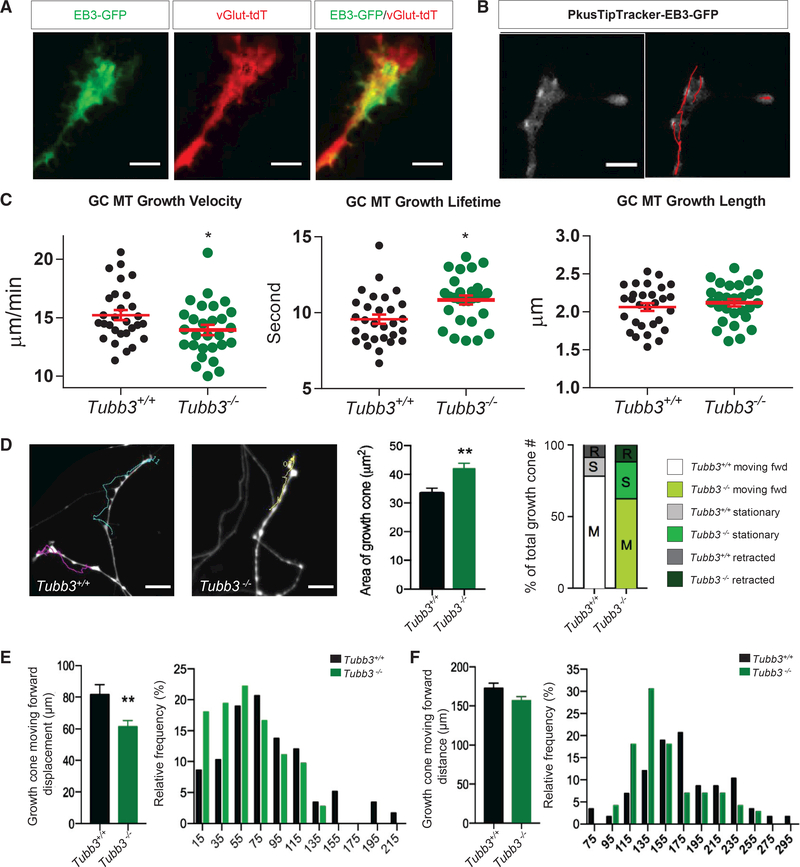
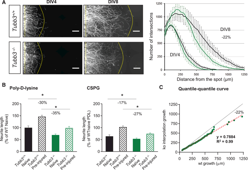
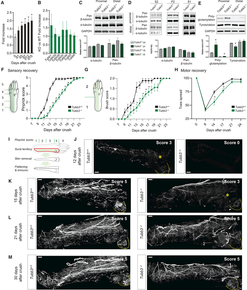
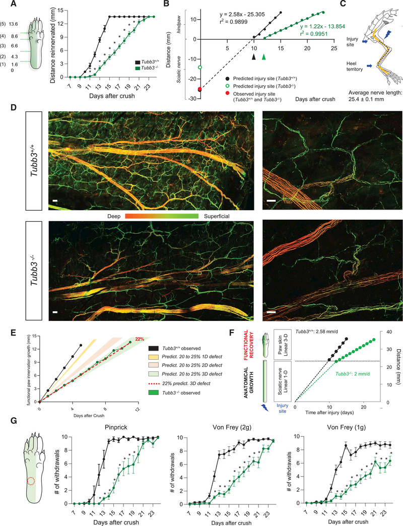
We generated a knockout mouse for the neuronal-specific β-tubulin isoform Tubb3 to investigate its role in nervous system formation and maintenance. Tubb3 −/− mice have no detectable neurobehavioral or neuropathological deficits, and upregulation of mRNA and protein of the remaining β-tubulin isotypes results in equivalent total β-tubulin levels in Tubb3 −/− and wild-type mice. Despite similar levels of total β-tubulin, adult dorsal root ganglia lacking TUBB3 have decreased growth cone microtubule dynamics and a decreased neurite outgrowth rate of 22% in vitro and in vivo . The effect of the 22% slower growth rate is exacerbated for sensory recovery, where fibers must reinnervate the full volume of the skin to recover touch function. Overall, these data reveal that, while TUBB3 is not required for formation of the nervous system, it has a specific role in the rate of peripheral axon regeneration that cannot be replaced by other β-tubulins.
Overview
The claims and statistics of this paper haven’t been checked yet.
Run the check — free, no account needed. It reads the open-access text and checks the sources it cites.
Integrity signals
Registry
Cites
Statistics
Figures
Verification
Claims the paper makes
The claims and statistics of this paper haven’t been checked yet.
Run the check — free, no account needed. It reads the open-access text and checks the sources it cites.
Claims other papers make about this one
Each is checked against this paper’s own text: does it say what the citing paper says it does?
In the citing paper
“TUBB3, a neuron-specific cytoskeletal protein required for neurite extension,51 was predominantly localized to the neurites in NC cells but was mainly confined to the soma in sgDennd10 cells (Figure 1F).”
From Endosomal protein DENND10 promotes developmental competence of neurite extension (2025) · cites this paper as [51]
Self-check
Percentages, sums and reported differences re-computed from the paper’s own numbers.
The self-check needs the paper’s full text.
Citations
Cites
Voxel-Based Morphometry—The Methods
Ashburner, J., & Friston, K. J. (2000) · NeuroImage · doi:10.1006/nimg.2000.0582 · Cited by 8,676
Avants, B., Epstein, C. L., Grossman, M., & Gee, J. C. (2007) · Medical Image Analysis · doi:10.1016/j.media.2007.06.004 · Cited by 5,967
(2000) · Cited by 919
Rapid Quantification of Adult and Developing Mouse Spatial Vision Using a Virtual Optomotor System
Prusky, G. T., Alam, N. M., Beekman, S., & Douglas, R. M. (2004) · Investigative Opthalmology & Visual Science · doi:10.1167/iovs.04-0541 · Cited by 772
Pajevic, S., & Pierpaoli, C. M. (2000) · Magnetic Resonance in Medicine · doi:10.1002/1522-2594(200006)43:6<921::aid-mrm23>3.0.co;2-i · Cited by 765
Self-citationSustained axon regeneration induced by co-deletion of PTEN and SOCS3
Sun, F., Park, K. K., Belin, S., Wang, D., Lu, T., Chen, G., Zhang, K., Yeung, C., Feng, G., Yankner, B. A., et al. (2011) · Nature · doi:10.1038/nature10594 · Cited by 754
Neumann, S., & Woolf, C. J. (1999) · Neuron · doi:10.1016/s0896-6273(00)80755-2 · Cited by 655
Self-citationHuman TUBB3 Mutations Perturb Microtubule Dynamics, Kinesin Interactions, and Axon Guidance
Tischfield, M. A., Baris, H. N., Wu, C., Rudolph, G., Van Maldergem, L., He, W., Chan, W.-M., Andrews, C., Demer, J. L., Robertson, R. L., et al. (2010) · Cell · doi:10.1016/j.cell.2009.12.011 · Cited by 595
Self-citationMicrotubule Stabilization Reduces Scarring and Causes Axon Regeneration After Spinal Cord Injury
Hellal, F., Hurtado, A., Ruschel, J., Flynn, K. C., Laskowski, C. J., Umlauf, M., Kapitein, L. C., Strikis, D., Lemmon, V., Bixby, J., et al. (2011) · Science · doi:10.1126/science.1201148 · Cited by 588
Ruijs, A. C. J., Jaquet, J.-B., Kalmijn, S., Giele, H., & Hovius, S. E. R. (2005) · Plastic and Reconstructive Surgery · doi:10.1097/01.prs.0000172896.86594.07 · Cited by 452
Systemic administration of epothilone B promotes axon regeneration after spinal cord injury
Ruschel, J., Hellal, F., Flynn, K. C., Dupraz, S., Elliott, D. A., Tedeschi, A., Bates, M., Sliwinski, C., Brook, G., Dobrindt, K., et al. (2015) · Science · doi:10.1126/science.aaa2958 · Cited by 432
ATF3 Increases the Intrinsic Growth State of DRG Neurons to Enhance Peripheral Nerve Regeneration
Seijffers, R., Mills, C. D., & Woolf, C. J. (2007) · The Journal of Neuroscience · doi:10.1523/jneurosci.5313-06.2007 · Cited by 425
Self-citationSullivan, K. F., & Cleveland, D. W. (1986) · Proceedings of the National Academy of Sciences · doi:10.1073/pnas.83.12.4327 · Cited by 400
Microtubule dynamics in vitro are regulated by the tubulin isotype composition.
Panda, D., Miller, H. P., Banerjee, A., Ludueña, R. F., & Wilson, L. (1994) · Proceedings of the National Academy of Sciences · doi:10.1073/pnas.91.24.11358 · Cited by 374
Ciaramitaro, P., Mondelli, M., Logullo, F., Grimaldi, S., Battiston, B., Sard, A., Scarinzi, C., Migliaretti, G., Faccani, G., Cocito, D., et al. (2010) · Journal of the Peripheral Nervous System · doi:10.1111/j.1529-8027.2010.00260.x · Cited by 371
The role of microtubule dynamics in growth cone motility and axonal growth.
Tanaka, E., Ho, T., & Kirschner, M. W. (1995) · The Journal of cell biology · doi:10.1083/jcb.128.1.139 · Cited by 367
A Systems-Level Analysis of the Peripheral Nerve Intrinsic Axonal Growth Program
Chandran, V., Coppola, G., Nawabi, H., Omura, T., Versano, R., Huebner, E. A., Zhang, A., Costigan, M., Yekkirala, A., Barrett, L., et al. (2016) · Neuron · doi:10.1016/j.neuron.2016.01.034 · Cited by 366
Self-citationInjury-Induced HDAC5 Nuclear Export Is Essential for Axon Regeneration
Cho, Y., Sloutsky, R., Naegle, K. M., & Cavalli, V. (2013) · Cell · doi:10.1016/j.cell.2013.10.004 · Cited by 327
The wide spectrum of tubulinopathies: what are the key features for the diagnosis?
Bahi-Buisson, N., Poirier, K., Fourniol, F., Saillour, Y., Valence, S., Lebrun, N., Hully, M., Fallet Bianco, C., Boddaert, N., Elie, C., et al. (2014) · Brain · doi:10.1093/brain/awu082 · Cited by 324
Poirier, K., Saillour, Y., Bahi-Buisson, N., Jaglin, X. H., Fallet-Bianco, C., Nabbout, R., Castelnau-Ptakhine, L., Roubertie, A., Attie-Bitach, T., Desguerre, I., et al. (2010) · Human Molecular Genetics · doi:10.1093/hmg/ddq377 · Cited by 271
Accelerating axonal growth promotes motor recovery after peripheral nerve injury in mice
Ma, C. H. E., Omura, T., Cobos, E. J., Latrémolière, A., Ghasemlou, N., Brenner, G. J., van Veen, J. E., Barrett, L. B., Sawada, T., Gao, F., et al. (2011) · Journal of Clinical Investigation · doi:10.1172/jci58675 · Cited by 263
Self-citationInjury-Induced Decline of Intrinsic Regenerative Ability Revealed by Quantitative Proteomics
Belin, S., Nawabi, H., Wang, C., Tang, S., Latremoliere, A., Warren, P., Schorle, H., Uncu, C., Woolf, C. J., He, Z., et al. (2015) · Neuron · doi:10.1016/j.neuron.2015.03.060 · Cited by 259
Self-citationAre tubulin isotypes functionally significant.
Ludueña, R. F. (1993) · Molecular Biology of the Cell · doi:10.1091/mbc.4.5.445 · Cited by 253
Distinct Roles for Secreted Semaphorin Signaling in Spinal Motor Axon Guidance
Huber, A. B., Kania, A., Tran, T. S., Gu, C., García, N. D. M., Lieberam, I., Johnson, D., Jessell, T. M., Ginty, D. D., & Kolodkin, A. L. (2005) · Neuron · doi:10.1016/j.neuron.2005.12.003 · Cited by 233
The multitubulin hypothesis revisited: what have we learned?
Cleveland, D. W. (1987) · The Journal of Cell Biology · doi:10.1083/jcb.104.3.381 · Cited by 215
Resolving bundled microtubules using anti-tubulin nanobodies
Mikhaylova, M., Cloin, B. M. C., Finan, K., van den Berg, R., Teeuw, J., Kijanka, M. M., Sokołowski, M., Katrukha, E. A., Maidorn, M., Opazo, F., et al. (2015) · Nature Communications · doi:10.1038/ncomms8933 · Cited by 206
Tubulin isoform composition tunes microtubule dynamics
Vemu, A., Atherton, J., Spector, J. O., Moores, C. A., & Roll‐Mecak, A. (2017) · Molecular Biology of the Cell · doi:10.1091/mbc.e17-02-0124 · Cited by 186
Irfanoglu, M. O., Modi, P., Nayak, A., Hutchinson, E. B., Sarlls, J. E., & Pierpaoli, C. M. (2014) · NeuroImage · doi:10.1016/j.neuroimage.2014.11.042 · Cited by 176
Self-citationWhat’s Wrong with My Mouse? Behavioral Phenotyping of Transgenic and Knockout Mice
Crusio, W. E. (2008) · Genes Brain & Behavior · doi:10.1111/j.1601-183x.2008.00424_1.x · Cited by 167
Trivedi, N., Marsh, P. J., Goold, R., Wood‐Kaczmar, A., & Gordon‐Weeks, P. R. (2005) · Journal of Cell Science · doi:10.1242/jcs.01697 · Cited by 166
Buchthal, F., & Kühl, V. (1979) · Journal of Neurology Neurosurgery & Psychiatry · doi:10.1136/jnnp.42.5.436 · Cited by 139
Expression of the Class III β‐tubulin isotype in developing neurons in culture
Ferreira, A., & Cáceres, A. (1992) · Journal of Neuroscience Research · doi:10.1002/jnr.490320407 · Cited by 136
Jiang, Y. Q., & Oblinger, M. M. (1992) · Journal of Cell Science · doi:10.1242/jcs.103.3.643 · Cited by 132
Tischfield, M. A., & Engle, E. C. (2010) · Bioscience Reports · doi:10.1042/bsr20100025 · Cited by 131
Self-citationA novel syndrome caused by the E410K amino acid substitution in the neuronal β-tubulin isotype 3
Chew, S., Balasubramanian, R., Chan, W. M., Kang, P. B., Andrews, C. V., Webb, B. D., MacKinnon, S. E., Oystreck, D. T., Rankin, J. K., Crawford, T. O., et al. (2013) · Brain · doi:10.1093/brain/aws345 · Cited by 126
Self-citationHuman CFEOM1 Mutations Attenuate KIF21A Autoinhibition and Cause Oculomotor Axon Stalling
Cheng, L., Desai, J. S., Miranda, C. J., Duncan, J. S., Qiu, W., Nugent, A. A., Kolpak, A., Wu, C. C., Drokhlyansky, E., DeLisle, M. M., et al. (2014) · Neuron · doi:10.1016/j.neuron.2014.02.038 · Cited by 118
Self-citationMicrotubule stability decreases axon elongation but not axoplasm production
Rochlin, M. W., Wickline, K. M., & Bridgman, P. C. (1996) · The Journal of Neuroscience · doi:10.1523/jneurosci.16-10-03236.1996 · Cited by 117
Nawabi, H., Belin, S., Cartoni, R., Williams, P. R., Wang, C., Latrémolière, A., Wang, X., Zhu, J., Taub, D. G., Fu, X., et al. (2015) · Neuron · doi:10.1016/j.neuron.2015.10.005 · Cited by 116
Self-citationTi, S.-C., Pamula, M. C., Howes, S. C., Duellberg, C., Cade, N. I., Kleiner, R. E., Forth, S. T., Surrey, T., Nogales, E., & Kapoor, T. M. (2016) · Developmental Cell · doi:10.1016/j.devcel.2016.03.003 · Cited by 109
β‐Tubulin mutations that cause severe neuropathies disrupt axonal transport
Niwa, S., Takahashi, H., & Hirokawa, N. (2013) · The EMBO Journal · doi:10.1038/emboj.2013.59 · Cited by 104
The structured core of human β tubulin confers isotype-specific polymerization properties
Pamula, M. C., Ti, S.-C., & Kapoor, T. M. (2016) · The Journal of Cell Biology · doi:10.1083/jcb.201603050 · Cited by 103
Recognizable cerebellar dysplasia associated with mutations in multiple tubulin genes
Oegema, R., Cushion, T. D., Phelps, I. G., Chung, S., Dempsey, J. C., Collins, S., Mullins, J. G. L., Dudding, T. E., Gill, H., Green, A., et al. (2015) · Human Molecular Genetics · doi:10.1093/hmg/ddv250 · Cited by 94
Sustained GSK3 activity markedly facilitates nerve regeneration
Gobrecht, P., Leibinger, M., Andreadaki, A., & Fischer, D. (2014) · Nature Communications · doi:10.1038/ncomms5561 · Cited by 92
Moskowitz, P. F., & Oblinger, M. (1995) · Journal of Neuroscience · doi:10.1523/jneurosci.15-02-01545.1995 · Cited by 86
Moskowitz, P. F., Smith, R. S., Pickett, J., Frankfurter, A. J., & Oblinger, M. M. (1993) · Journal of Neuroscience Research · doi:10.1002/jnr.490340113 · Cited by 83
Mass Spectrometry Analysis of C-Terminal Posttranslational Modifications of Tubulins
Redeker, V. (2010) · Methods in cell biology · doi:10.1016/s0091-679x(10)95006-1 · Cited by 82
Promotion of Functional Nerve Regeneration by Inhibition of Microtubule Detyrosination
Gobrecht, P., Andreadaki, A., Diekmann, H., Heskamp, A., Leibinger, M., & Fischer, D. (2016) · Journal of Neuroscience · doi:10.1523/jneurosci.4486-15.2016 · Cited by 82
Two unique TUBB3 mutations cause both CFEOM3 and malformations of cortical development
Whitman, M. C., Andrews, C. V., Chan, W. M., Tischfield, M. A., Stasheff, S. F., Brancati, F., Ortiz-Gonzalez, X. R., Nuovo, S., Garaci, F., MacKinnon, S. E., et al. (2015) · American Journal of Medical Genetics Part A · doi:10.1002/ajmg.a.37362 · Cited by 74
Self-citationLudueña, R. F., & Banerjee, A. K. (2009) · Humana Press eBooks · doi:10.1007/978-1-59745-336-3_6 · Cited by 67
Saillour, Y., Broix, L., Bruel‐Jungerman, E., Lebrun, N., Muraca, G., Rucci, J., Poirier, K., Belvindrah, R., Francis, F., & Chelly, J. (2013) · Human Molecular Genetics · doi:10.1093/hmg/ddt538 · Cited by 65
The GSK3–MAP1B pathway controls neurite branching and microtubule dynamics
Barnat, M., Benassy, M., Vincensini, L., Soares, S., Fassier, C., Propst, F., Andrieux, A., von Boxberg, Y., & Nothias, F. (2016) · Molecular and Cellular Neuroscience · doi:10.1016/j.mcn.2016.01.001 · Cited by 59
Song, W., Cho, Y., Watt, D., & Cavalli, V. (2015) · Journal of Biological Chemistry · doi:10.1074/jbc.m114.622753 · Cited by 48
Minoura, I., Takazaki, H., Ayukawa, R., Saruta, C., Hachikubo, Y., Uchimura, S., Hida, T., Kamiguchi, H., Shimogori, T., & Muto, E. (2016) · Nature Communications · doi:10.1038/ncomms10058 · Cited by 34
Using plusTipTracker Software to Measure Microtubule Dynamics in Xenopus laevis Growth Cones
Stout, A. C., D'Amico, S., Enzenbacher, T. B., Ebbert, P. T., & Lowery, L. A. (2014) · Journal of Visualized Experiments · doi:10.3791/52138 · Cited by 24
Injury-Induced HDAC5 Nuclear Export Is Essential for Axon Regeneration
Cho, Y., Sloutsky, R., Naegle, K. M., & Cavalli, V. (2015) · Cell · doi:10.1016/j.cell.2015.04.019 · Cited by 18
Hoffman, P. N., & Ludueña, R. F. (1996) · Brain Research · doi:10.1016/s0006-8993(96)00980-8 · Cited by 15
Cited by
The tubulin code and its role in controlling microtubule properties and functions
Janke, C., & Magiera, M. M. (2020) · Nature Reviews Molecular Cell Biology · doi:10.1038/s41580-020-0214-3 · Cited by 956
Global analysis of protein turnover dynamics in single cells
Sabatier, P., Lechner, M., Guzmán, U. H., Beusch, C. M., Zeng, X., Wang, L., Izaguirre, F., Seth, A., Gritsenko, O., Rodin, S., et al. (2025) · Cell · doi:10.1016/j.cell.2025.03.002 · Cited by 41
Doublecortin-like Kinase 1 Regulates α-Synuclein Levels and Toxicity
Vázquez-Vélez, G. E., Gonzales, K. A., Revelli, J.-P., Adamski, C. J., Alavi Naini, F., Bajić, A., Craigen, E., Richman, R., Heman-Ackah, S. M., Wood, M. J. A., et al. (2020) · The Journal of Neuroscience · doi:10.1523/jneurosci.1076-19.2019 · Cited by 22
Endosomal protein DENND10 promotes developmental competence of neurite extension
Li, A., Zhang, J., Ma, C., Qi, L., Hu, Q., Li, Q., Fang, Y., Song, J., Liu, Y., & Zhang, Y. (2025) · iScience · doi:10.1016/j.isci.2025.112385
Metadata
- Authors
- Latremoliere, A., Cheng, L., DeLisle, M., Wu, C., Chew, S., Hutchinson, E. B., Sheridan, A., Alexandre, C., Latremoliere, F., Sheu, S.-H., Golidy, S., Omura, T., Huebner, E. A., Fan, Y., Whitman, M. C., Nguyen, E., Hermawan, C., Pierpaoli, C., Tischfield, M. A., Woolf, C. J., & Engle, E. C.
- Venue
- Cell Reports
- Publisher
- Elsevier BV
- Published
- 2018-08
- Volume
- 24
- Issue
- 7
- Pages
- 1865-1879.e9
- ISSN
- 2211-1247
- PMC
- PMC6155462
- Cited by
- 183
- References
- 56
- Language
- en
- Licence
- www.elsevier.com/tdm/userlicense/1.0/ (tdm)
- www.elsevier.com/legal/tdmrep-license (tdm)
- creativecommons.org/licenses/by-nc-nd/4.0/ (vor)
- Funders
- Dr. Miriam and Sheldon G. Adelson Medical Research Foundation
- National Institutes of Health (5T32HL110852-05, K08EY027850)
- Congressionally Directed Medical Research Programs (W81XWH-13-2-0019)
- Howard Hughes Medical Institute
About these checks
Registry, retraction, Cites and statistics results are computed, not judged, with one exception: a printed reference that no index lists is read once by a model before it counts as a citation error. Claims are extracted by a model and kept only when their quote appears verbatim in the text; each cited source is then checked, with a model’s help, for whether it says what the claim relies on. Those verdicts can be wrong — read the claim’s quote and the cited source before relying on one.
Statistics and arithmetic are recomputed, not judged.
Confidence bands are shown only for fields calibrated on our annotated test set. None is calibrated yet, so no band is shown.
What counts as a claim ›How we measure confidence ›How we check this ›
Are you an author, or do you see a problem with this page? Request a review
Data sources
Bibliographic data: Semantic Scholar, OpenAlex, Crossref.
Contains information from the Semantic Scholar Academic Graph Datasets, made available under the ODC Attribution License (ODC-BY 1.0).
Semantic Scholar’s data is described in: Kinney, R., et al. (2023). The Semantic Scholar Open Data Platform. arXiv:2301.10140.
