Student or educator? Sign up with your school email and get 6 months of Pro free.
Cited by 21

The DOI matches its Crossref record; 73 of the 73 references it cites were found in a registry or an index.

Molecular basis of C-S bond cleavage in the glycyl radical enzyme isethionate sulfite-lyase

Dawson, Christopher D., Irwin, Stephania M., Backman, Lindsey R.F., Le, Chip, Wang, Jennifer X., Vennelakanti, Vyshnavi, Yang, Zhongyue, Kulik, Heather J., Drennan, Catherine L., Balskus, Emily P.·2021·Cell Chemical Biology

Abstract

Desulfonation of isethionate by the bacterial glycyl radical enzyme (GRE) isethionate sulfite-lyase (IslA) generates sulfite, a substrate for respiration that in turn produces the disease-associated metabolite hydrogen sulfide. Here, we present a 2.7 Å resolution X-ray structure of wild-type IslA from Bilophila wadsworthia with isethionate bound. In comparison with other GREs, alternate positioning of the active site β strands allows for distinct residue positions to contribute to substrate binding. These structural differences, combined with sequence variations, create a highly tailored active site for the binding of the negatively charged isethionate substrate. Through the kinetic analysis of 14 IslA variants and computational analyses, we probe the mechanism by which radical chemistry is used for C-S bond cleavage. This work further elucidates the structural basis of chemistry within the GRE superfamily and will inform structure-based inhibitor design of IsIA and thus of microbial hydrogen sulfide production.

Overview

The claims and statistics of this paper haven’t been checked yet.

Run the check — free, no account needed. It reads the open-access text and checks the sources it cites.

A few free checks a day · papers already verified open instantly · results are public

Integrity signals

Registry

Crossref · 10.1016/j.chembiol.2021.03.001

Cites

73 of 73 references verified0 citation errors · 0 not checkable

Statistics

Figures

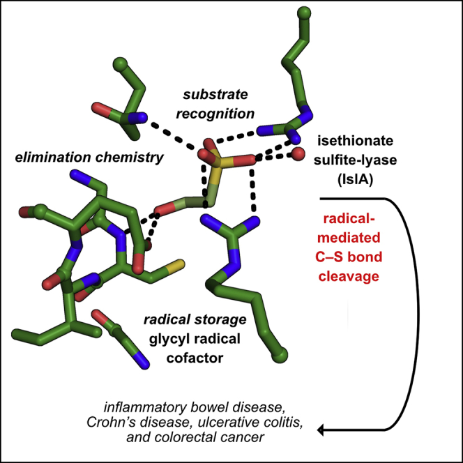
Graphical abstract
Graphical abstract CC BY · PubMed Central
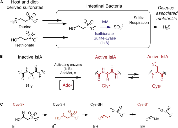
Figure 1 IslA-mediated anaerobic metabolism of organosulfonates by intestinal bacteria releases the disease-associated metabolite hydrogen sulfide (H2S) (A) Deamination of taurine by human gut microbes yields isethionate (Ise), which is cleaved and reduced to H2S in microbial respiration. (B) The activating enzyme for IslA, IslB, installs a glycyl radical on a particular glycine residue of IslA using radical SAM chemistry, i.e., the formation of a 5′-deoxyadenosyl radical (Ado⋅) species from the reductive cleavage of S-adenosylmethionine (AdoMet) using an one electron reduced [4Fe-4S] cluster. The glycyl radical (Gly⋅) transiently forms the catalytically essential thiyl radical species (Cys⋅). (C) Proposed reaction scheme for IslA. Radical species are shown in red.
Figure 1 IslA-mediated anaerobic metabolism of organosulfonates by intestinal bacteria releases the disease-associated metabolite hydrogen sulfide (H2S) (A) Deamination of taurine by human gut microbes yields isethionate (Ise), which is cleaved and reduced to H2S in microbial respiration. (B) The activating enzyme for IslA, IslB, installs a glycyl radical on a particular glycine residue of IslA using radical SAM chemistry, i.e., the formation of a 5′-deoxyadenosyl radical (Ado⋅) species from the reductive cleavage of S-adenosylmethionine (AdoMet) using an one electron reduced [4Fe-4S] cluster. The glycyl radical (Gly⋅) transiently forms the catalytically essential thiyl radical species (Cys⋅). (C) Proposed reaction scheme for IslA. Radical species are shown in red.CC BY · PubMed Central
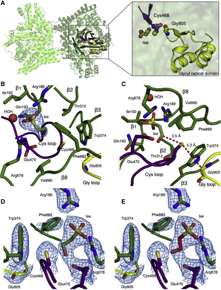
Figure 2 Overall architecture of Bilophila wadsworthia isethionate sulfite-lyase (IslA) (A) IslA dimer contains an active site comprised of two five-stranded half barrels enclosing substrate, the Cys loop (purple) and the Gly loop (yellow). Shown in spheres are Ise, and the catalytic cysteine and glycine residues. (B) Active site views of substrate-interacting residues with Fo − Fc composite omit map contoured to 1.5σ around Ise. (C) Active site view with proposed H atom abstraction route shown in red. (D) A different orientation of the Ise-bound IsIA structure and maps shown in (B). In this refined orientation of Ise (green carbons), it is the pro-R hydrogen on C2 that points toward Cys468. (E) When Ise (red carbons) is modeled into the map shown in (B) such that the pro-S hydrogen on C2 is pointing toward Cys468, the fit to the density is less good. (D) and (E) also show composite omit map contoured to 1.5σ (blue) for the side chains of residues that surround the substrate. Chain A was used to generate (D and E). Chain D was used for all other structure figures.
Figure 2 Overall architecture of Bilophila wadsworthia isethionate sulfite-lyase (IslA) (A) IslA dimer contains an active site comprised of two five-stranded half barrels enclosing substrate, the Cys loop (purple) and the Gly loop (yellow). Shown in spheres are Ise, and the catalytic cysteine and glycine residues. (B) Active site views of substrate-interacting residues with Fo − Fc composite omit map contoured to 1.5σ around Ise. (C) Active site view with proposed H atom abstraction route shown in red. (D) A different orientation of the Ise-bound IsIA structure and maps shown in (B). In this refined orientation of Ise (green carbons), it is the pro-R hydrogen on C2 that points toward Cys468. (E) When Ise (red carbons) is modeled into the map shown in (B) such that the pro-S hydrogen on C2 is pointing toward Cys468, the fit to the density is less good. (D) and (E) also show composite omit map contoured to 1.5σ (blue) for the side chains of residues that surround the substrate. Chain A was used to generate (D and E). Chain D was used for all other structure figures.CC BY · PubMed Central
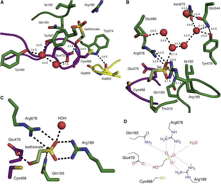
Figure 3 Ise binding mode in IslA (A) Residues and water molecules interacting with Ise hydroxyl group and Gly loop shown as dotted lines and waters shown as red spheres. (B) Residues and water molecules interacting with Ise sulfonate group. (C) Simplified active site view. (D) Active site hydrogen bond interaction scheme.
Figure 3 Ise binding mode in IslA (A) Residues and water molecules interacting with Ise hydroxyl group and Gly loop shown as dotted lines and waters shown as red spheres. (B) Residues and water molecules interacting with Ise sulfonate group. (C) Simplified active site view. (D) Active site hydrogen bond interaction scheme.CC BY · PubMed Central
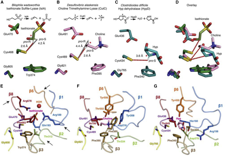
Figure 4 Substrate positioning and active site architecture differs among GRE eliminases (A) IslA shown in green with proposed radical transfer pathway shown as a red dotted line from the catalytic Cys (Cys468) to the closest substrate carbon marked with a red star. The gray dotted line indicates the distance between the more distant hydrogen of the substrate carbon. (B) CutC (PDB: 5FAU) shown in pink. (C) HypD (PDB: 6VXE) shown in teal. (D) Overlay of (A–C). (E) IslA with substrate, Gly loop, Cys loop, β1, β2, β3, β6, and β8 shown as pink, yellow, purple, blue, green, brown, orange, and red, respectively. Arrows highlight particularly different β strands. (F) CutC (PDB: 5FAU) colored as in (E). (G) HypD (PDB: 6VXE) colored as in (E).
Figure 4 Substrate positioning and active site architecture differs among GRE eliminases (A) IslA shown in green with proposed radical transfer pathway shown as a red dotted line from the catalytic Cys (Cys468) to the closest substrate carbon marked with a red star. The gray dotted line indicates the distance between the more distant hydrogen of the substrate carbon. (B) CutC (PDB: 5FAU) shown in pink. (C) HypD (PDB: 6VXE) shown in teal. (D) Overlay of (A–C). (E) IslA with substrate, Gly loop, Cys loop, β1, β2, β3, β6, and β8 shown as pink, yellow, purple, blue, green, brown, orange, and red, respectively. Arrows highlight particularly different β strands. (F) CutC (PDB: 5FAU) colored as in (E). (G) HypD (PDB: 6VXE) colored as in (E).CC BY · PubMed Central
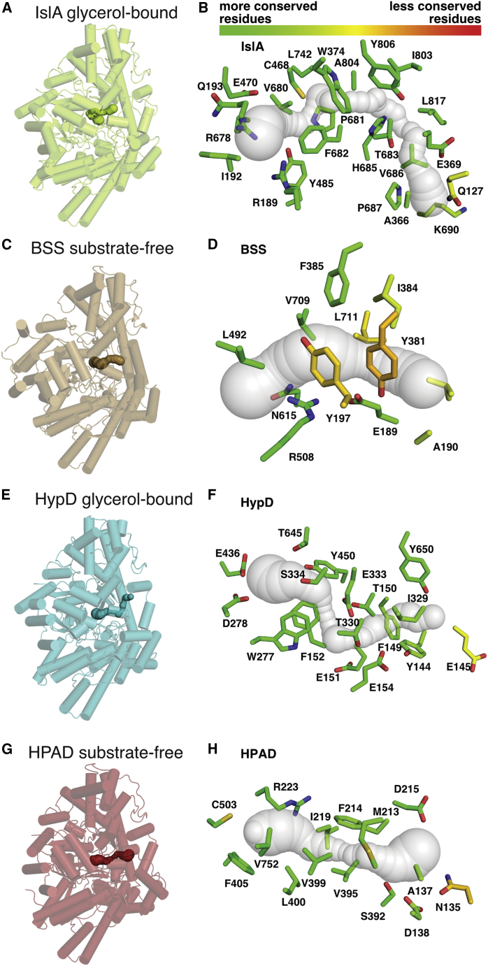
Figure 5 Several putative GRE substrate channels share a similar location (A) One monomer of the IslA dimer is shown in green and oriented similarly to other GRE enzyme monomers, which are shown transparent with channels identified using CAVER 3.0, shown opaque. (B) Conservation of residues of substrate channel (transparent surface) of glycerol-bound IslA. Residues within 4 Å of the putative substrate channel are shown as sticks and colored by conservation score as computed by ConSurf (Ashkenazy et al., 2016) with a gradient from lower scores (more conserved) show in green to higher scores (less conserved) shown in red. (C) BSS (PDB: 4PKC) α subunit (brown). (D) Same representation as (B), but for BSS α subunit (PDB: 4PKC). (E) HypD (PDB: 6VXE) (teal). (F) Same representation as (B), but for HypD (PDB: 6VXE). (G) HPAD (PDB: 2Y8N) α subunit (red). (H) Same representation as (B), but for HPAD α subunit (PDB: 2Y8N).
Figure 5 Several putative GRE substrate channels share a similar location (A) One monomer of the IslA dimer is shown in green and oriented similarly to other GRE enzyme monomers, which are shown transparent with channels identified using CAVER 3.0, shown opaque. (B) Conservation of residues of substrate channel (transparent surface) of glycerol-bound IslA. Residues within 4 Å of the putative substrate channel are shown as sticks and colored by conservation score as computed by ConSurf (Ashkenazy et al., 2016) with a gradient from lower scores (more conserved) show in green to higher scores (less conserved) shown in red. (C) BSS (PDB: 4PKC) α subunit (brown). (D) Same representation as (B), but for BSS α subunit (PDB: 4PKC). (E) HypD (PDB: 6VXE) (teal). (F) Same representation as (B), but for HypD (PDB: 6VXE). (G) HPAD (PDB: 2Y8N) α subunit (red). (H) Same representation as (B), but for HPAD α subunit (PDB: 2Y8N).CC BY · PubMed Central
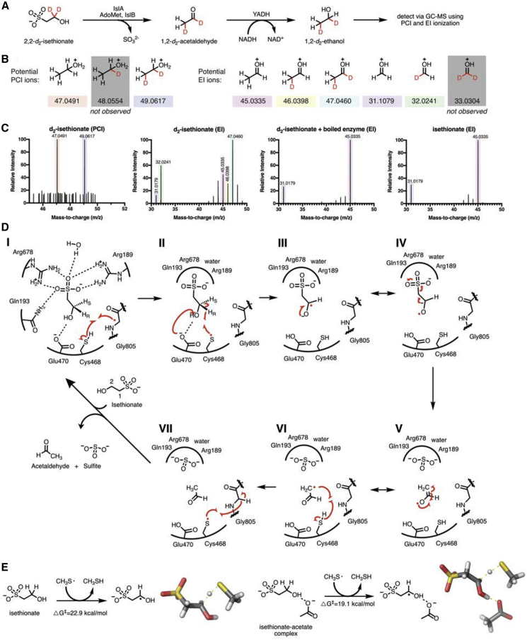
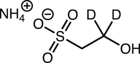
Figure 6 Biochemical and computational support for proposed IsIA mechanism (A) Schematic of IsIA GC-MS assay with stable-isotope-labeled Ise. (B) Potential positive chemical ionization (PCI) ions to determine total deuterium incorporation and potential electron impact (EI) ions used to identify the location of the deuterium atoms. (C) MS data from GC-MS assays. The enzymatic reaction with 2,2-d2-Ise generates 1,2-d2-ethanol as the only detectable deuterated product. Unlabeled ethanol (m/z = 47.0491) and d2-ethanol (m/z = 49.0617) are detected via PCI when IslA reacts with 2,2-d2-Ise. Using EI ionization and 2,2-d2-Ise as a substrate for IslA, the ion at 47.0460 represents di-deuterated ethanol. There is a +1 shift of the 31.0179 fragment ion of this product to 32.0241, indicating only a single deuterium is on the C1 fragment, as double-deuterated 33.0304 is not observed. We have located this deuterium to the C, rather than the O, since the O-H bond is generated via the yeast alcohol dehydrogenase-catalyzed reduction reaction that takes place in aqueous buffer. Thus, the 46.0398 ion arises from a loss of deuterium due to ionization and does not correspond to 1-d1-ethanol. The reaction of IslA with unlabeled isethionate generates unlabeled ethanol (m/z = 45.0335 and 31.0179). When boiled enzyme is mixed with d2-Ise, unlabeled ethanol is present in the background, likely due to our multi-user anaerobic chambers. These assays were performed in triplicate, and a representative spectrum is shown. (D) Proposed 1,2–elimination mechanism of IslA. During the catalytic cycle, the glycyl radical (Gly805) abstracts a hydrogen atom from Cys468, forming a thiyl radical (I, II). This thiyl radical abstracts the pro-R hydrogen atom from C2 of Ise. Glu470 deprotonates the hydroxyl group of Ise to form a transient substrate ketyl radical species (III, IV). This unstable intermediate decomposes, resulting in C-S bond cleavage and release of sulfite (V). The resulting radical species then abstracts a hydrogen atom from Cys468 to produce the second product, acetaldehyde (VI, VII). Arrows are shown in red. Hydrogen bonds are shown in dashed black. (E) Schematic of the local coupled cluster quantum mechanical calculations of C-H abstraction from Ise both with and without an acetate molecule present in a dielectric medium. Spatially resolved structures of the transition states are shown beside each schematic.
Figure 6 Biochemical and computational support for proposed IsIA mechanism (A) Schematic of IsIA GC-MS assay with stable-isotope-labeled Ise. (B) Potential positive chemical ionization (PCI) ions to determine total deuterium incorporation and potential electron impact (EI) ions used to identify the location of the deuterium atoms. (C) MS data from GC-MS assays. The enzymatic reaction with 2,2-d2-Ise generates 1,2-d2-ethanol as the only detectable deuterated product. Unlabeled ethanol (m/z = 47.0491) and d2-ethanol (m/z = 49.0617) are detected via PCI when IslA reacts with 2,2-d2-Ise. Using EI ionization and 2,2-d2-Ise as a substrate for IslA, the ion at 47.0460 represents di-deuterated ethanol. There is a +1 shift of the 31.0179 fragment ion of this product to 32.0241, indicating only a single deuterium is on the C1 fragment, as double-deuterated 33.0304 is not observed. We have located this deuterium to the C, rather than the O, since the O-H bond is generated via the yeast alcohol dehydrogenase-catalyzed reduction reaction that takes place in aqueous buffer. Thus, the 46.0398 ion arises from a loss of deuterium due to ionization and does not correspond to 1-d1-ethanol. The reaction of IslA with unlabeled isethionate generates unlabeled ethanol (m/z = 45.0335 and 31.0179). When boiled enzyme is mixed with d2-Ise, unlabeled ethanol is present in the background, likely due to our multi-user anaerobic chambers. These assays were performed in triplicate, and a representative spectrum is shown. (D) Proposed 1,2–elimination mechanism of IslA. During the catalytic cycle, the glycyl radical (Gly805) abstracts a hydrogen atom from Cys468, forming a thiyl radical (I, II). This thiyl radical abstracts the pro-R hydrogen atom from C2 of Ise. Glu470 deprotonates the hydroxyl group of Ise to form a transient substrate ketyl radical species (III, IV). This unstable intermediate decomposes, resulting in C-S bond cleavage and release of sulfite (V). The resulting radical species then abstracts a hydrogen atom from Cys468 to produce the second product, acetaldehyde (VI, VII). Arrows are shown in red. Hydrogen bonds are shown in dashed black. (E) Schematic of the local coupled cluster quantum mechanical calculations of C-H abstraction from Ise both with and without an acetate molecule present in a dielectric medium. Spatially resolved structures of the transition states are shown beside each schematic.CC BY · PubMed Central
Image 8
CC BY · PubMed Central
Image 9
CC BY · PubMed Central
Image 10
CC BY · PubMed Central

Verification

Claims the paper makes

The claims and statistics of this paper haven’t been checked yet.

Run the check — free, no account needed. It reads the open-access text and checks the sources it cites.

A few free checks a day · papers already verified open instantly · results are public

Claims other papers make about this one

Each is checked against this paper’s own text: does it say what the citing paper says it does?

Self-check

Percentages, sums and reported differences re-computed from the paper’s own numbers.

The self-check needs the paper’s full text.

Citations

Cites

Show all 73 references

Metadata

Authors
Dawson, C. D., Irwin, S. M., Backman, L. R. F., Le, C., Wang, J. X., Vennelakanti, V., Yang, Z., Kulik, H. J., Drennan, C. L., & Balskus, E. P.
Venue
Cell Chemical Biology
Publisher
Elsevier BV
Published
2021-09
Volume
28
Issue
9
Pages
1333-1346.e7
ISSN
2451-9456
Cited by
21
References
73
Language
en
Licence
Funders
  • Natural Sciences and Engineering Research Council of Canada
  • National Science Foundation (1122374)
  • Bill and Melinda Gates Foundation
  • National Institutes of Health (R35 GM126982, T32GM007287)
  • Office of Science
  • Howard Hughes Medical Institute
  • Argonne National Laboratory (DE-AC02-06CH11357)
  • U.S. Department of Energy
  • Massachusetts Institute of Technology
  • National Science Foundation Directorate for Engineering (CBET-1704266)
  • Office of Research Infrastructure Programs, National Institutes of Health (S10 RR029205, S10OD021527)
  • Burroughs Wellcome Fund

About these checks

Registry, retraction, Cites and statistics results are computed, not judged, with one exception: a printed reference that no index lists is read once by a model before it counts as a citation error. Claims are extracted by a model and kept only when their quote appears verbatim in the text; each cited source is then checked, with a model’s help, for whether it says what the claim relies on. Those verdicts can be wrong — read the claim’s quote and the cited source before relying on one.

Statistics and arithmetic are recomputed, not judged.

Confidence bands are shown only for fields calibrated on our annotated test set. None is calibrated yet, so no band is shown.

What counts as a claim ›How we measure confidence ›How we check this ›

Are you an author, or do you see a problem with this page? Request a review

Data sources

Bibliographic data: Semantic Scholar, OpenAlex, Crossref.

Contains information from the Semantic Scholar Academic Graph Datasets, made available under the ODC Attribution License (ODC-BY 1.0).

Semantic Scholar’s data is described in: Kinney, R., et al. (2023). The Semantic Scholar Open Data Platform. arXiv:2301.10140.