Student or educator? Sign up with your school email and get 6 months of Pro free.
Cited by 39

The DOI matches its Crossref record; 2 of the 2 references it cites were found in a registry or an index.

Systemic enzyme delivery by blood-brain barrier-penetrating SapC-DOPS nanovesicles for treatment of neuronopathic Gaucher disease

Sun, Ying, Liou, Benjamin, Chu, Zhengtao, Fannin, Venette, Blackwood, Rachel, Peng, Yanyan, Grabowski, Gregory A., Davis, Harold W., Qi, Xiaoyang·2020·eBioMedicine

Overview

The claims and statistics of this paper haven’t been checked yet.

Run the check — free, no account needed. It reads the open-access text and checks the sources it cites.

A few free checks a day · papers already verified open instantly · results are public

Integrity signals

Registry

Crossref · 10.1016/j.ebiom.2020.102735

Cites

2 of 2 references verified0 citation errors · 0 not checkable

Statistics

Figures

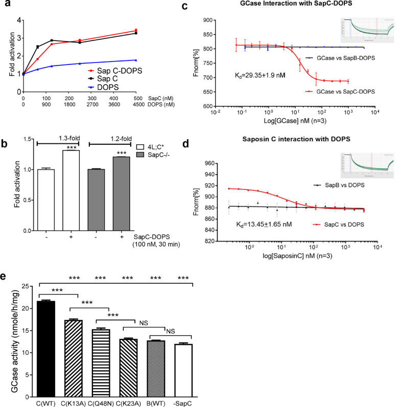
Fig. 1 SapC-DOPS activates GCase. (a) SapC-DOPS and SapC similarly activate GCase in a cell- free assay. DOPS is a control. SapC, 0 to 500 nM; DOPS, 0 - 4500 nM. Free GCase incubated with SapC-DOPS or SapC for 30 min before determining the GCase activity. (b) SapC-DOPS increases GCase activity in fibroblasts from SapC-deficient mice (4 L;C* and SapC-/-). The cultured cells were incubated with 100 nM SapC-DOPS for 30 min before harvesting for GCase activity assay. Student's t-test. ***, p<0.0001. (c) Kd of GCase interaction with SapC-DOPS compared to SapB-DOPS. (d) Kd of SapC interaction with DOPS compared to SapB with DOPS. The Kd values were determined by microscale thermophoresis (MST) using Affinity Analysis v2.1.3 software. (e) Functional domain of SapC. GCase activity in 4 L;C* fibroblasts treated with WT SapC, mutant SapCs or WT SapB formulated with DOPS-GCase. The cells were incubated for 5 days with two changes of fresh medium with the formulation. One-Way ANOVA with Tukey's multiple comparison test. ***, p <0.05. The samples were assayed in triplicates/experiment of 2–3 independent experiments.
Fig. 1 SapC-DOPS activates GCase. (a) SapC-DOPS and SapC similarly activate GCase in a cell- free assay. DOPS is a control. SapC, 0 to 500 nM; DOPS, 0 - 4500 nM. Free GCase incubated with SapC-DOPS or SapC for 30 min before determining the GCase activity. (b) SapC-DOPS increases GCase activity in fibroblasts from SapC-deficient mice (4 L;C* and SapC-/-). The cultured cells were incubated with 100 nM SapC-DOPS for 30 min before harvesting for GCase activity assay. Student's t-test. ***, p<0.0001. (c) Kd of GCase interaction with SapC-DOPS compared to SapB-DOPS. (d) Kd of SapC interaction with DOPS compared to SapB with DOPS. The Kd values were determined by microscale thermophoresis (MST) using Affinity Analysis v2.1.3 software. (e) Functional domain of SapC. GCase activity in 4 L;C* fibroblasts treated with WT SapC, mutant SapCs or WT SapB formulated with DOPS-GCase. The cells were incubated for 5 days with two changes of fresh medium with the formulation. One-Way ANOVA with Tukey's multiple comparison test. ***, p <0.05. The samples were assayed in triplicates/experiment of 2–3 independent experiments.CC BY-NC-ND · PubMed Central
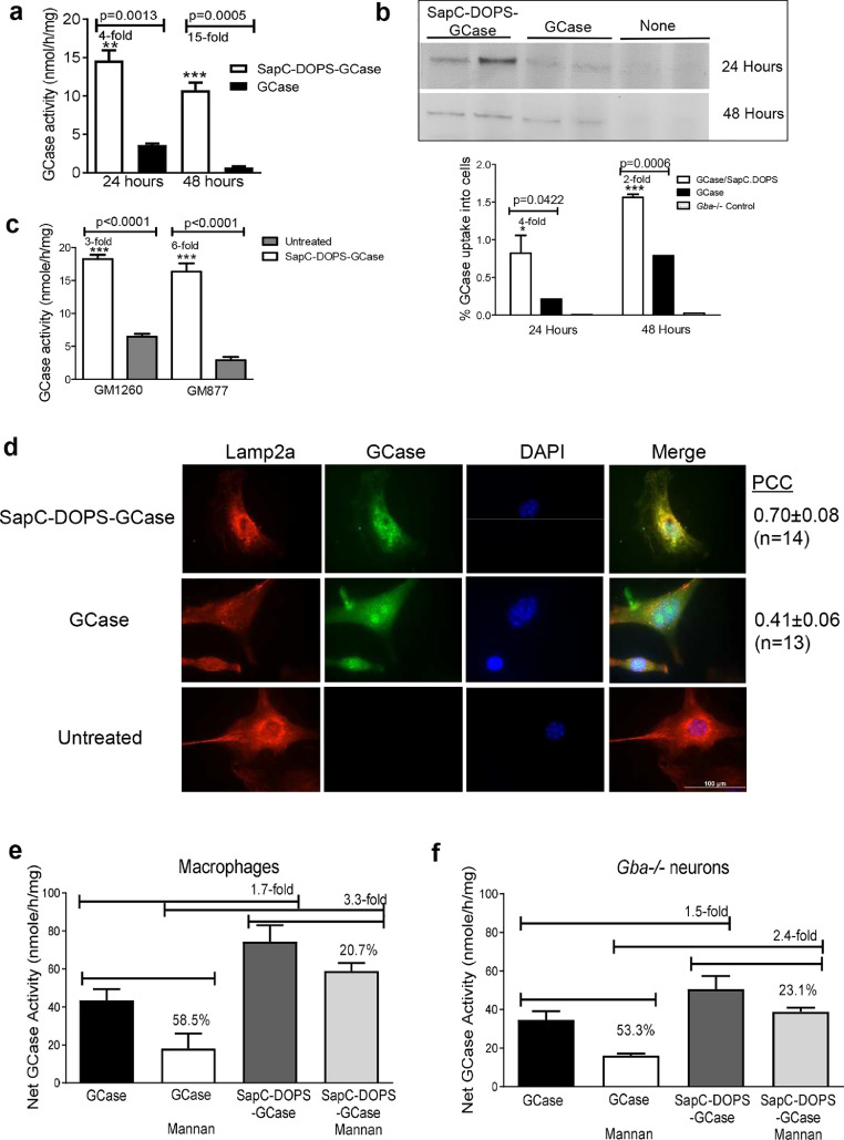
Fig. 2 SapC-DOPS nanovesicles preserve GCase. Cellular GCase activity (a) and GCase protein (b) of mouse Gba1-/- fibroblasts that were incubated for 24 or 48 h with SapC-DOPS-GCase and GCase, respectively. SapC-DOPS-GCase incubated cells showed higher levels of GCase activity and protein than GCase incubated cells. (c) GCase activity of human GD2 and GD3 fibroblasts, GM1260 (L444P/P415R) and GM877 (L444P/L444P), incubated with SapC-DOPS-GCase for 24 h. (d) Human GCase (green) detected in the lysosome (Lamp2a, red) of Gba1-/- fibroblasts incubated with SapC-DOPS- GCase and GCase for 24 h. The cells with SapC-DOPS-GCase showed higher PCC (Person correlation coefficient) compared to GCase incubated cells (n = 8–14 cells), indicating more GCase targeting to lysosomes in SapC-DOPS-GCase incubated cells compared to free GCase incubated cells. Scale bar =100 µm for all images. (e) Effect of mannan on GCase uptake. J774E macrophages and Gba1-/- neurons were incubated with 80 µg GCase/mL of SapC-DOPS-GCase or free GCase in the presence and absence of mannan (2 mg/mL). All the cells used for the assays were washed prior to collection. The data presents net GCase activity (increased GCase activity level minus basal/endogenous GCase activity level). The samples were assayed in triplicates/experiment of 2–3 independent experiments.
Fig. 2 SapC-DOPS nanovesicles preserve GCase. Cellular GCase activity (a) and GCase protein (b) of mouse Gba1-/- fibroblasts that were incubated for 24 or 48 h with SapC-DOPS-GCase and GCase, respectively. SapC-DOPS-GCase incubated cells showed higher levels of GCase activity and protein than GCase incubated cells. (c) GCase activity of human GD2 and GD3 fibroblasts, GM1260 (L444P/P415R) and GM877 (L444P/L444P), incubated with SapC-DOPS-GCase for 24 h. (d) Human GCase (green) detected in the lysosome (Lamp2a, red) of Gba1-/- fibroblasts incubated with SapC-DOPS- GCase and GCase for 24 h. The cells with SapC-DOPS-GCase showed higher PCC (Person correlation coefficient) compared to GCase incubated cells (n = 8–14 cells), indicating more GCase targeting to lysosomes in SapC-DOPS-GCase incubated cells compared to free GCase incubated cells. Scale bar =100 µm for all images. (e) Effect of mannan on GCase uptake. J774E macrophages and Gba1-/- neurons were incubated with 80 µg GCase/mL of SapC-DOPS-GCase or free GCase in the presence and absence of mannan (2 mg/mL). All the cells used for the assays were washed prior to collection. The data presents net GCase activity (increased GCase activity level minus basal/endogenous GCase activity level). The samples were assayed in triplicates/experiment of 2–3 independent experiments.CC BY-NC-ND · PubMed Central
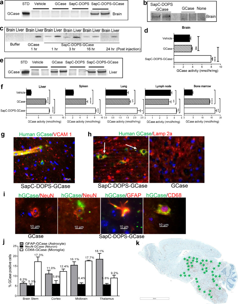
Fig. 3 Tissue distribution of SapC-DOPS-GCase. (a to e) 4 L;C* and WT mice at 38 days of age were i.v. infused with one bolus injection of SapC-DOPS-GCase, vehicle (CP buffer) or free GCase (54.6 mg/kg SapC-DOPS and 0.4 mg/kg GCase; 0.4 mg/kg free GCase). Tissues were collected 3 h post injection or as indicated. (a to c) GCase protein detected by immunoprecipitation followed with immunoblot in 4 L;C* (a) and WT (b) brains with SapC-DOPS-GCase. (c) GCase protein detected in the 4 L;C* brains and livers at 1 to 24 h post injection with SapC-DOPS-GCase. (d) GCase activity increased in SapC-DOPS-GCase treated brains. (e) GCase protein detected in the 4 L;C* liver. Human GCase (20 ng) is a loading control in A and E. (f) GCase activity in 4 L;C* mouse tissues at 49 days of age treated with SapC-DOPS-GCase, free GCase or vehicle-CP buffer (2 h post i.p. injection of, 2 x injection/2 h). SapC-DOPS-GCase was distributed to liver, spleen, lung, lymph nodes and bone marrow cells. SapC-DOPS-GCase-treated mice have significantly higher activity in those tissues than free GCase-treated mice. (g-k) Distribution of human GCase in brain regions of 4 L;C* mice at 38 days of age treated with SapC-DOPS-GCase or free GCase (3 h post one bolus i.v. injection). (g) Human GCase (green) co-stained with VCAM 1 (red) positive endothelium by immunofluorescence (IF) in SapC-DOPS-GCase treated 4 L;C* mouse brain. Scale bar is 20 μm. (h) Human GCase (green) detected by IF in the lysosome (Lamp2a, red) of brain cells. No GCase is detected in free GCase treated 4 L;C* brains. Image magnifications are 400x. (i) Panel: representative images of GCase protein (green) detected by IF in astrocytes (red, GFAP), neurons (red, NeuN) and microglia (red, CD68) in SapC- DOPS-GCase-treated 4 L;C* mouse brains. Scale bar = 10 µm for all images. (j) The graph shows the percentage of brain cells containing GCase. (k) Human GCase positive cells (green stars) determined by IF were distributed in 4 L;C* mouse brain regions that show inflammation stained with anti-CD68 (brown). Hematoxylin stains nuclei (blue). Scale bar is 3 mm. n = 3 mice, replicated assays of 2- 3 independent experiments, n = > 5 images/mouse. Student's t-test. *, p < 0.05; **, p < 0.01; ***, p < 0.001.
Fig. 3 Tissue distribution of SapC-DOPS-GCase. (a to e) 4 L;C* and WT mice at 38 days of age were i.v. infused with one bolus injection of SapC-DOPS-GCase, vehicle (CP buffer) or free GCase (54.6 mg/kg SapC-DOPS and 0.4 mg/kg GCase; 0.4 mg/kg free GCase). Tissues were collected 3 h post injection or as indicated. (a to c) GCase protein detected by immunoprecipitation followed with immunoblot in 4 L;C* (a) and WT (b) brains with SapC-DOPS-GCase. (c) GCase protein detected in the 4 L;C* brains and livers at 1 to 24 h post injection with SapC-DOPS-GCase. (d) GCase activity increased in SapC-DOPS-GCase treated brains. (e) GCase protein detected in the 4 L;C* liver. Human GCase (20 ng) is a loading control in A and E. (f) GCase activity in 4 L;C* mouse tissues at 49 days of age treated with SapC-DOPS-GCase, free GCase or vehicle-CP buffer (2 h post i.p. injection of, 2 x injection/2 h). SapC-DOPS-GCase was distributed to liver, spleen, lung, lymph nodes and bone marrow cells. SapC-DOPS-GCase-treated mice have significantly higher activity in those tissues than free GCase-treated mice. (g-k) Distribution of human GCase in brain regions of 4 L;C* mice at 38 days of age treated with SapC-DOPS-GCase or free GCase (3 h post one bolus i.v. injection). (g) Human GCase (green) co-stained with VCAM 1 (red) positive endothelium by immunofluorescence (IF) in SapC-DOPS-GCase treated 4 L;C* mouse brain. Scale bar is 20 μm. (h) Human GCase (green) detected by IF in the lysosome (Lamp2a, red) of brain cells. No GCase is detected in free GCase treated 4 L;C* brains. Image magnifications are 400x. (i) Panel: representative images of GCase protein (green) detected by IF in astrocytes (red, GFAP), neurons (red, NeuN) and microglia (red, CD68) in SapC- DOPS-GCase-treated 4 L;C* mouse brains. Scale bar = 10 µm for all images. (j) The graph shows the percentage of brain cells containing GCase. (k) Human GCase positive cells (green stars) determined by IF were distributed in 4 L;C* mouse brain regions that show inflammation stained with anti-CD68 (brown). Hematoxylin stains nuclei (blue). Scale bar is 3 mm. n = 3 mice, replicated assays of 2- 3 independent experiments, n = > 5 images/mouse. Student's t-test. *, p < 0.05; **, p < 0.01; ***, p < 0.001.CC BY-NC-ND · PubMed Central
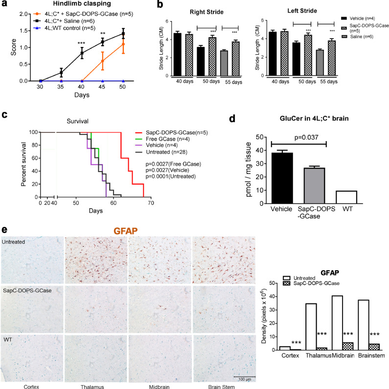
Fig. 4 Therapeutic efficacy of Sap-DOPS-GCase. 4 L;C* mice were treated with SapC-DOPS-GCase and vehicle or free GCase by daily i.p. injections of SapC-DOPS-GCase from day 21 to 27 followed by tail i.v. vein injection from day 28 to terminal age, 3 times per week . (a) Hindlimb clasping was significantly improved in SapC-DOPS-GCase treated 4 L;C* mice compared to saline treated 4 L;C* mice. 4 L;WT mice is the normal control. ANOVA test. **, p < 0.01; ***, p < 0.001. (b) Sap-DOPS-GCase treatment significantly improved right and left strides of 4 L;C* mice at 50 and 55 days of age compared to vehicle (CP buffer) or saline control. Student's t-test. ***, p < 0.001. (c) Compared to free GCase, vehicle and untreated 4 L;C* mice, SapC-DOPS-GCase treatment significantly prolonged survival of 4 L;C* mice. Log-Rank (Mantel-Cox) test (p<0.05). (d) GCase substrate, GluCer was significantly reduced in treated 4 L;C* brain (n = 3, 50 days of age). Student's t-test (p<0.05). (e) Treated 4 L;C* brain regions (cortex, thalamus, brain stem and midbrain) showed significantly reduced inflammation determined by anti-GFAP (astrocyte) staining (n = 3, 50 days of age). The signal densities of GFAP, were quantified with Image J software and presented as a bar graph. Scale bar is the same for all images. n = >5 images /mouse. Student's t-test. ***, p < 0.001.
Fig. 4 Therapeutic efficacy of Sap-DOPS-GCase. 4 L;C* mice were treated with SapC-DOPS-GCase and vehicle or free GCase by daily i.p. injections of SapC-DOPS-GCase from day 21 to 27 followed by tail i.v. vein injection from day 28 to terminal age, 3 times per week . (a) Hindlimb clasping was significantly improved in SapC-DOPS-GCase treated 4 L;C* mice compared to saline treated 4 L;C* mice. 4 L;WT mice is the normal control. ANOVA test. **, p < 0.01; ***, p < 0.001. (b) Sap-DOPS-GCase treatment significantly improved right and left strides of 4 L;C* mice at 50 and 55 days of age compared to vehicle (CP buffer) or saline control. Student's t-test. ***, p < 0.001. (c) Compared to free GCase, vehicle and untreated 4 L;C* mice, SapC-DOPS-GCase treatment significantly prolonged survival of 4 L;C* mice. Log-Rank (Mantel-Cox) test (p<0.05). (d) GCase substrate, GluCer was significantly reduced in treated 4 L;C* brain (n = 3, 50 days of age). Student's t-test (p<0.05). (e) Treated 4 L;C* brain regions (cortex, thalamus, brain stem and midbrain) showed significantly reduced inflammation determined by anti-GFAP (astrocyte) staining (n = 3, 50 days of age). The signal densities of GFAP, were quantified with Image J software and presented as a bar graph. Scale bar is the same for all images. n = >5 images /mouse. Student's t-test. ***, p < 0.001.CC BY-NC-ND · PubMed Central
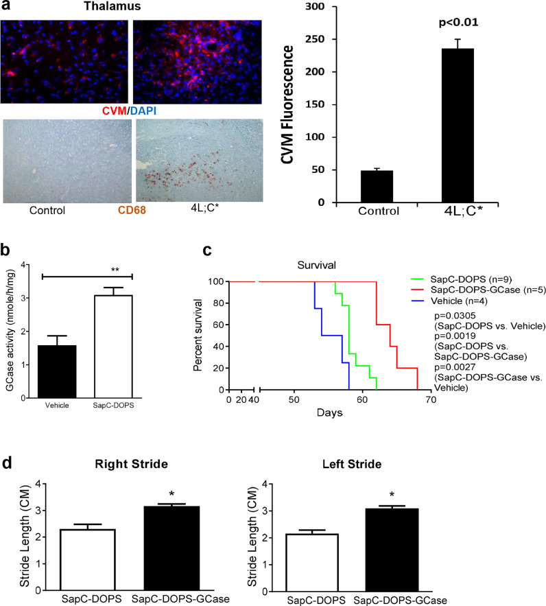
Fig. 5 SapC-DOPS in vitro and in vivo efficacy. (a) SapC-DOPS-CVM (red) detected in the thalamus region of 4 L;C* mouse brain that has inflammation stained positive by anti-CD68 (brown). SapC-DOPS-CVM also showed in control 4 L;WT mice brain that has no inflammation. Magnification of images are 100x. Bar graph showed CVM levels in 4 L;C* and control mice brain in thalamus region. 4 L;C* mice at 40 days of age were i.v. injected with 200 µL SapC-DOPS-CVM (SapC = 0.4 mg/mL, DOPS = 0.082 mg/mL, CVM = 0.04 µM). Tissues were collected 24 h post injection. (b-d) 4 L;C* mice were administered with SapC-DOPS, SapC-DOPS-GCase or vehicle (CP buffer) by daily i.p. injections of SapC-DOPS or SapC-DOPS-GCase from day 21 to 27, followed by tail i.v. vein injection of SapC-DOPS or SapC-DOPS-GCase, 3 times per week, from day 28 to terminal age. (b) Increased GCase activity in the brain of 4 L;C* mice treated with SapC-DOPS (n = 3, 40 days of age). Student's t-test. **, p < 0.01. (c) SapC-DOPS treated 4 L;C* mice showed significantly prolonged survival compared to vehicel-4 L;C*. The survival is longer in SapC-DOPS-GCase-treated 4 L;C* compared to SapC-DOPS-treated 4 L;C* mice. Log-Rank (Mantel-Cox) test (p<0.05). (d) In comparison to SapC-DOPS, SapC-DOPS-GCase treated 4 L;C* mice showed significant improved left stride and right stride at 45 days of age (n = 6). Student's t-test. *, p < 0.05.
Fig. 5 SapC-DOPS in vitro and in vivo efficacy. (a) SapC-DOPS-CVM (red) detected in the thalamus region of 4 L;C* mouse brain that has inflammation stained positive by anti-CD68 (brown). SapC-DOPS-CVM also showed in control 4 L;WT mice brain that has no inflammation. Magnification of images are 100x. Bar graph showed CVM levels in 4 L;C* and control mice brain in thalamus region. 4 L;C* mice at 40 days of age were i.v. injected with 200 µL SapC-DOPS-CVM (SapC = 0.4 mg/mL, DOPS = 0.082 mg/mL, CVM = 0.04 µM). Tissues were collected 24 h post injection. (b-d) 4 L;C* mice were administered with SapC-DOPS, SapC-DOPS-GCase or vehicle (CP buffer) by daily i.p. injections of SapC-DOPS or SapC-DOPS-GCase from day 21 to 27, followed by tail i.v. vein injection of SapC-DOPS or SapC-DOPS-GCase, 3 times per week, from day 28 to terminal age. (b) Increased GCase activity in the brain of 4 L;C* mice treated with SapC-DOPS (n = 3, 40 days of age). Student's t-test. **, p < 0.01. (c) SapC-DOPS treated 4 L;C* mice showed significantly prolonged survival compared to vehicel-4 L;C*. The survival is longer in SapC-DOPS-GCase-treated 4 L;C* compared to SapC-DOPS-treated 4 L;C* mice. Log-Rank (Mantel-Cox) test (p<0.05). (d) In comparison to SapC-DOPS, SapC-DOPS-GCase treated 4 L;C* mice showed significant improved left stride and right stride at 45 days of age (n = 6). Student's t-test. *, p < 0.05.CC BY-NC-ND · PubMed Central
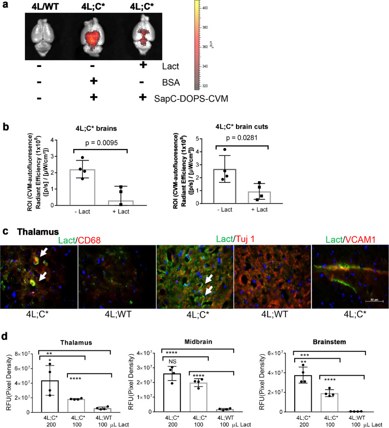
Fig. 6 Phosphatidylserine (PS)-mediated inflamed brain targeting determined by PS-binding protein, lactadherin (Lact). (a and b) Lact blocked SapC-DOPS-CVM targeting into 4 L;C* mouse brains. (a) Significantly diminished CVM signals were observed in 4 L;C* brains i.v. injected with Lact compared with BSA of whole brain and saggital brain cuts. Non-symptomatic, non-injected littermate, 4 L/WT, brains, used as controls, have no CVM signal. (b) Relative CVM signal levels are shown in the graph. CVM signals were acquired by ex-vivo IVIS imaging. Student's t-test (n = 4 mice). (c and d) Lact distributes in neural cells and inflamed brain regions. 4 L;C* and 4 L/WT control mice were injected with 100 or 200 µL of Lact (83 µg/mL) by i.v. The brain sections were stained with anti-lactadherin-FITC (green) in combination with anti-CD68 (microglia, red), anti-Tuj1 (Neuron, red) and anti-VCAM1 (endothelium, red). (c) Lact signals were around microglia and neuron cells, and on endothelium. (d) Signal levels (RFU, relative fluorescence unit) of Lact in inflamed 4 L;C* brain regions. Student's t-test (n = 4 mice). *, p < 0.05; **, p < 0.01; ***, p < 0.001; ****, p < 0.0001.
Fig. 6 Phosphatidylserine (PS)-mediated inflamed brain targeting determined by PS-binding protein, lactadherin (Lact). (a and b) Lact blocked SapC-DOPS-CVM targeting into 4 L;C* mouse brains. (a) Significantly diminished CVM signals were observed in 4 L;C* brains i.v. injected with Lact compared with BSA of whole brain and saggital brain cuts. Non-symptomatic, non-injected littermate, 4 L/WT, brains, used as controls, have no CVM signal. (b) Relative CVM signal levels are shown in the graph. CVM signals were acquired by ex-vivo IVIS imaging. Student's t-test (n = 4 mice). (c and d) Lact distributes in neural cells and inflamed brain regions. 4 L;C* and 4 L/WT control mice were injected with 100 or 200 µL of Lact (83 µg/mL) by i.v. The brain sections were stained with anti-lactadherin-FITC (green) in combination with anti-CD68 (microglia, red), anti-Tuj1 (Neuron, red) and anti-VCAM1 (endothelium, red). (c) Lact signals were around microglia and neuron cells, and on endothelium. (d) Signal levels (RFU, relative fluorescence unit) of Lact in inflamed 4 L;C* brain regions. Student's t-test (n = 4 mice). *, p < 0.05; **, p < 0.01; ***, p < 0.001; ****, p < 0.0001.CC BY-NC-ND · PubMed Central

Verification

Claims the paper makes

The claims and statistics of this paper haven’t been checked yet.

Run the check — free, no account needed. It reads the open-access text and checks the sources it cites.

A few free checks a day · papers already verified open instantly · results are public

Claims other papers make about this one

Each is checked against this paper’s own text: does it say what the citing paper says it does?

Self-check

Percentages, sums and reported differences re-computed from the paper’s own numbers.

The self-check needs the paper’s full text.

Citations

Cites

Research works printed but not linked: 70

  1. Sandhoff, 1998, Sphingolipid metabolism. sphingoid analogs, sphingolipid activator proteins, and the pathology of the cell, Ann N Y Acad Sci, 845, 139, 10.1111/j.1749-6632.1998.tb09667.x · DOI
  2. Gupta, 2011, Type 2 gaucher disease: phenotypic variation and genotypic heterogeneity, Blood Cells Mol Dis, 46, 75, 10.1016/j.bcmd.2010.08.012 · DOI
  3. Conradi, 1984, Neuropathology of the norrbottnian type of Gaucher disease. Morphological and biochemical studies, Acta Neuropathol, 65, 99, 10.1007/BF00690463 · DOI
  4. Goker-Alpan, 2006, Glucocerebrosidase mutations are an important risk factor for Lewy body disorders, Neurology, 67, 908, 10.1212/01.wnl.0000230215.41296.18 · DOI
  5. DePaolo, 2009, The association between mutations in the lysosomal protein glucocerebrosidase and parkinsonism, Movement disord, 24, 1571, 10.1002/mds.22538 · DOI
  6. Mazzulli, 2011, Gaucher disease glucocerebrosidase and alpha-synuclein form a bidirectional pathogenic loop in synucleinopathies, Cell, 146, 37, 10.1016/j.cell.2011.06.001 · DOI · Paper page
  7. Lachmann, 2011, Enzyme replacement therapy for lysosomal storage diseases, Curr Opin Pediatr, 23, 588, 10.1097/MOP.0b013e32834c20d9 · DOI
  8. Platt, 2008, Substrate reduction therapy, Acta Pædiatrica, 97, 88, 10.1111/j.1651-2227.2008.00656.x · DOI
  9. Bennett, 2013, Gaucher disease and its treatment options, Ann Pharmacother, 47, 1182, 10.1177/1060028013500469 · DOI
  10. Burrow, 2015, CNS, lung, and lymph node involvement in Gaucher disease type 3 after 11 years of therapy: clinical, histopathologic, and biochemical findings, Mol Genet Metab, 114, 233, 10.1016/j.ymgme.2014.08.011 · DOI
  11. Lukina, 2014, Eliglustat, an investigational oral therapy for Gaucher disease type 1: phase 2 trial results after 4 years of treatment, Blood Cells Mol Dis, 53, 274, 10.1016/j.bcmd.2014.04.002 · DOI
  12. Kuter, 2013, Miglustat therapy in type 1 Gaucher disease: clinical and safety outcomes in a multicenter retrospective cohort study, Blood Cells Mol Dis, 51, 116, 10.1016/j.bcmd.2013.04.005 · DOI
  13. Marshall, 2016, CNS-accessible inhibitor of glucosylceramide synthase for substrate reduction therapy of neuronopathic gaucher disease, Mol Ther, 24, 1019, 10.1038/mt.2016.53 · DOI
  14. Sands, 2006, Gene therapy for lysosomal storage diseases, Mol Ther, 13, 839, 10.1016/j.ymthe.2006.01.006 · DOI · Paper page
  15. Sardi, 2011, CNS expression of glucocerebrosidase corrects alpha-synuclein pathology and memory in a mouse model of Gaucher-related synucleinopathy, Proc Natl Acad Sci U S A, 108, 12101, 10.1073/pnas.1108197108 · DOI
  16. Massaro, 2018, Fetal gene therapy for neurodegenerative disease of infants, Nat Med, 24, 1317, 10.1038/s41591-018-0106-7 · DOI
  17. Lieberman, 2007, Structure of acid beta-glucosidase with pharmacological chaperone provides insight into Gaucher disease, Nat Chem Biol, 3, 101, 10.1038/nchembio850 · DOI
  18. Maegawa, 2009, Identification and characterization of ambroxol as an enzyme enhancement agent for Gaucher disease, J Biol Chem, 284, 23502, 10.1074/jbc.M109.012393 · DOI
  19. Aflaki, 2016, A new glucocerebrosidase chaperone reduces alpha-Synuclein and glycolipid levels in iPSC-Derived dopaminergic neurons from patients with Gaucher disease and parkinsonism, J Neurosci, 36, 7441, 10.1523/JNEUROSCI.0636-16.2016 · DOI
  20. Qi, 1996, Functional organization of saposin C. Definition of the neurotrophic and acid beta-glucosidase activation regions, J Biol Chem, 271, 6874, 10.1074/jbc.271.12.6874 · DOI
  21. Sun, 2003, Saposin c is required for normal resistance of acid beta-glucosidase to proteolytic degradation, J Biol Chem, 278, 31918, 10.1074/jbc.M302752200 · DOI
  22. Yap, 2013, Saposin C protects glucocerebrosidase against alpha-synuclein inhibition, Biochemistry, 52, 7161, 10.1021/bi401191v · DOI
  23. Qi, 1994, Functional human saposins expressed in Escherichia coli. Evidence for binding and activation properties of saposins c with acid beta-glucosidase, J Biol Chem, 269, 16746, 10.1016/S0021-9258(19)89454-1 · DOI
  24. Pampols, 1999, Neuronopathic juvenile glucosylceramidosis due to sap-C deficiency: clinical course, neuropathology and brain lipid composition in this Gaucher disease variant, Acta Neuropathol (Berl), 97, 91, 10.1007/s004010050960 · DOI
  25. Vaccaro, 2010, Saposin c mutations in Gaucher disease patients resulting in lysosomal lipid accumulation, saposin c deficiency, but normal prosaposin processing and sorting, Hum Mol Genet, 19, 2987, 10.1093/hmg/ddq204 · DOI
  26. Qi, 2012, Saposin C coupled lipid nanovesicles specifically target arthritic mouse joints for optical imaging of disease severity, PLoS ONE, 7, e33966, 10.1371/journal.pone.0033966 · DOI
  27. Blanco, 2014, Phosphatidylserine-selective targeting and anticancer effects of SAPC-dops nanovesicles on brain tumors, Oncotarget, 5, 7105, 10.18632/oncotarget.2214 · DOI
  28. Wojton, 2013, Systemic delivery of SAPC-dops has antiangiogenic and antitumor effects against glioblastoma, Mol Ther, 21, 1517, 10.1038/mt.2013.114 · DOI
  29. Qi, 2009, Cancer-selective targeting and cytotoxicity by liposomal-coupled lysosomal saposin C protein, Clin Cancer Res, 15, 5840, 10.1158/1078-0432.CCR-08-3285 · DOI
  30. Liou, 2006, Analyses of variant acid beta-glucosidases: effects of Gaucher disease mutations, J Biol Chem, 281, 4242, 10.1074/jbc.M511110200 · DOI
  31. Blonder, 1976, Effects of detergents and choline-containing phospholipids on human spleen glucocerebrosidase, Biochim Biophys Acta, 431, 45, 10.1016/0005-2760(76)90258-7 · DOI
  32. Tybulewicz, 1992, Animal model of Gaucher's disease from targeted disruption of the mouse glucocerebrosidase gene, Nature, 357, 407, 10.1038/357407a0 · DOI
  33. Sun, 2010, Neuronopathic Gaucher disease in the mouse: viable combined selective saposin C deficiency and mutant glucocerebrosidase (V394L) mice with glucosylsphingosine and glucosylceramide accumulation and progressive neurological deficits, Hum Mol Genet, 19, 1088, 10.1093/hmg/ddp580 · DOI
  34. Sun, 2010, Specific saposin C deficiency: CNS impairment and acid beta-glucosidase effects in the mouse, Hum Mol Genet, 19, 634, 10.1093/hmg/ddp531 · DOI
  35. Westbroek, 2016, A new glucocerebrosidase-deficient neuronal cell model provides a tool to probe pathophysiology and therapeutics for Gaucher disease, Dis Model Mech, 9, 769
  36. Du, 2005, The role of mannosylated enzyme and the mannose receptor in enzyme replacement therapy, Am J Hum Genet, 77, 1061, 10.1086/498652 · DOI
  37. Fabbro, 1987, Gaucher disease: genetic heterogeneity within and among the subtypes detected by immunoblotting, Am J Hum Genet, 40, 15
  38. Makinen, 2001, Inhibition of lymphangiogenesis with resulting lymphedema in transgenic mice expressing soluble VEGF receptor-3, Nat Med, 7, 199, 10.1038/84651 · DOI
  39. Sun, 2013, Substrate compositional variation with tissue/region and GBA1 mutations in mouse models–implications for Gaucher disease, PLoS ONE, 8, e57560, 10.1371/journal.pone.0057560 · DOI
  40. Schindelin, 2012, Fiji: an open-source platform for biological-image analysis, Nat Methods, 9, 676, 10.1038/nmeth.2019 · DOI · Paper page
  41. Schmued, 2005, Fluoro-Jade C results in ultra high resolution and contrast labeling of degenerating neurons, Brain Res, 1035, 24, 10.1016/j.brainres.2004.11.054 · DOI
  42. Liou, 2016, Modulating ryanodine receptors with dantrolene attenuates neuronopathic phenotype in Gaucher disease mice, Hum Mol Genet, 25, 5126
  43. Qi, 2001, Differential membrane interactions of saposins A and C: implications for the functional specificity, J Biol Chem, 276, 27010, 10.1074/jbc.M101075200 · DOI
  44. Qi, 1998, Acid beta-glucosidase: intrinsic fluorescence and conformational changes induced by phospholipids and saposin C, Biochemistry, 37, 11544, 10.1021/bi980785+ · DOI
  45. Xu, 1996, Turnover and distribution of intravenously administered mannose-terminated human acid beta-glucosidase in murine and human tissues, Pediatr Res, 39, 313, 10.1203/00006450-199602000-00021 · DOI
  46. Asmari, 2018, Thermophoresis for characterizing biomolecular interaction, Methods, 10.1016/j.ymeth.2018.02.003 · DOI
  47. Qi, 2004, Fusogenic domain and lysines in saposin C, Arch Biochem Biophys, 424, 210, 10.1016/j.abb.2004.02.023 · DOI
  48. Wigderson, 1989, Characterization of mutations in Gaucher patients by cDNA cloning, Am J Hum Genet, 44, 365
  49. Brady, 1994, Modifying exogenous glucocerebrosidase for effective replacement therapy in Gaucher disease, J Inherit Metab Dis, 17, 510, 10.1007/BF00711365 · DOI
  50. Van Patten, 2007, Effect of mannose chain length on targeting of glucocerebrosidase for enzyme replacement therapy of Gaucher disease, Glycobiology, 17, 467, 10.1093/glycob/cwm008 · DOI
  51. Schueler, 2002, Uptake of mannose-terminal glucocerebrosidase in cultured human cholinergic and dopaminergic neuron cell lines, Neurochem Res, 27, 325, 10.1023/A:1014915430398 · DOI
  52. Xu, 2010, Comparative therapeutic effects of velaglucerase alfa and imiglucerase in a Gaucher disease mouse model, PLoS ONE, 5, e10750, 10.1371/journal.pone.0010750 · DOI
  53. Dasgupta, 2015, Neuronopathic Gaucher disease: dysregulated mRNAs and miRNAs in brain pathogenesis and effects of pharmacologic chaperone treatment in a mouse model, Hum Mol Genet, 24, 7031
  54. Yeung, 2008, Membrane phosphatidylserine regulates surface charge and protein localization, Science, 319, 210, 10.1126/science.1152066 · DOI
  55. Shao, 2008, Crystal structure of lactadherin C2 domain at 1.7A resolution with mutational and computational analyses of its membrane-binding motif, J Biol Chem, 283, 7230, 10.1074/jbc.M705195200 · DOI
  56. Shi, 2004, Lactadherin binds selectively to membranes containing phosphatidyl-L-serine and increased curvature, Biochim Biophys Acta, 1667, 82, 10.1016/j.bbamem.2004.09.006 · DOI
  57. Raper, 2016, How do meningeal lymphatic vessels drain the CNS?, Trends Neurosci, 39, 581, 10.1016/j.tins.2016.07.001 · DOI
  58. Botto, 2004, Phosphatidylserine receptor and apoptosis: consequences of a non-ingested meal, Arthritis Res Ther, 6, 147, 10.1186/ar1191 · DOI
  59. Brouckaert, 2004, Phagocytosis of necrotic cells by macrophages is phosphatidylserine dependent and does not induce inflammatory cytokine production, Mol Biol Cell, 15, 1089, 10.1091/mbc.e03-09-0668 · DOI
  60. Dixon, 2010, Lymphatic lipid transport: sewer or subway?, Trends Endocrinol Metab, 21, 480, 10.1016/j.tem.2010.04.003 · DOI
  61. Iqbal, 2009, Intestinal lipid absorption, Am J Physiol Endocrinol Metab, 296, E1183, 10.1152/ajpendo.90899.2008 · DOI · Paper page
  62. Qi, 2002, Ex vivo localization of the mouse saposin C activation region for acid beta-glucosidase, Mol Genet Metab, 76, 189, 10.1016/S1096-7192(02)00040-9 · DOI
  63. Christomanou, 1986, Immunochemical characterization of two activator proteins stimulating enzymic sphingomyelin degradation in vitro. absence of one of them in a human Gaucher disease variant, Biol Chem Hoppe Seyler., 367, 879, 10.1515/bchm3.1986.367.2.879 · DOI
  64. Christomanou, 1989, Activator protein deficient Gaucher's disease. A second patient with the newly identified lipid storage disorder, Klin Wochenschr, 67, 999, 10.1007/BF01716064 · DOI
  65. Kang, 2017, A rare form of Gaucher disease resulting from saposin C deficiency, Blood Cells Mol Dis
  66. Pandey, 2017, Complement drives glucosylceramide accumulation and tissue inflammation in Gaucher disease, Nature, 543, 108, 10.1038/nature21368 · DOI
  67. Sun, 2011, Isofagomine in vivo effects in a neuronopathic Gaucher disease mouse, PLoS ONE, 6, e19037, 10.1371/journal.pone.0019037 · DOI
  68. Rixe, 2018, First-in-human, first-in-class phase 1a study of BXQ-350 for solid tumors and gliomas, J Clin Oncol, 36, 2517, 10.1200/JCO.2018.36.15_suppl.2517 · DOI
  69. Cruze, 2018, Absence of indicators of hypercoagulability and antiphospholipid syndrome in bxq-350 first in human study, J Clin Oncol, 36, e14533, 10.1200/JCO.2018.36.15_suppl.e14533 · DOI
  70. Alcalay, 2015, Glucocerebrosidase activity in Parkinson's disease with and without GBA mutations, Brain: J Neurol, 138, 2648, 10.1093/brain/awv179 · DOI

Cited by

See all 39

Metadata

Authors
Sun, Y., Liou, B., Chu, Z., Fannin, V., Blackwood, R., Peng, Y., Grabowski, G. A., Davis, H. W., & Qi, X.
Venue
eBioMedicine
Publisher
Elsevier BV
Published
2020-05
Volume
55
Pages
102735
ISSN
2352-3964
Cited by
39
References
2
Language
en
Funders
National Institutes of Health (R21NS 095047, R01 NS 086134, UH2 NS092981)

About these checks

Registry, retraction, Cites and statistics results are computed, not judged, with one exception: a printed reference that no index lists is read once by a model before it counts as a citation error. Claims are extracted by a model and kept only when their quote appears verbatim in the text; each cited source is then checked, with a model’s help, for whether it says what the claim relies on. Those verdicts can be wrong — read the claim’s quote and the cited source before relying on one.

Statistics and arithmetic are recomputed, not judged.

Confidence bands are shown only for fields calibrated on our annotated test set. None is calibrated yet, so no band is shown.

What counts as a claim ›How we measure confidence ›How we check this ›

Are you an author, or do you see a problem with this page? Request a review