

The DOI matches its Crossref record; 215 of the 215 references it cites were found in a registry or an index.
Advancing cancer radiotherapy: Harnessing radiosensitizers and nanotechnology for enhanced tumor control
Open accessAbstract
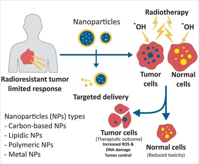
Cancer affects millions of individuals annually, with radiotherapy playing a crucial role in the treatment of over 70% of cases. Nevertheless, the effectiveness of radiotherapy is frequently limited by tumor radioresistance and the associated toxicity to normal tissues. Radiosensitizers enhance tumor sensitivity to ionizing radiation, optimizing doses and reducing damage. This review evaluates diverse radiosensitizers, including natural compounds (curcumin, resveratrol), chemotherapeutics (cisplatin, doxorubicin), and high-Z nanoparticles (gold, hafnium), which amplify DNA damage and ROS production. Advanced nanocarriers-carbon-, lipid-, and polymer-based-improve targeted delivery, specificity, and bioavailability. Various molecular mechanisms involving proteins such as p53, PARP1, miRNAs (miR-21, miR-155), and siRNAs are discussed in relation to their roles in modulating cell cycle progression, apoptosis, and gene expression to overcome resistance. Radioprotectors like antioxidants and amifostine safeguard normal tissues. Ongoing trials with hafnium oxide and cisplatin regimens show promise. Key findings highlight nanotechnology's role in enhancing radiosensitization via dose enhancement factors and synergistic therapies. Implications include personalized treatments tailored to tumor biology, the reduction of radioresistance through precision medicine and data-driven strategies, and ultimately, the improvement of radiotherapy efficacy and patient outcomes.
Overview
The claims and statistics of this paper haven’t been checked yet.
Run the check — free, no account needed. It reads the open-access text and checks the sources it cites.
Integrity signals
Registry
Cites
Statistics
Figures
Verification
Claims the paper makes
The claims and statistics of this paper haven’t been checked yet.
Run the check — free, no account needed. It reads the open-access text and checks the sources it cites.
Claims other papers make about this one
Each is checked against this paper’s own text: does it say what the citing paper says it does?
The source backs part of the claim, not all of it — or backs it for a different population, condition or comparison.
In the citing paper
“Early studies predominantly focused on high-atomic-number (high-Z) materials, including gold, silver, gadolinium, and hafnium-based nanoparticles, with the primary objective of maximizing radiation energy deposition and secondary electron generation [9,85,95,96].”
From Nanoparticle-Mediated Radiosensitization in Breast Cancer: A Systematic Review of Preclinical Evidence and Translational Challenges (2026) · cites this paper as [96]
Experimental — accuracy not yet measuredThe source backs part of the claim, not all of it — or backs it for a different population, condition or comparison.
In the citing paper
“Across the included studies, nanoparticle-assisted radiotherapy was frequently associated with enhanced therapeutic activity compared with radiotherapy alone, including reduced cell viability, decreased clonogenic survival, increased apoptosis, enhanced DNA damage, delayed tumor growth, and, in selected studies, improved survival outcomes [14,23,26,87,96,104,105].”
From Nanoparticle-Mediated Radiosensitization in Breast Cancer: A Systematic Review of Preclinical Evidence and Translational Challenges (2026) · cites this paper as [96]
Experimental — accuracy not yet measuredThe source backs part of the claim, not all of it — or backs it for a different population, condition or comparison.
In the citing paper
“Although these combinatorial systems often demonstrate enhanced efficacy experimentally, their clinical development is challenged by issues related to reproducibility, manufacturing consistency, regulatory approval, scalability, and long-term safety [25,96,97].”
From Nanoparticle-Mediated Radiosensitization in Breast Cancer: A Systematic Review of Preclinical Evidence and Translational Challenges (2026) · cites this paper as [96]
Experimental — accuracy not yet measured
Self-check
Percentages, sums and reported differences re-computed from the paper’s own numbers.
The self-check needs the paper’s full text.
Citations
Cites
Gene regulation by long non-coding RNAs and its biological functions
Statello, L., Guo, C.-J., Chen, L.-L., & Huarte, M. (2021) · Nature Reviews Molecular Cell Biology · doi:10.1038/s41580-020-00315-9 · Cited by 4,991
Genome Regulation by Long Noncoding RNAs
Rinn, J. L., & Chang, H. Y. (2012) · Annual Review of Biochemistry · doi:10.1146/annurev-biochem-051410-092902 · Cited by 4,091
Lipid nanoparticles for mRNA delivery
Hou, X., Zaks, T., Langer, R., & Dong, Y. (2021) · Nature Reviews Materials · doi:10.1038/s41578-021-00358-0 · Cited by 4,076
Impact of Nanotechnology on Drug Delivery
Farokhzad, O. C., & Langer, R. (2009) · ACS Nano · doi:10.1021/nn900002m · Cited by 3,443
Danhier, F., Féron, O., & Véronique Préat, A. (2010) · Journal of Controlled Release · doi:10.1016/j.jconrel.2010.08.027 · Cited by 2,891
Liposomes as nanomedical devices
Bozzuto, G., & Molinari, A. (2015) · International Journal of Nanomedicine · doi:10.2147/ijn.s68861 · Cited by 2,382
Guidelines and definitions for research on epithelial-mesenchymal transition
Yang, J., Antin, P., Berx, G., Blanpain, C., Brabletz, T., Bronner, M., Campbell, K., Cano, A., Casanova, J., Christofori, G., et al. (2020) · Nature Reviews Molecular Cell Biology · doi:10.1038/s41580-020-0237-9 · Cited by 2,226
Progress and challenges towards targeted delivery of cancer therapeutics
Rosenblum, D., Joshi, N., Tao, W., Karp, J. M., & Peer, D. (2018) · Nature Communications · doi:10.1038/s41467-018-03705-y · Cited by 2,220
Joudeh, N., & Linke, D. (2022) · Journal of Nanobiotechnology · doi:10.1186/s12951-022-01477-8 · Cited by 2,045
The Onpattro story and the clinical translation of nanomedicines containing nucleic acid-based drugs
Akinc, A., Maier, M. A., Manoharan, M., Fitzgerald, K., Jayaraman, M., Barros, S., Ansell, S., Du, X., Hope, M. J., Madden, T. D., et al. (2019) · Nature Nanotechnology · doi:10.1038/s41565-019-0591-y · Cited by 1,636
Research works printed but not linked: 35
- Akiyama, 2019, Abnormal accumulation of p53 predicts radioresistance leading to poor survival in patients with endometrial carcinoma, Oncol. Lett., 18, 5952
- Aung, 2007, Crocin from Crocus sativus possesses significant anti-proliferation effects on human colorectal cancer cells, Exp. Oncol., 29, 175
- Babaye Abdollahi, 2021, Main approaches to enhance radiosensitization in cancer cells by nanoparticles: a systematic review, Adv. Pharm. Bull., 11, 212
- Boateng, 2019, Novel bioerodable eluting-spacers for radiotherapy applications with in situ dose painting, Br. J. Radiol., 92
- Boateng, 2017, New designed radiotherapy biomaterials with reservoir and coated spacers for radiotherapy applications: WE-AB-605-10, Med. Phys., 44, 3194
- Bray, 2024, Global cancer statistics 2022: GLOBOCAN estimates of incidence and mortality worldwide for 36 cancers in 185 countries, CA Cancer J. Clin., 74, 229
- Chryssanthi, 2007, Inhibition of breast cancer cell proliferation by style constituents of different Crocus species, Anticancer Res., 27, 357
- Chu, 2022, Upconversion nanoparticles@AgBiS2 core-shell nanoparticles with cancer-cell-specific cytotoxicity for combined photothermal and photodynamic therapy of cancers, Bioact. Mater., 17, 71
- Dadashi, 2024, Enhancing ovarian cancer treatment: Synergistic effects of methotrexate (Methotrexate)- and quercetin-loaded chitosan nanoparticles, Carbohydr. Polym. Technol. Appl., 8
- Gong, 2018, Application of multifunctional nanomaterials in tumor radiosensitization, Acta Phys.-Chim. Sin., 34, 140
- Guo, 2019, Comprehensive analysis of key genes and microRNAs in radioresistant nasopharyngeal carcinoma, BMC Med. Genet., 12, 1
- Haque, 2023, The promise of nanoparticles-based radiotherapy in cancer treatment, Cancers (Basel), 15
- Hsieh, 2005, Biodegradable polymeric nanoparticles bearing stealth PEG shell and lipophilic polyester core, J. Chin. Inst. Chem. Eng., 36, 609
- Kris, 2014, Chemotherapy for lung cancers: here to stay, J. Clin. Oncol., 34, e375
- Kumar, 2013, Third generation gold nanoplatform optimized for radiation therapy, Transl. Cancer Res., 2
- Kwatra, 2013, Nanoparticles in radiation therapy: a summary of various approaches to enhance radiosensitization in cancer, Transl. Cancer Res., 2
- McDermott, 2017, Profiling of a panel of radioresistant prostate cancer cells identifies deregulation of key miRNAs, Clin. Transl. Radiat. Oncol., 2, 63
- Mikami, 2019, Resveratrol-induced REG III expression enhances chemo-and radiosensitivity in head and neck cancer in xenograft mice, Oncol. Rep., 42, 436
- Mostafavi, 2024, State-of-the-art application of nanoparticles in radiotherapy: a platform for synergistic effects in cancer treatment, Strahlenther. Onkol., 1
- Prabhu, 2015, Polymeric nanoparticles for targeted treatment in oncology: current insights, Int. J. Nanomedicine, 10, 1001
- Rouhi, 2024, Targeted therapy of gastric cancer with gingerol-loaded hyaluronic acid/PEG-coated PLGA nanoparticles: development and physicochemical evaluation, J. Drug Deliv. Sci. Technol., 97
- Shin, 2008, Sensitization of the apoptotic effect of gamma-irradiation in genistein-pretreated CaSki cervical cancer cells, J. Microbiol. Biotechnol., 18, 523
- Sisin, 2022, Natural baicalein-rich fraction as radiosensitizer in combination with bismuth oxide nanoparticles and cisplatin for clinical radiotherapy, Int. J. Nanomedicine, 17, 3853, 10.2147/IJN.S370478 · DOI
- Song, 2023, Application of nanomedicine in radiotherapy sensitization, Front. Oncol., 13
- Sung, 2021, Global cancer statistics 2020: GLOBOCAN estimates of incidence and mortality worldwide for 36 cancers in 185 countries, CA Cancer J. Clin., 71, 209
- Tarighi, 2025, Optimizing cancer treatment: a comprehensive review of active and passive drug delivery strategies, Iran. Biomed. J., 29, 173, 10.61882/ibj.4960 · DOI
- Thakor, 2013, Nanooncology: the future of cancer diagnosis and therapy, CA Cancer J. Clin., 63, 395
- Thakur, 2017, Utilization of polymeric nanoparticle in cancer treatment: a review, J. Pharm. Care Health Syst., 4, 1
- Vaezi, 2011, ERCC1 and XRCC1 as biomarkers for lung and head and neck cancer, Pharmacogenomics Pers. Med., 47
- Wu, 2022, The mechanism of long non-coding RNA in cancer radioresistance/radiosensitivity: a systematic review, Front. Pharmacol., 13
- Xie, 2021, Application of new radiosensitizer based on nano-biotechnology in the treatment of glioma, Front. Oncol., 11
- Yang, 2021, Iron-palladium decorated carbon nanotubes achieve radiosensitization via reactive oxygen species burst, Front. Bioeng. Biotechnol., 9
- Yao, 2020, Nanoparticle-based drug delivery in cancer therapy and its role in overcoming drug resistance, Front. Mol. Biosci., 7
- Zamanlu, 2019, Enhanced thrombolysis using tissue plasminogen activator (tPA)-loaded PEGylated PLGA nanoparticles for ischemic stroke, J. Drug Deliv. Sci. Technol., 53
- Zhao, 2015, Codelivery of doxorubicin and curcumin with lipid nanoparticles results in improved efficacy of chemotherapy in liver cancer, Int. J. Nanomedicine, 257
Cited by
Sorted by citations
Nanotechnology-based Strategies in Breast Cancer Diagnosis and Therapy: A Mini-review
Kasaai, M. R. (2026) · Oncology Advances · doi:10.14218/ona.2025.00027 · Cited by 1
Lunca, S., Morarasu, S., & Dimofte, G. M. (2026) · International Journal of Molecular Sciences · doi:10.3390/ijms27146522 · Cited by 2
Díaz, Y. A., Qi, Z., Yang, B., Cianciosi, D., Mazzoni, L., Gasparrini, M., Forbes-Hernández, T. Y., Quiles, J. L., Iglesias, R. C., Battino, M. A., et al. (2026) · Journal of Food Biochemistry · doi:10.1155/jfbc/9961129
Li, P., Wang, X., He, S., & Ma, H. (2026) · MedComm – Biomaterials and Applications · doi:10.1002/mba2.70058
Fontinele, D. O., Reiniger, K. A. R., de Azevedo, A. H. D., Lima, J. P. M. S., & Gupta, S. (2026) · BMC Bioinformatics · doi:10.1186/s12859-026-06592-z
Metadata
- Authors
- Shiridokht, F., Kehtari, P., Eskandani, M., Farajollahi, A., & Vandghanooni, S.
- Affiliations
- Tabriz University of Medical Sciences (IR)
- Corresponding author
- Vandghanooni, Somayeh
- Venue
- International Journal of Pharmaceutics: X
- Publisher
- Elsevier BV
- Published
- 2025-12
- Volume
- 10
- Pages
- 100419
- ISSN
- 2590-1567 (ISSN-L)
- PMC
- PMC12581699
- OpenAlex
- W4415958691
- Cited by
- 4
- References
- 215
- Citations by year
- 2026: 4
- FWCI
- 0.70
- Type
- journal article
- Language
- en
- Open access
- gold
- doi.org
- Licence
- Locations
- International Journal of Pharmaceutics X (publishedVersion, cc-by-nc)
- Europe PMC (PubMed Central) (publishedVersion, cc-by-nc)
- PubMed (publishedVersion)
- Funders
- Tabriz University of Medical Sciences
- Topics
- Physical Sciences › Physics and Astronomy › Radiation › Radiation Therapy and Dosimetry
- Health Sciences › Medicine › Radiology, Nuclear Medicine and Imaging › Effects of Radiation Exposure
- Physical Sciences › Physics and Astronomy › Radiation › Advanced Radiotherapy Techniques
- Keywords
- cancer radiotherapy, radiosensitizers, tumor radioresistance, nanocarriers, high-Z nanoparticles, gold nanoparticles, hafnium oxide nanoparticles, cisplatin, curcumin, resveratrol, reactive oxygen species, DNA damage, p53, PARP1, microRNA, siRNA, radiosensitization, radiotherapy toxicity, antioxidants, amifostine, targeted drug delivery, precision medicine
About these checks
Registry, retraction, Cites and statistics results are computed, not judged, with one exception: a printed reference that no index lists is read once by a model before it counts as a citation error. Claims are extracted by a model and kept only when their quote appears verbatim in the text; each cited source is then checked, with a model’s help, for whether it says what the claim relies on. Those verdicts can be wrong — read the claim’s quote and the cited source before relying on one.
Statistics and arithmetic are recomputed, not judged.
Confidence bands are shown only for fields calibrated on our annotated test set. None is calibrated yet, so no band is shown.
What counts as a claim ›How we measure confidence ›How we check this ›
Are you an author, or do you see a problem with this page? Request a review
