

The DOI matches its Crossref record; 60 of the 60 references it cites were found in a registry or an index.
Analysis of Plasmodium falciparum myosin B ATPase activity and structure in complex with the calmodulin-like domain of its light chain MLC-B
Open accessPublished version of a preprint
Abstract
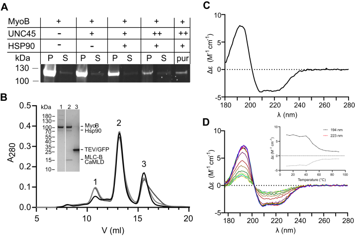
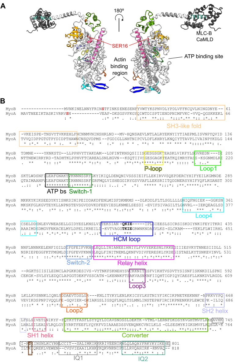
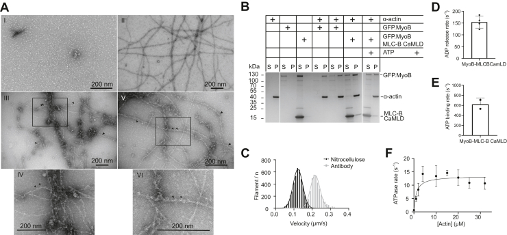
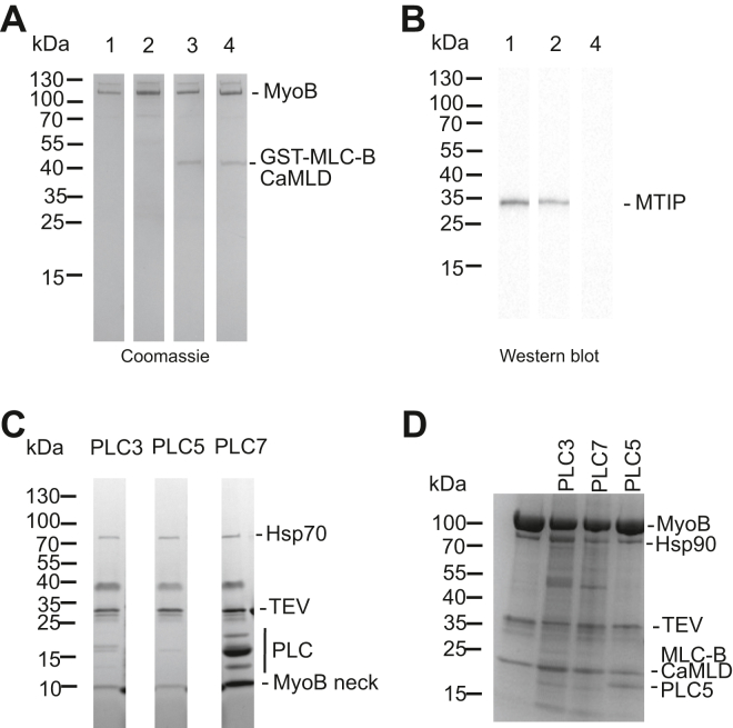
Myosin B (MyoB) is a class 14 myosin expressed in all invasive stages of the malaria parasite, Plasmodium falciparum. It is not associated with the glideosome complex that drives motility and invasion of host cells. During red blood cell invasion, MyoB remains at the apical tip of the merozoite but is no longer observed once invasion is completed. MyoB is not essential for parasite survival, but when it is knocked out, merozoites are delayed in the initial stages of red blood cell invasion, giving rise to a growth defect that correlates with reduced invasion success. Therefore, further characterization is needed to understand how MyoB contributes to parasite invasion. Here, we have expressed and purified functional MyoB with the help of parasite-specific chaperones Hsp90 and Unc45, characterized its binding to actin and its known light chain MLC-B using biochemical and biophysical methods and determined its low-resolution structure in solution using small angle X-ray scattering. In addition to MLC-B, we found that four other putative regulatory light chains bind to the MyoB IQ2 motif in vitro. The purified recombinant MyoB adopted the overall shape of a myosin, exhibited actin-activated ATPase activity, and moved actin filaments in vitro. Additionally, we determined that the ADP release rate was faster than the ATP turnover number, and thus, does not appear to be rate limiting. This, together with the observed high affinity to actin and the specific localization of MyoB, may point toward a role in tethering and/or force sensing during early stages of invasion.
Overview
The claims and statistics of this paper haven’t been checked yet.
Run the check — free, no account needed. It reads the open-access text and checks the sources it cites.
Integrity signals
Registry
Cites
Statistics
Figures
Verification
Claims the paper makes
The claims and statistics of this paper haven’t been checked yet.
Run the check — free, no account needed. It reads the open-access text and checks the sources it cites.
Self-check
Percentages, sums and reported differences re-computed from the paper’s own numbers.
The self-check needs the paper’s full text.
Citations
Cites
Highly accurate protein structure prediction with AlphaFold
Jumper, J., Evans, R., Pritzel, A., Green, T., Figurnov, M., Ronneberger, O., Tunyasuvunakool, K., Bates, R., Žídek, A., Potapenko, A., et al. (2021) · Nature · doi:10.1038/s41586-021-03819-2 · Cited by 43,764
UCSF ChimeraX: Structure visualization for researchers, educators, and developers
Pettersen, E. F., Goddard, T. D., Huang, C. C., Meng, E. C., Couch, G. S., Croll, T. I., Morris, J. H., & Ferrin, T. E. (2021) · Protein Science · doi:10.1002/pro.3943 · Cited by 9,514
Huang, D. W., Sherman, B. T., Tan, Q., Collins, J., Alvord, W. G., Roayaei, J., Stephens, R. D., Baseler, M., Lane, H. C., & Lempicki, R. A. (2007) · Genome biology · doi:10.1186/gb-2007-8-9-r183 · Cited by 2,482
Svergun, D. I. (1999) · Biophysical Journal · doi:10.1016/s0006-3495(99)77443-6 · Cited by 2,221
Accurate secondary structure prediction and fold recognition for circular dichroism spectroscopy
Micsonai, A., Wien, F., Kernya, L., Lee, Y.-H., Goto, Y., Réfrégiers, M., & Kardos, J. (2015) · Proceedings of the National Academy of Sciences · doi:10.1073/pnas.1500851112 · Cited by 1,703
Determination of Domain Structure of Proteins from X-Ray Solution Scattering
Svergun, D. I., Petoukhov, M. V., & Koch, M. H. J. (2001) · Biophysical Journal · doi:10.1016/s0006-3495(01)76260-1 · Cited by 1,420
Franke, D., Petoukhov, M. V., Konarev, P. V., Panjkovich, A., Tuukkanen, A., Mertens, H. D. T., Kikhney, A. G., Hajizadeh, N. R., Franklin, J. M., Jeffries, C. M., et al. (2017) · Journal of Applied Crystallography · doi:10.1107/s1600576717007786 · Cited by 1,329
[18] Purification of muscle actin
Pardee, J. D., & Aspudich, J. (1982) · Methods in Enzymology · doi:10.1016/0076-6879(82)85020-9 · Cited by 1,186
Di Tommaso, P., Moretti, S., Xenarios, I., Orobitg, M., Montanyola, A., Chang, J.-M., Taly, J.-F., & Notredame, C. (2011) · Nucleic Acids Research · doi:10.1093/nar/gkr245 · Cited by 1,165
New insights into myosin evolution and classification
Foth, B. J., Goedecke, M. C., & Soldati, D. (2006) · Proceedings of the National Academy of Sciences · doi:10.1073/pnas.0506307103 · Cited by 498
Metadata
- Authors
- Pires, I., Hung, Y.-F., Bergmann, U., Molloy, J. E., & Kursula, I.
- Affiliations
- University of Oulu (FI)
- The Francis Crick Institute (GB)
- University of Bergen (NO)
- Corresponding author
- Pires, Isa
- Kursula, Inari
- Venue
- Journal of Biological Chemistry
- Publisher
- Elsevier BV
- Published
- 2022-12
- Volume
- 298
- Issue
- 12
- Pages
- 102634
- ISSN
- 0021-9258 (ISSN-L)
- PMC
- PMC9692044
- OpenAlex
- W4306958619
- Cited by
- 1
- References
- 60
- Citations by year
- 2024: 1
- FWCI
- 0.22
- Type
- journal article
- Language
- en
- Open access
- gold
- doi.org
- Licence
- cc-by
- www.elsevier.com/tdm/userlicense/1.0/ (tdm)
- www.elsevier.com/legal/tdmrep-license (tdm)
- creativecommons.org/licenses/by/4.0/ (vor)
- Locations
- Journal of Biological Chemistry (publishedVersion, cc-by)
- bioRxiv (Cold Spring Harbor Laboratory) (acceptedVersion, cc-by-nc-nd)
- PubMed Central (publishedVersion, cc-by-nc-nd)
- PubMed (publishedVersion)
- Desy publication database (The Deutsches Elektronen-Synchrotron) (submittedVersion, other-oa)
- NORA - Norwegian Open Research Archives (submittedVersion, cc-by)
- Figshare (submittedVersion, cc-by-nc-nd)
- The Francis Crick Institute
- Funders
- Academy of Finland
- Francis Crick Institute
- Emil Aaltosen Säätiö
- Medical Research Council
- Norges Forskningsråd
- Sigrid Juséliuksen Säätiö
- Wellcome Trust
- Cancer Research UK (FC001119)
- Topics
- Health Sciences › Medicine › Public Health, Environmental and Occupational Health › Malaria Research and Control
- Health Sciences › Medicine › Cardiology and Cardiovascular Medicine › Cardiomyopathy and Myosin Studies
- Health Sciences › Medicine › Physiology (medical) › Erythrocyte Function and Pathophysiology
- Keywords
- Plasmodium falciparum, myosin B, calmodulin-like domain, ATPase activity, actin binding, merozoite invasion, erythrocyte invasion, HSP90, molecular chaperones, small-angle X-ray scattering, actin filament motility, ADP release, IQ motif, parasite motility
- MeSH terms
- Actins, Calmodulin, Myosins, Plasmodium falciparum, Protozoan Proteins, Nonmuscle Myosin Type IIB
About these checks
Registry, retraction, Cites and statistics results are computed, not judged, with one exception: a printed reference that no index lists is read once by a model before it counts as a citation error. Claims are extracted by a model and kept only when their quote appears verbatim in the text; each cited source is then checked, with a model’s help, for whether it says what the claim relies on. Those verdicts can be wrong — read the claim’s quote and the cited source before relying on one.
Statistics and arithmetic are recomputed, not judged.
Confidence bands are shown only for fields calibrated on our annotated test set. None is calibrated yet, so no band is shown.
What counts as a claim ›How we measure confidence ›How we check this ›
Are you an author, or do you see a problem with this page? Request a review
How to cite
Cite CoChat for the checks on this page, and the paper for its own findings.
- This page
- CoChat. (2026). Citation check: Analysis of Plasmodium falciparum myosin B ATPase activity and structure in complex with the calmodulin-like domain of its light chain MLC-B. https://app.cochat.ai/papers/10.1016/j.jbc.2022.102634
- The paper
- Pires, I., Hung, Y.-F., Bergmann, U., Molloy, J. E., & Kursula, I. (2022). Analysis of Plasmodium falciparum myosin B ATPase activity and structure in complex with the calmodulin-like domain of its light chain MLC-B. Journal of Biological Chemistry. https://doi.org/10.1016/j.jbc.2022.102634
Data sources
Bibliographic data: Semantic Scholar, OpenAlex, Crossref.
Contains information from the Semantic Scholar Academic Graph Datasets, made available under the ODC Attribution License (ODC-BY 1.0).
Semantic Scholar’s data is described in: Kinney, R., et al. (2023). The Semantic Scholar Open Data Platform. arXiv:2301.10140.
