Cited by 0Heat shock proteins researchProtein Structure and DynamicsBacterial Genetics and Biotechnology

The DOI matches its Crossref record; the references it cites are not checked yet.

The nucleotide exchange factor, GrpE, modulates substrate affinity by interaction of its N-terminal tails with the DnaK substrate–binding domain

Open access
Maqtedar, Akshitha, Rossi, Maria-Agustina, Clerico, Eugenia M., Williams, Robert V., Gierasch, Lila M.·2026·Journal of Biological Chemistry

Published version of a preprint

Abstract

Abstract The 70-kDa heat shock proteins (Hsp70s) assist in protein folding through allosteric communication between their nucleotide-binding domains (NBDs) and substrate-binding domains (SBDs), which are connected by an interdomain linker. Their nucleotide-dependent allosteric cycle is modulated by ligand binding and co-chaperones, including nucleotide exchange factors (NEFs). GrpE, the NEF for the E. coli Hsp70, DnaK, has been proposed to have a dual effect on the chaperone, facilitating the exchange of ADP for ATP in the NBD in a temperature-dependent fashion and promoting substrate release from the SBD. We recently reported NMR-based evidence that GrpE binding to DnaK has a direct structural effect on the SBD. Here, we expanded on these findings and obtained new evidence for a model in which the disordered N-terminal tails of GrpE facilitate peptide dissociation from the nucleotide-free DnaK/GrpE complex by transiently binding to the canonical substrate-binding site in the SBD. This GrpE/SBD interaction, while weak, is favored by the high local concentration of the tails around the SBD after complex formation. Moreover, we identified the DnaK binding motif in GrpE’s N-terminal disordered tails as 17 IIM 19 , which is conserved across many bacterial species. Excitingly, our data further suggest a mechanism for the temperature-dependence of GrpE’s modulation of DnaK’s refolding activity: as the temperature increases, unfolding of GrpE’s coiled-coil weakens its contacts with the SBD, reducing N-terminal tail binding, and thus increasing DnaK affinity to substrates.

Overview

The claims and statistics of this paper haven’t been checked yet.

Run the check — free, no account needed. It reads the open-access text and checks the sources it cites.

A few free checks a day · papers already verified open instantly · results are public

Integrity signals

Registry

Crossref · 10.1016/j.jbc.2026.111176

Cites

Statistics

Figures

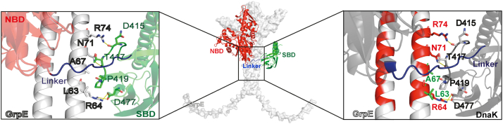
Figure 1 AlphaFold2-predicted Escherichia coli DnaK–GrpE complex reveals conserved interaction modes. The AlphaFold2-predicted structure of the E. coli DnaK–GrpE complex is shown in the center, and a close-up view of the interaction interface between DnaK SBD and GrpE coiled-coil domain is illustrated on the right and left. GrpE is highlighted in white, and in the Hsp70, the NBD is in red, the SBD is in green, and the interdomain linker is in blue. The left panel shows a zoomed view highlighting contacting residues between the GrpE coiled-coil (light gray) and DnaK SBD (green), whereas the right panel shows the same view with conserved GrpE residues indicated in red. DnaK, 70-kDa heat shock protein of prokaryote; GrpE, nucleotide exchange factor of prokaryote; Hsp70, 70-kDa heat shock protein; NBD, nucleotide-binding domain; SBD, substrate-binding domain.
Figure 1 AlphaFold2-predicted Escherichia coli DnaK–GrpE complex reveals conserved interaction modes. The AlphaFold2-predicted structure of the E. coli DnaK–GrpE complex is shown in the center, and a close-up view of the interaction interface between DnaK SBD and GrpE coiled-coil domain is illustrated on the right and left. GrpE is highlighted in white, and in the Hsp70, the NBD is in red, the SBD is in green, and the interdomain linker is in blue. The left panel shows a zoomed view highlighting contacting residues between the GrpE coiled-coil (light gray) and DnaK SBD (green), whereas the right panel shows the same view with conserved GrpE residues indicated in red. DnaK, 70-kDa heat shock protein of prokaryote; GrpE, nucleotide exchange factor of prokaryote; Hsp70, 70-kDa heat shock protein; NBD, nucleotide-binding domain; SBD, substrate-binding domain.CC BY · PubMed Central
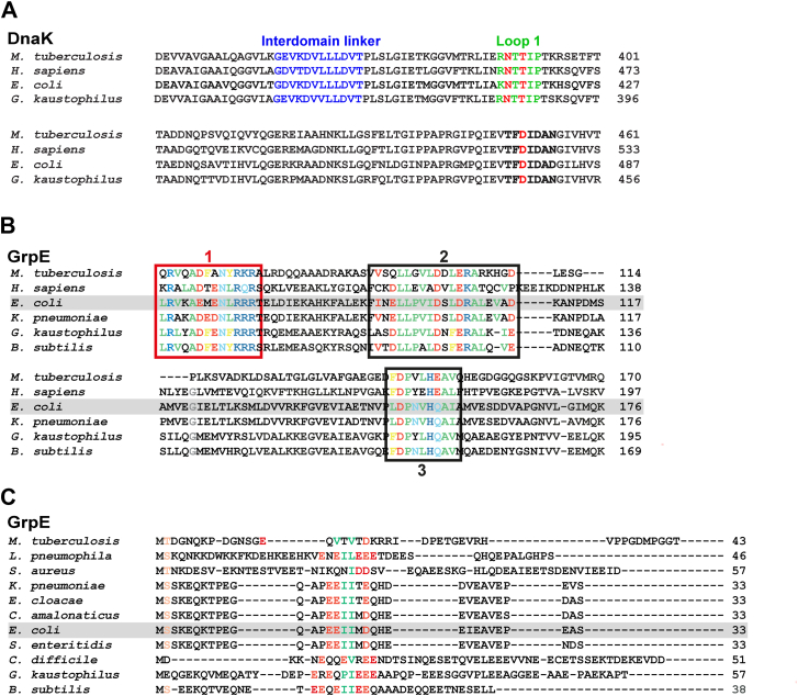
Figure 2 DnaK and GrpE homologs exhibit sequence conservation in the interaction interfaces of the DnaK–GrpE complex. A, sequence alignment of DnaK from Mycobacterium tuberculosis, human (mortalin, HSPA9), Escherichia coli, and Geobacillus kaustophilus. Residues in the interdomain linker sequence are highlighted in blue. SBD loop 1 residues are shown in green (E. coli K414–P419, G. kaustophilus R382–P388, H. sapiens mortalin HSPA9 R460–P465, and M. tuberculosis R388–P393). The residues that contact the GrpE coiled-coil domain are highlighted in red: Asn (E. coli N415, G. kaustophilus N383, mortalin HSPA9 N461, and M. tuberculosis N389), Thr (E. coli T417, G. kaustophilus T385, mortalin HSPA9 T463, and M. tuberculosis N391), and Asp (E. coli D477, G. kaustophilus D446, mortalin HSPA9 D523, and M. tuberculosis D451). B, sequence alignment of GrpE homologs where conserved residues are highlighted (hydrophobic in green, polar in light blue, negatively charged in red, positively charged in blue, and aromatic in yellow). The (1) L63–R75 region in the coiled-coil domain that interacts with DnaK is highlighted with a red box; the (2) I91–D110 region that contacts DnaK NBD subdomain IB; and the (3) L148–I157 region that interacts with DnaK NBD subdomain IIB are highlighted with black boxes. The numbering in this caption corresponds to the E. coli GrpE. C, sequence alignment for the N-terminal disordered tails of GrpE homologs. The only sequence similarity region in the N-terminal tails is highlighted (hydrophobic residues in green, negatively charged ones in red). DnaK, 70-kDa heat shock protein of prokaryote; GrpE, nucleotide exchange factor of prokaryote; NBD, nucleotide-binding domain; SBD, substrate-binding domain.
Figure 2 DnaK and GrpE homologs exhibit sequence conservation in the interaction interfaces of the DnaK–GrpE complex. A, sequence alignment of DnaK from Mycobacterium tuberculosis, human (mortalin, HSPA9), Escherichia coli, and Geobacillus kaustophilus. Residues in the interdomain linker sequence are highlighted in blue. SBD loop 1 residues are shown in green (E. coli K414–P419, G. kaustophilus R382–P388, H. sapiens mortalin HSPA9 R460–P465, and M. tuberculosis R388–P393). The residues that contact the GrpE coiled-coil domain are highlighted in red: Asn (E. coli N415, G. kaustophilus N383, mortalin HSPA9 N461, and M. tuberculosis N389), Thr (E. coli T417, G. kaustophilus T385, mortalin HSPA9 T463, and M. tuberculosis N391), and Asp (E. coli D477, G. kaustophilus D446, mortalin HSPA9 D523, and M. tuberculosis D451). B, sequence alignment of GrpE homologs where conserved residues are highlighted (hydrophobic in green, polar in light blue, negatively charged in red, positively charged in blue, and aromatic in yellow). The (1) L63–R75 region in the coiled-coil domain that interacts with DnaK is highlighted with a red box; the (2) I91–D110 region that contacts DnaK NBD subdomain IB; and the (3) L148–I157 region that interacts with DnaK NBD subdomain IIB are highlighted with black boxes. The numbering in this caption corresponds to the E. coli GrpE. C, sequence alignment for the N-terminal disordered tails of GrpE homologs. The only sequence similarity region in the N-terminal tails is highlighted (hydrophobic residues in green, negatively charged ones in red). DnaK, 70-kDa heat shock protein of prokaryote; GrpE, nucleotide exchange factor of prokaryote; NBD, nucleotide-binding domain; SBD, substrate-binding domain.CC BY · PubMed Central
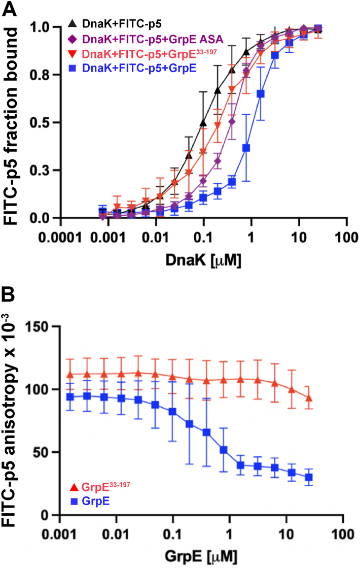
Figure 3 The binding of GrpE reduces the affinity of the DnaK SBD for a model peptide. A, apparent affinity of DnaK for the model peptide FITC-p5 in the absence or presence of 1 μM GrpE, GrpE33–197, and GrpE ASA at 22 °C. B, apparent affinity of DnaK for the model peptide FITC-p5 when the concentration of GrpE and GrpE33–197 was varied at 22 °C. DnaK, 70-kDa heat shock protein of prokaryote; GrpE, nucleotide exchange factor of prokaryote; SBD, substrate-binding domain.
Figure 3 The binding of GrpE reduces the affinity of the DnaK SBD for a model peptide. A, apparent affinity of DnaK for the model peptide FITC-p5 in the absence or presence of 1 μM GrpE, GrpE33–197, and GrpE ASA at 22 °C. B, apparent affinity of DnaK for the model peptide FITC-p5 when the concentration of GrpE and GrpE33–197 was varied at 22 °C. DnaK, 70-kDa heat shock protein of prokaryote; GrpE, nucleotide exchange factor of prokaryote; SBD, substrate-binding domain.CC BY · PubMed Central
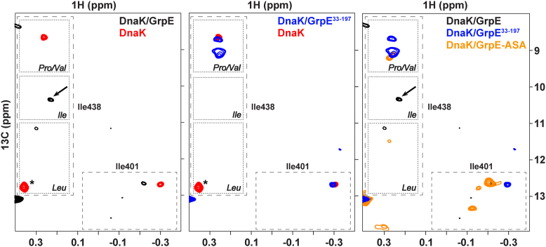
Figure 4 DnaK SBD reporter NMR signals, I401 and I438, indicate which residues bind to the central pocket when complexed with GrpE and its variants. Ile resonances of the substrate-binding pocket on the HMQC at 25 °C of (left) 13C-methyl-labeled ILV-DnaK–GrpE complex (black) and isolated apo-DnaK (red), (middle) 13C-methyl-labeled ILV-DnaK–GrpE33–197 complex (blue) and isolated apo-DnaK (red), and (right)13C-methyl-labeled ILV-DnaK–GrpE complex (black), 13C-methyl-labeled ILV-DnaK–GrpE33–197 complex (blue), and 13C-methyl-labeled ILV-DnaK–GrpE ASA complex (orange). An arrow marks the I438 peak in the ILV-DnaK–GrpE complex spectrum. The residue marked with an asterisk in the apo-DnaK spectrum corresponds to residue I338. DnaK, 70-kDa heat shock protein of prokaryote; GrpE, nucleotide exchange factor of prokaryote; HMQC, heteronuclear multiple quantum correlation; SBD, substrate-binding domain.
Figure 4 DnaK SBD reporter NMR signals, I401 and I438, indicate which residues bind to the central pocket when complexed with GrpE and its variants. Ile resonances of the substrate-binding pocket on the HMQC at 25 °C of (left) 13C-methyl-labeled ILV-DnaK–GrpE complex (black) and isolated apo-DnaK (red), (middle) 13C-methyl-labeled ILV-DnaK–GrpE33–197 complex (blue) and isolated apo-DnaK (red), and (right)13C-methyl-labeled ILV-DnaK–GrpE complex (black), 13C-methyl-labeled ILV-DnaK–GrpE33–197 complex (blue), and 13C-methyl-labeled ILV-DnaK–GrpE ASA complex (orange). An arrow marks the I438 peak in the ILV-DnaK–GrpE complex spectrum. The residue marked with an asterisk in the apo-DnaK spectrum corresponds to residue I338. DnaK, 70-kDa heat shock protein of prokaryote; GrpE, nucleotide exchange factor of prokaryote; HMQC, heteronuclear multiple quantum correlation; SBD, substrate-binding domain.CC BY · PubMed Central
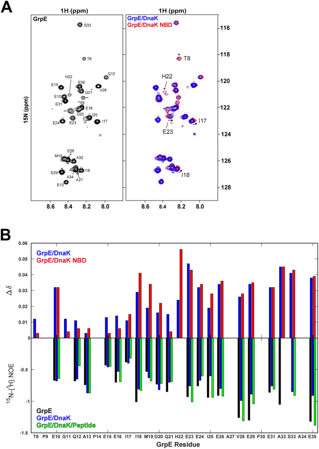
Figure 5 15N-GrpE NMR analysis supports transient binding of the GrpE N-terminal tails to the DnaK SBD. A, the 1H,15N-HSQC spectrum of 15N-GrpE (left panel) shows backbone amide resonances corresponding to disordered GrpE N-terminal tail residues. Resonance assignments are indicated, whereas unassigned peaks are labeled as ∗∗. An overlay (right) comparing equivalent spectra collected for the GrpE–DnaK complex (blue) and GrpE–DnaK–NBD (red) shows that the largest differences in peak position between the two complexes are observed for I17, I18, and E23 (indicated with arrows), whereas T8 and H22 experience more broadening in the GrpE–DnaK spectrum (indicated with asterisks). B, CSPs were measured for GrpE N-terminal amide signals in complex with DnaK (blue) or a truncated NBD (red) relative to those of isolated 15N-GrpE. B, top, bar plot comparing CSPs (Δδ) for 15N-GrpE in complex with full-length DnaK (blue) or the DnaK NBD1–392 (red) relative to 15N-GrpE alone. Missing CSPs correspond to proline residues (P9, P14, and P30) or shifts that could not be measured because of resonance overlap (A27, A34). Bottom,15N-{1H}-NOE values for GrpE (black), GrpE–DnaK (blue), and GrpE–DnaK–Ash2 peptide (green) complexes. Several NOEs are not indicated as there is a proline residue in the sequence (P9, P14, and P30), or there was resonance overlap resonance (A27 and A34), or there was insufficient intensity in the saturated spectrum (H22). CSP, chemical shift perturbation; DnaK, 70-kDa heat shock protein of prokaryote; GrpE, nucleotide exchange factor of prokaryote; HSQC, heteronuclear single quantum coherence; SBD, substrate-binding domain.
Figure 5 15N-GrpE NMR analysis supports transient binding of the GrpE N-terminal tails to the DnaK SBD. A, the 1H,15N-HSQC spectrum of 15N-GrpE (left panel) shows backbone amide resonances corresponding to disordered GrpE N-terminal tail residues. Resonance assignments are indicated, whereas unassigned peaks are labeled as ∗∗. An overlay (right) comparing equivalent spectra collected for the GrpE–DnaK complex (blue) and GrpE–DnaK–NBD (red) shows that the largest differences in peak position between the two complexes are observed for I17, I18, and E23 (indicated with arrows), whereas T8 and H22 experience more broadening in the GrpE–DnaK spectrum (indicated with asterisks). B, CSPs were measured for GrpE N-terminal amide signals in complex with DnaK (blue) or a truncated NBD (red) relative to those of isolated 15N-GrpE. B, top, bar plot comparing CSPs (Δδ) for 15N-GrpE in complex with full-length DnaK (blue) or the DnaK NBD1–392 (red) relative to 15N-GrpE alone. Missing CSPs correspond to proline residues (P9, P14, and P30) or shifts that could not be measured because of resonance overlap (A27, A34). Bottom,15N-{1H}-NOE values for GrpE (black), GrpE–DnaK (blue), and GrpE–DnaK–Ash2 peptide (green) complexes. Several NOEs are not indicated as there is a proline residue in the sequence (P9, P14, and P30), or there was resonance overlap resonance (A27 and A34), or there was insufficient intensity in the saturated spectrum (H22). CSP, chemical shift perturbation; DnaK, 70-kDa heat shock protein of prokaryote; GrpE, nucleotide exchange factor of prokaryote; HSQC, heteronuclear single quantum coherence; SBD, substrate-binding domain.CC BY · PubMed Central
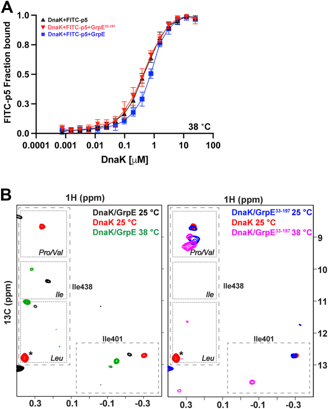
Figure 6 GrpE modulates peptide release from apo-DnaK less at elevated temperatures. A, apparent affinity of DnaK for the model peptide FITC-p5 in the absence or presence of 1 μM GrpE and GrpE33–197 at 38 °C. B, Ile resonances of the substrate binding pocket on the HMQC of (left) 13C-methyl-labeled ILV-DnaK–GrpE complex (black) at 25 °C, 13C-methyl-labeled ILV-DnaK–GrpE complex (green) at 38 °C, and isolated apo-DnaK (red) at 25 °C and (right) 13C-methyl-labeled ILV-DnaK–GrpE33–197 complex (blue) at 25 °C, 13C-methyl-labeled ILV-DnaK–GrpE33–197 complex (pink) at 38 °C, and isolated apo-DnaK (red) at 25 °C. The residue marked with an asterisk in the apo-DnaK spectrum corresponds to residue I338. DnaK, 70-kDa heat shock protein of prokaryote; GrpE, nucleotide exchange factor of prokaryote; HMQC, heteronuclear multiple quantum correlation.
Figure 6 GrpE modulates peptide release from apo-DnaK less at elevated temperatures. A, apparent affinity of DnaK for the model peptide FITC-p5 in the absence or presence of 1 μM GrpE and GrpE33–197 at 38 °C. B, Ile resonances of the substrate binding pocket on the HMQC of (left) 13C-methyl-labeled ILV-DnaK–GrpE complex (black) at 25 °C, 13C-methyl-labeled ILV-DnaK–GrpE complex (green) at 38 °C, and isolated apo-DnaK (red) at 25 °C and (right) 13C-methyl-labeled ILV-DnaK–GrpE33–197 complex (blue) at 25 °C, 13C-methyl-labeled ILV-DnaK–GrpE33–197 complex (pink) at 38 °C, and isolated apo-DnaK (red) at 25 °C. The residue marked with an asterisk in the apo-DnaK spectrum corresponds to residue I338. DnaK, 70-kDa heat shock protein of prokaryote; GrpE, nucleotide exchange factor of prokaryote; HMQC, heteronuclear multiple quantum correlation.CC BY · PubMed Central

Verification

Claims the paper makes

The claims and statistics of this paper haven’t been checked yet.

Run the check — free, no account needed. It reads the open-access text and checks the sources it cites.

A few free checks a day · papers already verified open instantly · results are public

Self-check

Percentages, sums and reported differences re-computed from the paper’s own numbers.

The self-check needs the paper’s full text.

Citations

The citation lists haven’t been fetched yet

References 37

Research works printed but not linked: 33

  1. Kim, 2013, Molecular chaperone functions in protein folding and proteostasis, Annu. Rev. Biochem., 82, 323, 10.1146/annurev-biochem-060208-092442 · DOI
  2. Rosenzweig, 2019, The Hsp70 chaperone network, Nat. Rev. Mol. Cell Biol., 20, 665, 10.1038/s41580-019-0133-3 · DOI · Paper page
  3. Clerico, 2019, Hsp70 molecular chaperones: multifunctional allosteric holding and unfolding machines, Biochem. J., 476, 1653, 10.1042/BCJ20170380 · DOI
  4. Melero, 2015, Modulation of the chaperone DnaK allosterism by the nucleotide exchange factor GrpE, J. Biol. Chem., 290, 10083, 10.1074/jbc.M114.623371 · DOI
  5. Moro, 2007, GrpE N-terminal domain contributes to the interaction with Dnak and modulates the dynamics of the chaperone substrate binding domain, J. Mol. Biol., 374, 1054, 10.1016/j.jmb.2007.10.002 · DOI
  6. Harrison, 1997, Crystal structure of the nucleotide exchange factor GrpE bound to the ATPase domain of the molecular chaperone DnaK, Science, 276, 431, 10.1126/science.276.5311.431 · DOI
  7. Xiao, 2024, Structure of the M. tuberculosis DnaK−GrpE complex reveals how key DnaK roles are controlled, Nat. Commun., 15, 660, 10.1038/s41467-024-44933-9 · DOI
  8. Brehmer, 2004, Influence of GrpE on DnaK-substrate interactions, J. Biol. Chem., 279, 27957, 10.1074/jbc.M403558200 · DOI
  9. Bracher, 2023, Nucleotide exchange factors for Hsp70 molecular chaperones: GrpE, Hsp110/Grp170, HspBP1/Sil1, and BAG domain proteins, Subcell. Biochem., 101, 1, 10.1007/978-3-031-14740-1_1 · DOI
  10. Schönfeld, 1995, The DnaK chaperone system of Escherichia coli: quaternary structures and interactions of the DnaK and GrpE components, J. Biol. Chem., 270, 2183, 10.1074/jbc.270.5.2183 · DOI
  11. Gelinas, 2003, Thermodynamic linkage in the GrpE nucleotide exchange factor, a molecular thermosensor, Biochemistry, 42, 9050, 10.1021/bi034416b · DOI
  12. Gelinas, 2002, A structure-based interpretation of E.coli GrpE thermodynamic properties, J. Mol. Biol., 323, 131, 10.1016/S0022-2836(02)00915-4 · DOI
  13. Grimshaw, 2003, Thermosensor action of GrpE. The DnaK chaperone system at heat shock temperatures, J. Biol. Chem., 278, 19048, 10.1074/jbc.M300924200 · DOI
  14. Diamant, 1998, Temperature-controlled activity of DnaK-DnaJ-GrpE chaperones: protein-folding arrest and recovery during and after heat shock depends on the substrate protein and the GrpE concentration, Biochemistry, 37, 9688, 10.1021/bi980338u · DOI
  15. Groemping, 2001, Folding properties of the nucleotide exchange factor GrpE from thermus thermophilus: GrpE is a thermosensor that mediates heat shock response, J. Mol. Biol., 314, 167, 10.1006/jmbi.2001.5116 · DOI
  16. Rossi, 2024, New insights into the structure and function of the complex between the Escherichia coli Hsp70, DnaK, and its nucleotide-exchange factor, GrpE, J. Biol. Chem., 300, 10.1016/j.jbc.2023.105574 · DOI
  17. Morizono, 2024, Structural insights into GrpEL1-mediated nucleotide and substrate release of human mitochondrial Hsp70, Nat. Commun., 15, 10.1038/s41467-024-54499-1 · DOI
  18. Wu, 2012, Crystal structure of DnaK protein complexed with nucleotide exchange factor GrpE in DnaK chaperone system, J. Biol. Chem., 287, 21461, 10.1074/jbc.M112.344358 · DOI
  19. Siegenthaler, 2005, The importance of having thermosensor control in the DnaK chaperone system, J. Biol. Chem., 280, 14395, 10.1074/jbc.M413803200 · DOI
  20. Bertelsen, 2009, Solution conformation of wild-type E. coli Hsp70 (DnaK) chaperone complexed with ADP and substrate, Proc. Natl. Acad. Sci. U. S. A., 106, 8471, 10.1073/pnas.0903503106 · DOI
  21. Grimshaw, 2005, The heat-sensitive Escherichia coli grpE280 phenotype: impaired interaction of GrpE(G122D) with DnaK, J. Mol. Biol., 353, 888, 10.1016/j.jmb.2005.08.069 · DOI
  22. Rüdiger, 1997, Substrate specificity of the DnaK chaperone determined by screening cellulose-bound peptide libraries, EMBO J., 16, 1501, 10.1093/emboj/16.7.1501 · DOI
  23. Davis, 1999, Intragenic suppressors of Hsp70 mutants: interplay between the ATPase- and peptide-binding domains, Proc. Natl. Acad. Sci. U. S. A., 96, 9269, 10.1073/pnas.96.16.9269 · DOI
  24. Clerico, 2021, Selective promiscuity in the binding of E. coli Hsp70 to an unfolded protein, Proc. Natl. Acad. Sci. U. S. A., 118, 10.1073/pnas.2016962118 · DOI
  25. Montgomery, 1999, Mutations in the substrate binding domain of the Escherichia coli 70 kDa molecular chaperone, DnaK, which alter substrate affinity or interdomain coupling, J. Mol. Biol., 286, 915, 10.1006/jmbi.1998.2514 · DOI
  26. Swain, 2007, Hsp70 chaperone ligands control domain association via an allosteric mechanism mediated by the interdomain linker, Mol. Cell., 26, 27, 10.1016/j.molcel.2007.02.020 · DOI
  27. Zhuravleva, 2011, Allosteric signal transmission in the nucleotide-binding domain of 70-kDa heat shock protein (Hsp70) molecular chaperones, Proc. Natl. Acad. Sci. U. S. A., 108, 6987, 10.1073/pnas.1014448108 · DOI
  28. Zhuravleva, 2012, An interdomain energetic tug-of-war creates the allosterically active state in Hsp70 molecular chaperones, Cell, 151, 1296, 10.1016/j.cell.2012.11.002 · DOI
  29. Li, 2022, A simple protocol for the production of highly deuterated proteins for biophysical studies, J. Biol. Chem., 298, 10.1016/j.jbc.2022.102253 · DOI
  30. Tugarinov, 2006, Isotope labeling strategies for the study of high-molecular-weight proteins by solution NMR spectroscopy, Nat. Protoc., 1, 749, 10.1038/nprot.2006.101 · DOI
  31. Madeira, 2024, The EMBL-EBI job dispatcher sequence analysis tools framework in 2024, Nucleic Acids Res., 52, W521, 10.1093/nar/gkae241 · DOI
  32. Altschul, 1997, Gapped BLAST and PSI-BLAST: a new generation of protein database search programs, Nucleic Acids Res., 25, 3389, 10.1093/nar/25.17.3389 · DOI · Paper page
  33. Altschul, 1990, Basic local alignment search tool, J. Mol. Biol., 215, 403, 10.1016/S0022-2836(05)80360-2 · DOI · Paper page

Metadata

Authors
Maqtedar, A., Rossi, M.-A., Clerico, E. M., Williams, R. V., & Gierasch, L. M.
Affiliations
University of Massachusetts Amherst (US)
Corresponding author
Gierasch, Lila M.
Venue
Journal of Biological Chemistry
Publisher
Elsevier BV
Published
2026-03
Volume
302
Issue
3
Pages
111176
ISSN
0021-9258 (ISSN-L)
OpenAlex
W7125431941
Cited by
0
References
37
FWCI
0.00
Type
journal article
Language
en
Open access
Locations
  • Journal of Biological Chemistry (publishedVersion, cc-by)
  • bioRxiv (Cold Spring Harbor Laboratory) (acceptedVersion, cc-by-nc-nd)
  • PubMed (publishedVersion)
  • PubMed Central (publishedVersion, cc-by)
  • Europe PMC (PubMed Central) (submittedVersion, cc-by-nc-nd)
Funders
  • National Institute of General Medical Sciences (F32 GM156016)
  • National Science Foundation (1946970, 2529058, S10-OD034297, S10-RR023041)
  • Institute for Applied Life Sciences, University of Massachusetts Amherst
  • National Institutes of Health (S10-RR023041, S10-OD034297, R35 GM18161)
Topics
  • Life Sciences › Biochemistry, Genetics and Molecular Biology › Molecular Biology › Heat shock proteins research
  • Life Sciences › Biochemistry, Genetics and Molecular Biology › Molecular Biology › Protein Structure and Dynamics
  • Life Sciences › Biochemistry, Genetics and Molecular Biology › Genetics › Bacterial Genetics and Biotechnology
Keywords
GrpE, DnaK, HSP70, nucleotide exchange factor, substrate-binding domain, substrate release, allosteric communication, temperature dependence, protein folding, Escherichia coli

About these checks

Registry, retraction, Cites and statistics results are computed, not judged, with one exception: a printed reference that no index lists is read once by a model before it counts as a citation error. Claims are extracted by a model and kept only when their quote appears verbatim in the text; each cited source is then checked, with a model’s help, for whether it says what the claim relies on. Those verdicts can be wrong — read the claim’s quote and the cited source before relying on one.

Statistics and arithmetic are recomputed, not judged.

Confidence bands are shown only for fields calibrated on our annotated test set. None is calibrated yet, so no band is shown.

What counts as a claim ›How we measure confidence ›How we check this ›

Are you an author, or do you see a problem with this page? Request a review

How to cite

Cite CoChat for the checks on this page, and the paper for its own findings.

This page
CoChat. (2026). Citation check: The nucleotide exchange factor, GrpE, modulates substrate affinity by interaction of its N-terminal tails with the DnaK substrate–binding domain. https://app.cochat.ai/papers/10.1016/j.jbc.2026.111176
The paper
Maqtedar, A., Rossi, M.-A., Clerico, E. M., Williams, R. V., & Gierasch, L. M. (2026). The nucleotide exchange factor, GrpE, modulates substrate affinity by interaction of its N-terminal tails with the DnaK substrate–binding domain. Journal of Biological Chemistry. https://doi.org/10.1016/j.jbc.2026.111176