

The DOI matches its Crossref record; 1 of the 1 references it cites were found in a registry or an index.
Oxidative Stress-mediated Sprouty-related Protein with an EVH1 Domain 1 Down-regulation Contributes to Resisting Oxidative Injury in Microglia
Overview
The claims and statistics of this paper haven’t been checked yet.
Run the check — free, no account needed. It reads the open-access text and checks the sources it cites.
Integrity signals
Registry
Cites
Statistics
Figures
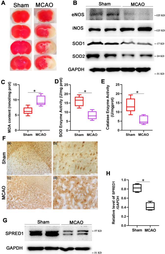
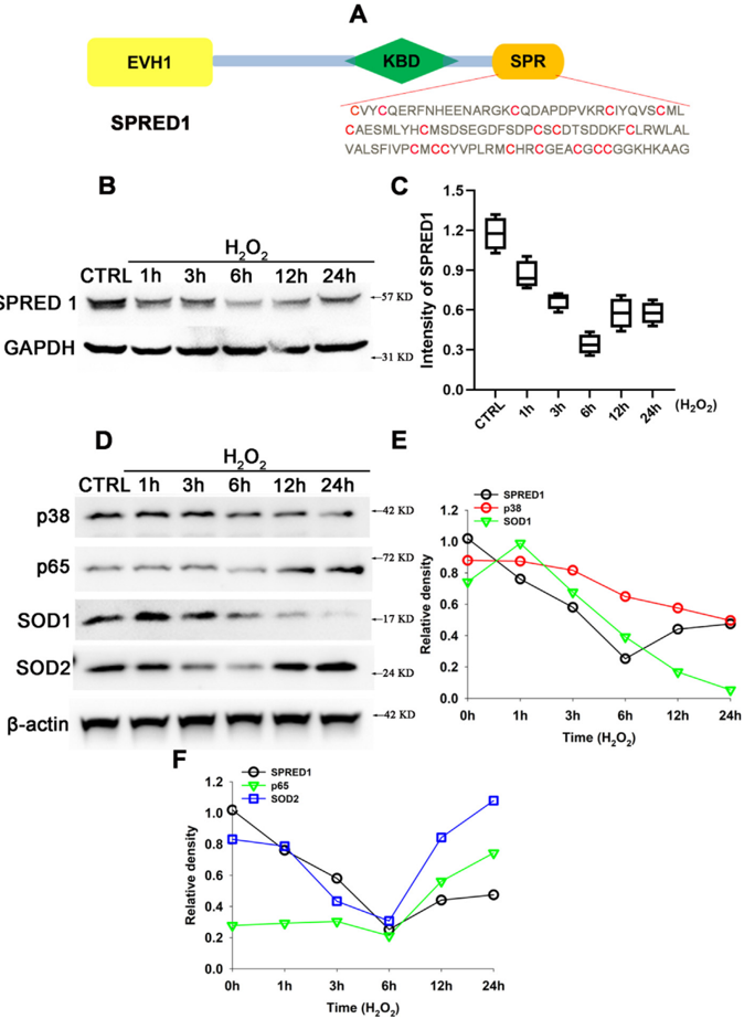
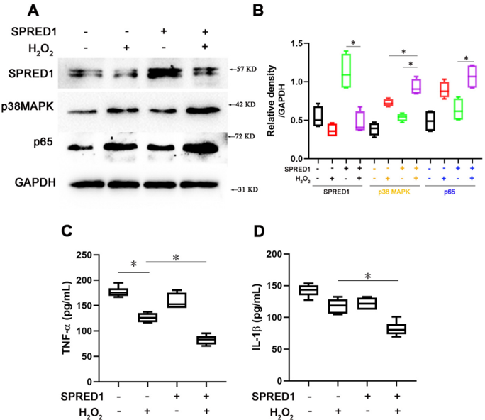
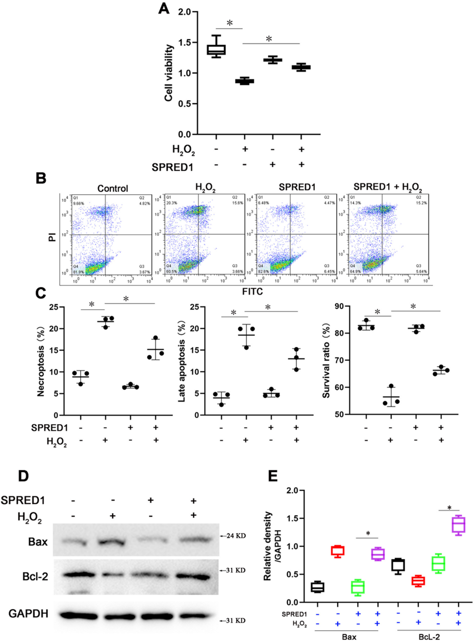
Verification
Claims the paper makes
The claims and statistics of this paper haven’t been checked yet.
Run the check — free, no account needed. It reads the open-access text and checks the sources it cites.
Claims other papers make about this one
Each is checked against this paper’s own text: does it say what the citing paper says it does?
The cited source says what the claim attributes to it.
In the citing paper
“In addition to these critical effectors, several members of the AD-specific miR-132 predicted targetome that we identified, e.g. TRIM44, NUCKS1, NFE2L2, LYN, ZEB2, SPRED1, ABHD5, RASA1, and NAP1L1 [143, 147–154], have known roles in inflammation and/or microglial cell states/function.”
From microRNA-132 attenuates inflammation in induced pluripotent stem cell-derived microglia from Alzheimer’s disease patients (2026) · cites this paper as [151]
Self-check
Percentages, sums and reported differences re-computed from the paper’s own numbers.
The self-check needs the paper’s full text.
Citations
Cites
Casas, A. I., Geuss, E., Kleikers, P. W. M., Mencl, S., Herrmann, A. M., Buendia, I., Egea, J., Meuth, S. G., Lopez, M. G., Kleinschnitz, C., et al. (2017) · Proceedings of the National Academy of Sciences · doi:10.1073/pnas.1705034114 · Cited by 152
Research works printed but not linked: 25
- Ablain, 2018, Human tumor genomics and zebrafish modeling identify SPRED1 loss as a driver of mucosal melanoma, Science, 362, 1055, 10.1126/science.aau6509 · DOI
- Barone, 2001, Inhibition of p38 mitogen-activated protein kinase provides neuroprotection in cerebral focal ischemia, Med Res Rev, 21, 129, 10.1002/1098-1128(200103)21:2<129::AID-MED1003>3.0.CO;2-H · DOI
- Chen, 2011, Oxidative stress in ischemic brain damage: Mechanisms of cell death and potential molecular targets for neuroprotection, Antioxid Redox Sign, 14, 1505, 10.1089/ars.2010.3576 · DOI
- Chen, 2020, Therapeutic targets of oxidative/nitrosative stress and neuroinflammation in ischemic stroke: Applications for natural product efficacy with omics and systemic biology, Pharmacol Res, 158, 10.1016/j.phrs.2020.104877 · DOI
- Dunzendorfer-Matt, 2016, The neurofibromin recruitment factor Spred1 binds to the GAP related domain without affecting Ras inactivation, Proc Natl Acad Sci USA, 113, 7497, 10.1073/pnas.1607298113 · DOI
- Ferrer, 2003, Early modifications in the expression of mitogen-activated protein kinase (MAPK/ERK), stress-activated kinases SAPK/JNK and p38, and their phosphorylated substrates following focal cerebral ischemia, Acta Neuropathol, 105, 425, 10.1007/s00401-002-0661-2 · DOI
- Gallo, 2002, Mixed-lineage kinase control of JNK and p38 MAPK pathways, Nat Rev Mol Cell Bio, 3, 663, 10.1038/nrm906 · DOI
- He, 2020, Highly bioactive zeolitic imidazolate framework-8-capped nanotherapeutics for efficient reversal of reperfusion-induced injury in ischemic stroke, Sci Adv, 6, 10.1126/sciadv.aay9751 · DOI
- Hu, 2015, miR-126 promotes angiogenesis and attenuates inflammation after contusion spinal cord injury in rats, Brain Res, 1608, 191, 10.1016/j.brainres.2015.02.036 · DOI
- Li, 2016, Neuron-derived FGF10 ameliorates cerebral ischemia injury via inhibiting NF-kappa B-dependent neuroinflammation and activating PI3K/Akt survival signaling pathway in mice, Sci Rep-Uk, 6
- Liang, 2019, Preactivation of Notch1 in remote ischemic preconditioning reduces cerebral ischemia-reperfusion injury through crosstalk with the NF-kappa B pathway, J Neuroinflamm, 16, 10.1186/s12974-019-1570-9 · DOI
- Park, 2018, RPM-1 and DLK-1 regulate pioneer axon outgrowth by controlling Wnt signaling, Development, 145, 10.1242/dev.164897 · DOI
- Phoenix, 2010, Spred1, a negative regulator of Ras-MAPK-ERK, is enriched in CNS germinal zones, dampens NSC proliferation, and maintains ventricular zone structure, Gene Dev, 24, 45, 10.1101/gad.1839510 · DOI
- Ridder, 2009, NF-kappa B signaling in cerebral ischemia, Neuroscience, 158, 995, 10.1016/j.neuroscience.2008.07.007 · DOI
- Sanderson, 2013, Molecular mechanisms of ischemia-reperfusion injury in brain: Pivotal role of the mitochondrial membrane potential in reactive oxygen species generation, Mol Neurobiol, 47, 9, 10.1007/s12035-012-8344-z · DOI
- Sun, 2017, Dyrk2 involved in regulating LPS-induced neuronal apoptosis, Int J Biol Macromol, 104, 979, 10.1016/j.ijbiomac.2017.06.087 · DOI
- Sun, 2022, High-performance SOD mimetic enzyme Au@Ce for arresting cell cycle and proliferation of acute myeloid leukemia, Bioact Mater, 10, 117
- Sun, 2022, Coronal relay reactor Fe3O4@CeO2 for accelerating ROS axial conversion through enhanced Enzyme-like effect and relay effect, Chem Eng J, 429, 10.1016/j.cej.2021.132303 · DOI
- Tadokoro, 2018, Spred1 safeguards hematopoietic homeostasis against diet-induced systemic stress, Cell Stem Cell, 22, 713, 10.1016/j.stem.2018.04.002 · DOI
- Wakioka, 2001, Spred is a Sprouty-related suppressor of Ras signalling, Nature, 412, 647, 10.1038/35088082 · DOI
- Xu, 2018, Dyrk2 mediated the release of proinflammatory cytokines in LPS-induced BV2 cells, Int J Biol Macromol, 109, 1115, 10.1016/j.ijbiomac.2017.11.095 · DOI
- Yan, 2020, Structural insights into the SPRED1-neurofibromin-KRAS complex and disruption of SPRED1-neurofibromin interaction by oncogenic EGFR, Cell Rep, 32, 10.1016/j.celrep.2020.107909 · DOI
- Yang, 2018, Diverse roles of mitochondria in ischemic stroke, Redox Biol, 16, 263, 10.1016/j.redox.2018.03.002 · DOI
- Yang, 2020, Neuroprotective and anti-inflammatory effect of tangeretin against cerebral ischemia-reperfusion injury in rats, Inflammation, 43, 2332, 10.1007/s10753-020-01303-z · DOI
- Zhao, 2017, Regulation of microglial activation in stroke, Acta Pharmacol Sin, 38, 445, 10.1038/aps.2016.162 · DOI
Cited by
Sorted by citations
Penning, A., Snoeck, S., Ormaechea, O. R., Ayyildiz, D., Polzer, O., Buitrago-Arango, M., Capobianco, R., de Winter, F., Balusu, S., Verhaagen, J., et al. (2026) · Acta Neuropathologica Communications · doi:10.1186/s40478-026-02228-8 · Cited by 6
Metadata
- Authors
- Wei, L., Li, X., Wei, Q., Chen, L., Xu, L., & Zhou, P.
- Venue
- Neuroscience
- Publisher
- Elsevier BV
- Published
- 2023-08
- Volume
- 526
- Pages
- 13-20
- ISSN
- 0306-4522
- Cited by
- 3
- References
- 1
- Language
- en
About these checks
Registry, retraction, Cites and statistics results are computed, not judged, with one exception: a printed reference that no index lists is read once by a model before it counts as a citation error. Claims are extracted by a model and kept only when their quote appears verbatim in the text; each cited source is then checked, with a model’s help, for whether it says what the claim relies on. Those verdicts can be wrong — read the claim’s quote and the cited source before relying on one.
Statistics and arithmetic are recomputed, not judged.
Confidence bands are shown only for fields calibrated on our annotated test set. None is calibrated yet, so no band is shown.
What counts as a claim ›How we measure confidence ›How we check this ›
Are you an author, or do you see a problem with this page? Request a review
