

The DOI matches its Crossref record; 42 of the 42 references it cites were found in a registry or an index.
Efficient Delivery of Antisense Oligonucleotides Using Bioreducible Lipid Nanoparticles In Vitro and In Vivo
Open accessAbstract
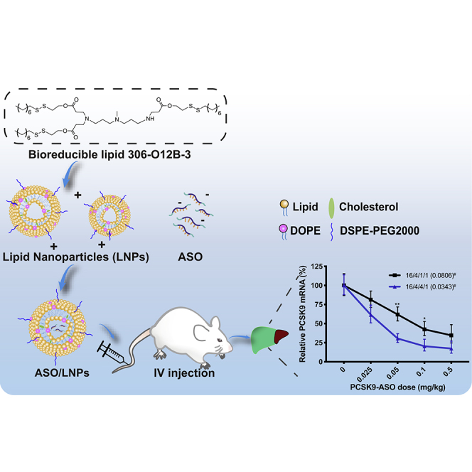
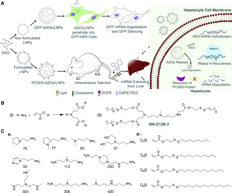
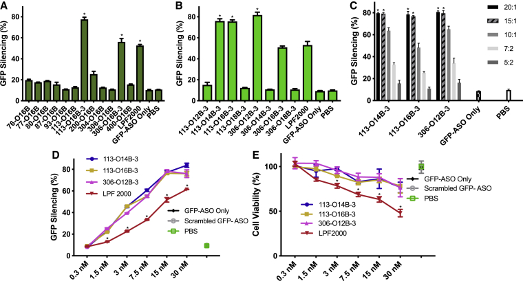
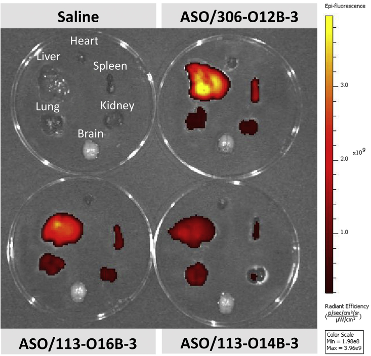
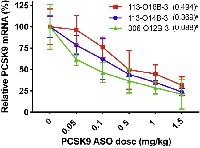
The efficient delivery of antisense oligonucleotides (ASOs) to the targeted cells and organs remains a challenge, in particular, in vivo . Here, we investigated the ability of a library of biodegradable lipid nanoparticles (LNPs) in delivering ASO to both cultured human cells and animal models. We first identified three top-performing lipids through in vitro screening using GFP-expressing HEK293 cells. Next, we explored these three candidates for delivering ASO to target proprotein convertase subtilisin/kexin type 9 (PCSK9) mRNA in mice. We found that lipid 306-O12B-3 showed efficiency with the median effective dose (ED 50 ) as low as 0.034 mg·kg -1 , which is a notable improvement over the efficiency reported in the literature. No liver or kidney toxicity was observed with a dose up to 5 mg·kg -1 of this ASO/LNP formulation. The biodegradable LNPs are efficient and safe in the delivery of ASO and pave the way for clinical translation.
Overview
The claims and statistics of this paper haven’t been checked yet.
Run the check — free, no account needed. It reads the open-access text and checks the sources it cites.
Integrity signals
Registry
Cites
Statistics
Figures
Verification
Claims the paper makes
The claims and statistics of this paper haven’t been checked yet.
Run the check — free, no account needed. It reads the open-access text and checks the sources it cites.
Claims other papers make about this one
Each is checked against this paper’s own text: does it say what the citing paper says it does?
The source backs part of the claim, not all of it — or backs it for a different population, condition or comparison.
In the citing paper
“However, this lipid formulation mainly delivered the ASO into the liver and could not cross the BBB through intravenous injection.”
From Neurotransmitter-derived lipidoids (NT-lipidoids) for enhanced brain delivery through intravenous injection (2020) · cites this paper as [30]
Experimental — accuracy not yet measuredThe cited source says what the claim attributes to it.
In the citing paper
“In a recent study, we identified lipidoid 306-O12B-3 that can efficiently deliver ASO both in vitro and in vivo (30).”
From Neurotransmitter-derived lipidoids (NT-lipidoids) for enhanced brain delivery through intravenous injection (2020) · cites this paper as [30]
Self-check
Percentages, sums and reported differences re-computed from the paper’s own numbers.
The self-check needs the paper’s full text.
Citations
Cites
Non-viral vectors for gene-based therapy
Yin, H., Kanasty, R. L., Eltoukhy, A. A., Vegas, A. J., Dorkin, J. R., & Anderson, D. G. (2014) · Nature Reviews Genetics · doi:10.1038/nrg3763 · Cited by 3,123
Patisiran, an RNAi Therapeutic, for Hereditary Transthyretin Amyloidosis
Adams, D., Gonzalez-Duarte, A., O’Riordan, W. D., Yang, C.-C., Ueda, M., Kristen, A. V., Tournev, I., Schmidt, H. H., Coelho, T., Berk, J. L., et al. (2018) · New England Journal of Medicine · doi:10.1056/nejmoa1716153 · Cited by 3,047
Self-citationRational design of cationic lipids for siRNA delivery
Semple, S. C., Akinc, A., Chen, J., Sandhu, A. P., Mui, B. L., Cho, C. K., Sah, D. W. Y., Stebbing, D., Crosley, E. J., Yaworski, E., et al. (2010) · Nature Biotechnology · doi:10.1038/nbt.1602 · Cited by 1,822
Self-citationMaximizing the potency of siRNA lipid nanoparticles for hepatic gene silencing in vivo
Jayaraman, M., Ansell, S. M., Mui, B. L., Tam, Y. K., Chen, J., Du, X., Butler, D., Eltepu, L., Matsuda, S., Narayanannair, J. K., et al. (2012) · Angewandte Chemie International Edition · doi:10.1002/anie.201203263 · Cited by 1,267
Self-citationLipid Nanoparticle Systems for Enabling Gene Therapies
Cullis, P. R., & Hope, M. J. (2017) · Molecular Therapy · doi:10.1016/j.ymthe.2017.03.013 · Cited by 1,208
A combinatorial library of lipid-like materials for delivery of RNAi therapeutics
Akinc, A., Zumbuehl, A., Goldberg, M., Leshchiner, E. S., Busini, V., Hossain, N., Bacallado, S. A., Nguyen, D. N., Fuller, J., Alvarez, R., et al. (2008) · Nature Biotechnology · doi:10.1038/nbt1402 · Cited by 1,150
Lipid-like materials for low-dose, in vivo gene silencing
Love, K. T., Mahon, K. P., Levins, C. G., Whitehead, K. A., Querbes, W., Dorkin, J. R., Qin, J., Cantley, W., Qin, L. L., Racie, T., et al. (2010) · Proceedings of the National Academy of Sciences · doi:10.1073/pnas.0910603106 · Cited by 969
Antisense oligonucleotides: the next frontier for treatment of neurological disorders
Rinaldi, C., & Wood, M. J. A. (2018) · Nature Reviews Neurology · doi:10.1038/nrneurol.2017.148 · Cited by 788
Chemistry, mechanism and clinical status of antisense oligonucleotides and duplex RNAs
Shen, X., & Corey, D. R. (2017) · Nucleic Acids Research · doi:10.1093/nar/gkx1239 · Cited by 654
Frank-Kamenetsky, M. D., Grefhorst, A., Anderson, N. N., Racie, T., Bramlage, B., Akinc, A., Butler, D., Charissé, K., Dorkin, J. R., Fan, Y., et al. (2008) · Proceedings of the National Academy of Sciences · doi:10.1073/pnas.0805434105 · Cited by 645
Maier, M. A., Jayaraman, M., Matsuda, S., Liu, J., Barros, S., Querbes, W., Tam, Y. K., Ansell, S. M., Kumar, V., Qin, J., et al. (2013) · Molecular Therapy · doi:10.1038/mt.2013.124 · Cited by 625
Efficient delivery of genome-editing proteins using bioreducible lipid nanoparticles
Wang, M., Zuris, J. A., Meng, F., Rees, H., Sun, S., Deng, P., Han, Y., Gao, X., Pouli, D., Wu, Q., et al. (2016) · Proceedings of the National Academy of Sciences · doi:10.1073/pnas.1520244113 · Cited by 624
Self-citationDesigning Chemically Modified Oligonucleotides for Targeted Gene Silencing
Deleavey, G. F., & Damha, M. J. (2012) · Chemistry & Biology · doi:10.1016/j.chembiol.2012.07.011 · Cited by 605
Fitzgerald, K. J., Frank-Kamenetsky, M. D., Shulga‐Morskaya, S., Liebow, A., Bettencourt, B. R., Sutherland, J. E., Hutabarat, R. M., Clausen, V. A., Karsten, V., Cehelsky, J. E., et al. (2013) · The Lancet · doi:10.1016/s0140-6736(13)61914-5 · Cited by 586
Cellular uptake and trafficking of antisense oligonucleotides
Crooke, S. T., Wang, S., Vickers, T. A., Shen, W., & Liang, X.-H. (2017) · Nature Biotechnology · doi:10.1038/nbt.3779 · Cited by 582
Molecular Mechanisms of Antisense Oligonucleotides
Crooke, S. T. (2017) · Nucleic Acid Therapeutics · doi:10.1089/nat.2016.0656 · Cited by 415
Molecular mechanisms of action of antisense drugs
Crooke, S. T. (1999) · Biochimica et Biophysica Acta (BBA) - Gene Structure and Expression · doi:10.1016/s0167-4781(99)00148-7 · Cited by 356
Biological Barriers to Therapy with Antisense and siRNA Oligonucleotides
Juliano, R. L., Bauman, J. A., Kang, H. M., & Ming, X. (2009) · Molecular Pharmaceutics · doi:10.1021/mp900093r · Cited by 306
Wang, M., Alberti, K. A., Sun, S., Arellano, C. L., & Xu, Q. (2014) · Angewandte Chemie International Edition · doi:10.1002/anie.201311245 · Cited by 250
Self-citationRNA therapeutics: RNAi and antisense mechanisms and clinical applications
Chery, J. (2016) · Postdoc Journal · doi:10.14304/surya.jpr.v4n7.5 · Cited by 242
The Role of the Helper Lipid on the DNA Transfection Efficiency of Lipopolyplex Formulations
Du, Z., Munye, M. M., Tagalakis, A. D., Manunta, M., & Hart, S. L. (2014) · Scientific Reports · doi:10.1038/srep07107 · Cited by 200
How does RNase H recognize a DNA.RNA hybrid?
Nakamura, H., Oda, Y., Iwai, S., Inoue, H., Ohtsuka, E., Kanaya, S., Kimura, S., Katsuda, C., Katayanagi, K., & Morikawa, K. (1991) · Proceedings of the National Academy of Sciences · doi:10.1073/pnas.88.24.11535 · Cited by 193
Basha, G., Novobrantseva, T. I., Rosin, N. L., Tam, Y. Y. C., Hafez, I. M., Wong, M., Sugo, T., Ruda, V. M., Qin, J., Klebanov, B., et al. (2011) · Molecular Therapy · doi:10.1038/mt.2011.190 · Cited by 182
Chaparro‐Riggers, J. F., Hong, L., DeVay, R. M., Bai, L., Sutton, J. E., Chen, W., Geng, T., Lindquist, K. C., Casas, M. G., Boustany, L. M., et al. (2012) · Journal of Biological Chemistry · doi:10.1074/jbc.m111.319764 · Cited by 168
Lipid Conjugated Oligonucleotides: A Useful Strategy for Delivery
Raouane, M., Desmaële, D., Urbinati, G., Massaad-Massade, L., & Couvreur, P. (2012) · Bioconjugate Chemistry · doi:10.1021/bc200422w · Cited by 161
Chang, J., Chen, X., Glass, Z., Gao, F., Mao, L., Wang, M., & Xu, Q. (2018) · Accounts of Chemical Research · doi:10.1021/acs.accounts.8b00493 · Cited by 130
Self-citationMochizuki, S., Kanegae, N., Nishina, K., Kamikawa, Y., Koiwai, K., Masunaga, H., & Sakurai, K. (2013) · Biochimica et Biophysica Acta · doi:10.1016/j.bbamem.2012.10.017 · Cited by 129
Shi, B., Keough, E., Matter, A., Leander, K., Young, S., Carlini, E., Sachs, A. B., Tao, W., Abrams, M., Howell, B., et al. (2011) · Journal of Histochemistry & Cytochemistry · doi:10.1369/0022155411410885 · Cited by 125
The Chemistry and Biology of Oligonucleotide Conjugates
Juliano, R. L., Ming, X., & Nakagawa, O. (2012) · Accounts of Chemical Research · doi:10.1021/ar2002123 · Cited by 125
Non-viral delivery of genome-editing nucleases for gene therapy
Wang, M., Glass, Z., & Xu, Q. (2016) · Gene Therapy · doi:10.1038/gt.2016.72 · Cited by 113
Self-citationEnhanced Intracellular siRNA Delivery using Bioreducible Lipid‐Like Nanoparticles
Wang, M., Alberti, K. A., Varone, A., Pouli, D., Georgakoudi, I., & Xu, Q. (2014) · Advanced Healthcare Materials · doi:10.1002/adhm.201400039 · Cited by 101
Self-citationAntisense Oligonucleotides for the Treatment of Spinal Muscular Atrophy
Porensky, P. N., & Burghes, A. H. M. (2013) · Human Gene Therapy · doi:10.1089/hum.2012.225 · Cited by 89
Vickers, T. A., & Crooke, S. T. (2015) · Nucleic Acids Research · doi:10.1093/nar/gkv920 · Cited by 84
Shirazi, R. S., Ewert, K., Leal, C., Majzoub, R. N., Bouxsein, N. F., & Safinya, C. (2011) · Biochimica et Biophysica Acta · doi:10.1016/j.bbamem.2011.04.020 · Cited by 82
Altınoglu, S., Wang, M., & Xu, Q. (2015) · Nanomedicine · doi:10.2217/nnm.14.192 · Cited by 70
Self-citationEteplirsen Approved for Duchenne Muscular Dystrophy: The FDA Faces a Difficult Choice
Stein, C. A. (2016) · Molecular Therapy · doi:10.1038/mt.2016.188 · Cited by 67
Falzarano, M. S., Passarelli, C., & Ferlini, A. (2014) · Nucleic Acid Therapeutics · doi:10.1089/nat.2013.0450 · Cited by 53
Defining the Factors That Contribute to On-Target Specificity of Antisense Oligonucleotides
Lima, W. F., Vickers, T. A., Nichols, J. G., Li, C., & Crooke, S. T. (2014) · PLoS ONE · doi:10.1371/journal.pone.0101752 · Cited by 48
Mercatante, D. R., & Kole, R. (2002) · Biochimica et Biophysica Acta (BBA) - Molecular Basis of Disease · doi:10.1016/s0925-4439(02)00075-3 · Cited by 46
DOPE facilitates quaternized lipidoids (QLDs) for in vitro DNA delivery
Sun, S., Wang, M., Alberti, K. A., Choy, A., & Xu, Q. (2013) · Nanomedicine Nanotechnology Biology and Medicine · doi:10.1016/j.nano.2013.01.006 · Cited by 35
Self-citationTakeda, Y. S., Wang, M., Deng, P. J., & Xu, Q. (2016) · Bioengineering & Translational Medicine · doi:10.1002/btm2.10021 · Cited by 21
Self-citationAnti-PCSK9 Antibodies: A New Era in the Treatment of Dyslipidemia
Schmitz, J., & Gouni‐Berthold, I. (2017) · Current Pharmaceutical Design · doi:10.2174/1381612823666170130155036 · Cited by 8
Research works printed but not linked: 1
- 2016, FDA approves first drug for spinal muscular atrophy, Press Anouncement
Cited by
Sorted by citations
Mendes, B. B., Conniot, J., Avital, A., Yao, D., Jiang, X., Zhou, X., Sharf-Pauker, N., Xiao, Y., Adir, O., Liang, H., et al. (2022) · Nature Reviews Methods Primers · doi:10.1038/s43586-022-00104-y · Cited by 719
Dilliard, S. A., & Siegwart, D. J. (2023) · Nature Reviews Materials · doi:10.1038/s41578-022-00529-7 · Cited by 744
Lipids and Lipid Derivatives for RNA Delivery
Zhang, Y., Sun, C., Wang, C., Jankovic, K. E., & Dong, Y. (2021) · Chemical Reviews · doi:10.1021/acs.chemrev.1c00244 · Cited by 649
An ionizable lipid toolbox for RNA delivery
Han, X., Zhang, H., Butowska, K., Swingle, K. L., Alameh, M.-G., Weissman, D., & Mitchell, M. J. (2021) · Nature Communications · doi:10.1038/s41467-021-27493-0 · Cited by 628
Jeong, M., Lee, Y., Park, J., Jung, H., & Lee, H. (2023) · Advanced Drug Delivery Reviews · doi:10.1016/j.addr.2023.114990 · Cited by 232
Metadata
- Authors
- Yang, L., Ma, F., Liu, F., Chen, J., Zhao, X., & Xu, Q.
- Affiliations
- Tufts University (US)
- Corresponding author
- Xu, Qiaobing
- Venue
- Molecular Therapy - Nucleic Acids
- Publisher
- Elsevier BV
- Published
- 2020-03
- Volume
- 19
- Pages
- 1357-1367
- ISSN
- 2162-2531 (ISSN-L)
- PMC
- PMC7036716
- OpenAlex
- W3002543944
- Cited by
- 103
- References
- 42
- Citations by year
- 2026: 14 · 2025: 16 · 2024: 21 · 2023: 18 · 2022: 15 · 2021: 12 · 2020: 7
- FWCI
- 4.22
- Type
- journal article
- Language
- en
- Open access
- gold
- doi.org
- Licence
- Locations
- Molecular Therapy — Nucleic Acids (publishedVersion, cc-by-nc-nd)
- PubMed Central (publishedVersion, cc-by-nc-nd)
- PubMed (publishedVersion)
- DOAJ (DOAJ: Directory of Open Access Journals) (submittedVersion, cc-by-sa)
- Funders
- National Institutes of Health (R01 EB027170-01, 1R21EB024041-01)
- Topics
- Life Sciences › Biochemistry, Genetics and Molecular Biology › Molecular Biology › RNA Interference and Gene Delivery
- Physical Sciences › Materials Science › Biomaterials › Nanoparticle-Based Drug Delivery
- Life Sciences › Pharmacology, Toxicology and Pharmaceutics › Pharmaceutical Science › Advancements in Transdermal Drug Delivery
- Keywords
- antisense oligonucleotides, lipid nanoparticles, biodegradable lipids, nucleic acid delivery, PCSK9, lipid screening, in vivo delivery, toxicity, gene silencing
About these checks
Registry, retraction, Cites and statistics results are computed, not judged, with one exception: a printed reference that no index lists is read once by a model before it counts as a citation error. Claims are extracted by a model and kept only when their quote appears verbatim in the text; each cited source is then checked, with a model’s help, for whether it says what the claim relies on. Those verdicts can be wrong — read the claim’s quote and the cited source before relying on one.
Statistics and arithmetic are recomputed, not judged.
Confidence bands are shown only for fields calibrated on our annotated test set. None is calibrated yet, so no band is shown.
What counts as a claim ›How we measure confidence ›How we check this ›
Are you an author, or do you see a problem with this page? Request a review
