

The DOI matches its Crossref record.
Bowel necrosis associated with COVID-19 pneumonia: A report of two cases
Open accessAbstract
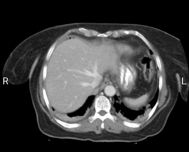
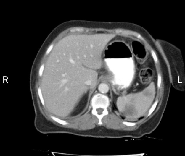
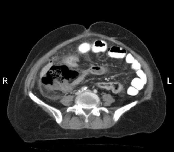
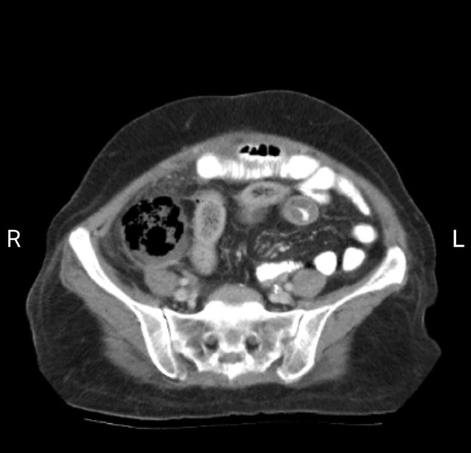
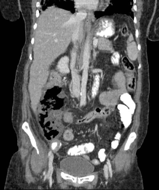
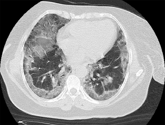
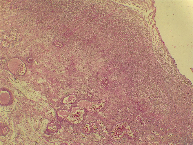
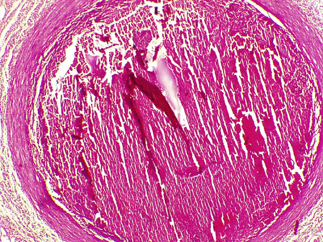
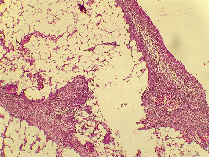
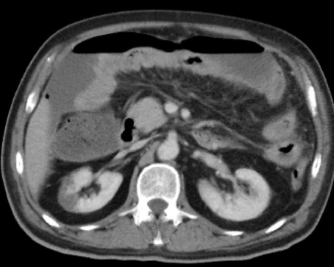
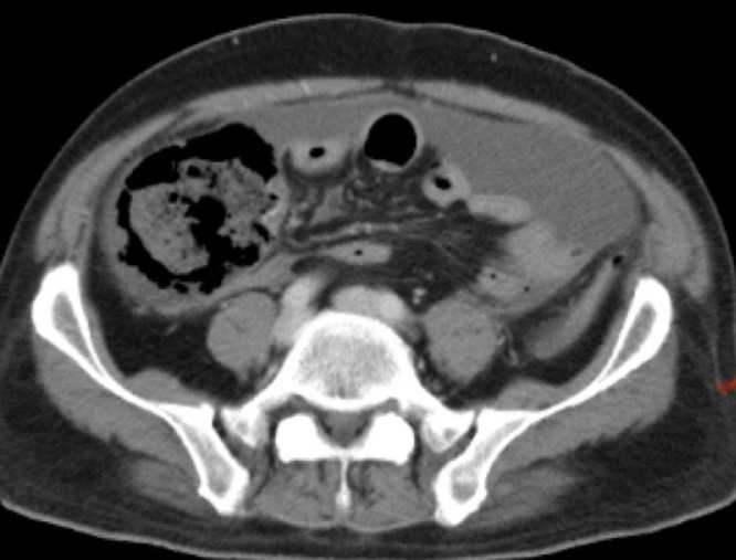
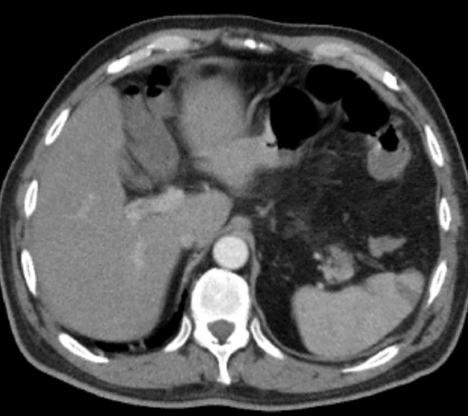
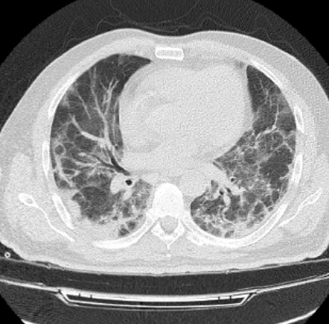
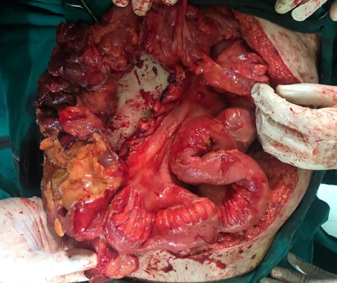
A novel coronavirus, called as SARS-CoV-2, caused multiple typical and atypical respiratory symptoms with several reported abdominal findings. We have described 2 cases of an unusual presentation of Covid-19. For patient 1, a woman aged 59-year-old had pneumonia and new onset abdominal pain. In patient 2, a 60-year-old man admitted with positive polymerase chain reaction test and abdominal pain from 2 weeks ago. Both lung and abdominal computed tomography (CT) were achieved in a few days. Acute ischemic mesenteritis and other abdominal complications were observed in the CT scan. Bowel necrosis in combination with peritonitis founded by laparotomy. Early diagnosis of abdominal complications in Covid-19 pneumonia can improve patient outcomes.
Overview
The claims and statistics of this paper haven’t been checked yet.
Run the check — free, no account needed. It reads the open-access text and checks the sources it cites.
Integrity signals
Registry
Cites
Statistics
Figures
Verification
Claims the paper makes
The claims and statistics of this paper haven’t been checked yet.
Run the check — free, no account needed. It reads the open-access text and checks the sources it cites.
Claims other papers make about this one
Each is checked against this paper’s own text: does it say what the citing paper says it does?
The cited source says what the claim attributes to it.
In the citing paper
“The case reported by Mir et al.44 described a 59-year-old, diabetic patient who developed mesenteric venous thrombosis.”
From Portal-splenic-mesenteric venous thrombosis in COVID-19 patients: a systematic review (2025) · cites this paper as Mir et al.44
Self-check
Percentages, sums and reported differences re-computed from the paper’s own numbers.
The self-check needs the paper’s full text.
Citations
Cites
No references could be resolved yet.
Research works printed but not linked: 11
- Vernuccio, 2020, Thromboembolic complications of COVID-19: the combined effect of a pro-coagulant pattern and an endothelial thrombo-inflammatory syndrome, Clin Radiol, 10.1016/j.crad.2020.07.019 · DOI
- Nopp, 2020, Risk of venous thromboembolism in patients with COVID-19: a systematic review and meta-analysis, Res Pract Thromb Haemost, 10.1002/rth2.12439 · DOI
- Ribes A, Vardon-Bounes F, Mémier V, Poette M, Au-Duong J, Garcia C, et al. Advances in biological regulation, 2020. doi:10.1016/j.jbior.2020.100735. · DOI
- Bilaloglu, 2020, Thrombosis in hospitalized patients with COVID-19 in a New York City health system, Jama, 324, 10.1001/jama.2020.13372 · DOI
- Porfidia, 2020, Venous thromboembolism in patients with COVID-19: systematic review and meta-analysis, Thrombosis Res, 10.1016/j.thromres.2020.08.020 · DOI
- Singhal, 2020, A review of coronavirus disease-2019 (COVID-19), Indian J Pediatr, 10.1007/s12098-020-03263-6 · DOI
- Harapan, 2020, Coronavirus disease 2019 (COVID-19): a literature review, J Infect Publ Health, 10.1016/j.jiph.2020.03.019 · DOI
- Shi, 2020, Radiological findings from 81 patients with COVID-19 pneumonia in Wuhan, China: a descriptive study, Lancet Infect Dis, 10.1016/S1473-3099(20)30086-4 · DOI · Paper page
- Hani, 2020, COVID-19 pneumonia: a review of typical CT findings and differential diagnosis, Diagn Intervent Imaging, 10.1016/j.diii.2020.03.014 · DOI
- Salehi, 2020, Coronavirus disease 2019 (COVID-19): a systematic review of imaging findings in 919 patients, AJR, 10.2214/AJR.20.23034 · DOI · Paper page
- G. Taourel, 1996, Acute mesenteric ischemia: diagnosis with contrast-enhanced CT, RSNA
Cited by
Sorted by citations
Portal-splenic-mesenteric venous thrombosis in COVID-19 patients: a systematic review
Gomes, R. Z., Salina, M. V. J., Pironatto, V. H. M., Lepinski, J. K., Gueiber, T. D., Juliatto, B. M. S., Prestes, M. Z., & Martins, C. M. (2025) · Jornal Vascular Brasileiro · doi:10.1590/1677-5449.202301282 · Cited by 2
Metadata
- Authors
- Mir, M. Z., Mashhadi, A., Jahantigh, M., & Seyedi, S. J.
- Affiliations
- Zahedan University of Medical Sciences (IR)
- ORCID
- Mashhadi, Amin
- Venue
- Radiology Case Reports
- Publisher
- Elsevier BV
- Published
- 2021-04
- Volume
- 16
- Issue
- 4
- Pages
- 819-823
- ISSN
- 1930-0433 (ISSN-L)
- PMC
- PMC7826008
- OpenAlex
- W3123777525
- Cited by
- 7
- References
- 0
- Citations by year
- 2025: 1 · 2023: 4 · 2022: 1 · 2021: 1
- FWCI
- 0.48
- Type
- journal article
- Language
- en
- Update notices
- erratum (2023-03-01)
- Open access
- gold
- doi.org
- Licence
- Locations
- Radiology Case Reports (publishedVersion, cc-by-nc-nd)
- PubMed Central (publishedVersion, cc-by-nc-nd)
- PubMed (publishedVersion)
- DOAJ (DOAJ: Directory of Open Access Journals) (submittedVersion, cc-by-sa)
- Topics
- Health Sciences › Medicine › Infectious Diseases › COVID-19 Clinical Research Studies
- Health Sciences › Medicine › Surgery › Abdominal vascular conditions and treatments
- Health Sciences › Medicine › Clinical Neurology › Long-Term Effects of COVID-19
- Keywords
- COVID-19 pandemic, SARS-CoV-2, intestinal necrosis, mesenteric ischemia, peritonitis, abdominal complications, abdominal pain, pneumonia, laparotomy, computed tomography findings
About these checks
Registry, retraction, Cites and statistics results are computed, not judged, with one exception: a printed reference that no index lists is read once by a model before it counts as a citation error. Claims are extracted by a model and kept only when their quote appears verbatim in the text; each cited source is then checked, with a model’s help, for whether it says what the claim relies on. Those verdicts can be wrong — read the claim’s quote and the cited source before relying on one.
Statistics and arithmetic are recomputed, not judged.
Confidence bands are shown only for fields calibrated on our annotated test set. None is calibrated yet, so no band is shown.
What counts as a claim ›How we measure confidence ›How we check this ›
Are you an author, or do you see a problem with this page? Request a review
How to cite
Cite CoChat for the checks on this page, and the paper for its own findings.
- This page
- CoChat. (2026). Citation check: Bowel necrosis associated with COVID-19 pneumonia: A report of two cases. https://app.cochat.ai/papers/10.1016/j.radcr.2021.01.043
- The paper
- Mir, M. Z., Mashhadi, A., Jahantigh, M., & Seyedi, S. J. (2021). Bowel necrosis associated with COVID-19 pneumonia: A report of two cases. Radiology Case Reports. https://doi.org/10.1016/j.radcr.2021.01.043
