

The DOI matches its Crossref record; 24 of the 24 references it cites were found in a registry or an index.
A cardiac contouring atlas for radiotherapy
Open accessAbstract
Background and purpose The heart is a complex anatomical organ and contouring the cardiac substructures is challenging. This study presents a reproducible method for contouring left ventricular and coronary arterial segments on radiotherapy CT-planning scans. Material and methods Segments were defined from cardiology models and agreed by two cardiologists. Reference atlas contours were delineated and written guidelines prepared. Six radiation oncologists tested the atlas. Spatial variation was assessed using the DICE similarity coefficient (DSC) and the directed Hausdorff average distance ( d → H , avg ). The effect of spatial variation on doses was assessed using six different breast cancer regimens. Results The atlas enabled contouring of 15 cardiac segments. Inter-observer contour overlap (mean DSC) was 0.60–0.73 for five left ventricular segments and 0.10–0.53 for ten coronary arterial segments. Inter-observer contour separation (mean d → H , avg ) was 1.5–2.2mm for left ventricular segments and 1.3–5.1mm for coronary artery segments. This spatial variation resulted in <1Gy dose variation for most regimens and segments, but 1.2–21.8Gy variation for segments close to a field edge. Conclusions This cardiac atlas enables reproducible contouring of segments of the left ventricle and main coronary arteries to facilitate future studies relating cardiac radiation doses to clinical outcomes.
Overview
The claims and statistics of this paper haven’t been checked yet.
Run the check — free, no account needed. It reads the open-access text and checks the sources it cites.
Integrity signals
Registry
Cites
Statistics
Figures
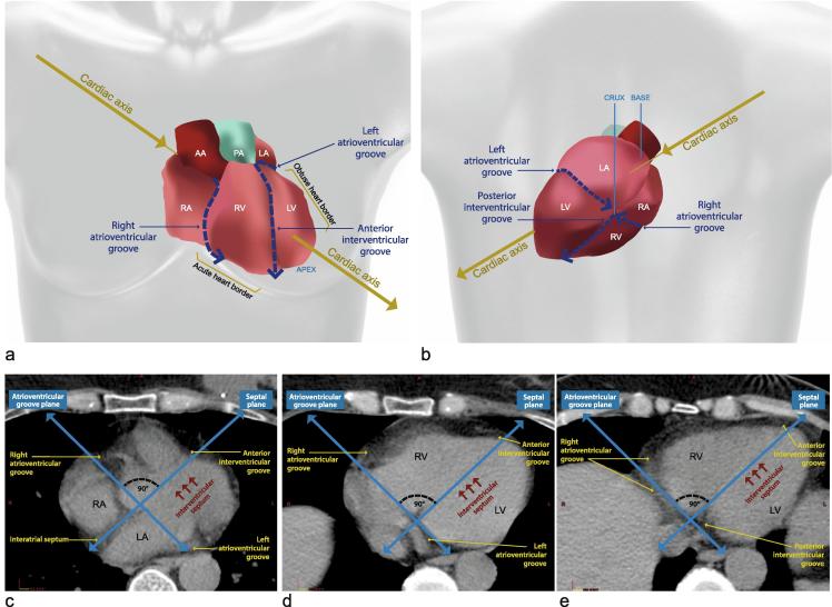
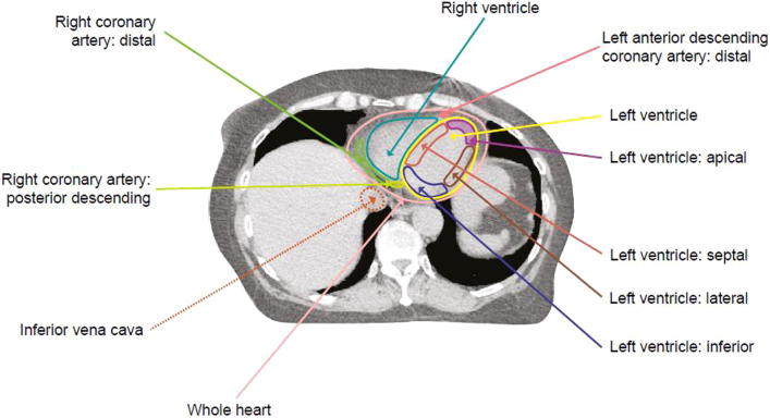
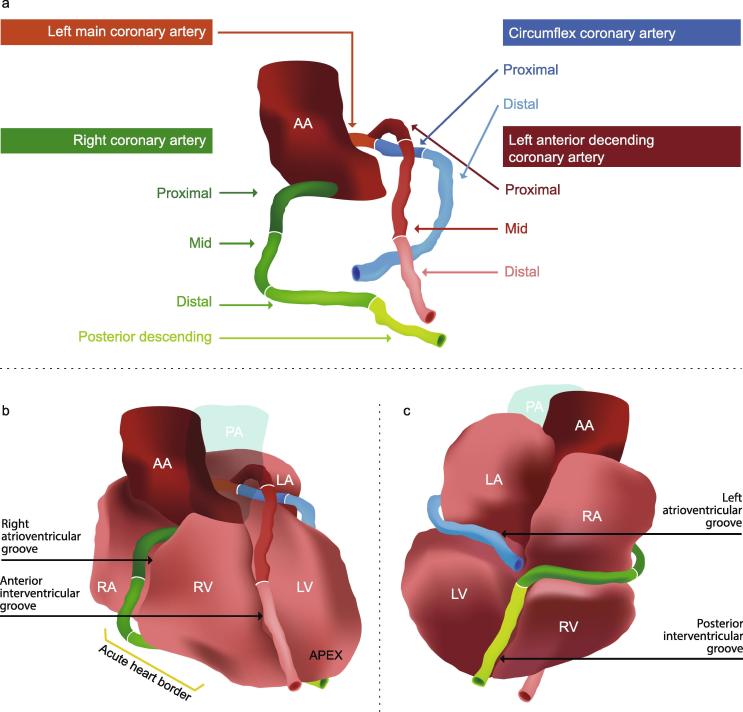
Verification
Claims the paper makes
The claims and statistics of this paper haven’t been checked yet.
Run the check — free, no account needed. It reads the open-access text and checks the sources it cites.
Claims other papers make about this one
Each is checked against this paper’s own text: does it say what the citing paper says it does?
The source backs part of the claim, not all of it — or backs it for a different population, condition or comparison.
In the citing paper
“This makes sense: it is the hardest of the three structures to segment in breast radiotherapy data because it is not always fully visible in the CT scan and contouring practices vary across treatment lateralities. 27”
From Uncertainty quantification of U‐Net based segmentation tool using conformal prediction (2026) · cites this paper as [27]
Experimental — accuracy not yet measured
Self-check
Percentages, sums and reported differences re-computed from the paper’s own numbers.
The self-check needs the paper’s full text.
Citations
Cites
Standardized Myocardial Segmentation and Nomenclature for Tomographic Imaging of the Heart
Cerqueira, M. D., Weissman, N. J., Dilsizian, V., Jacobs, A. K., Kaul, S., Laskey, W. K., Pennell, D. J., Rumberger, J. A., Ryan, T., & Verani, M. S. (2002) · Circulation · doi:10.1161/hc0402.102975 · Cited by 7,043
Risk of Ischemic Heart Disease in Women after Radiotherapy for Breast Cancer
Darby, S. C., Ewertz, M., McGale, P., Bennet, A. M., Blom-Goldman, U., Brønnum, D., Correa, C., Cutter, D., Gagliardi, G., Gigante, B., et al. (2013) · New England Journal of Medicine · doi:10.1056/nejmoa1209825 · Cited by 3,974
Self-citationWg, A., Edwards, J., Frye, R., Gg, G., Gott, V. L., Griffith, L., McGoon, D. C., Murphy, M., & Roe, B. B. (1975) · Circulation · doi:10.1161/01.cir.51.4.5 · Cited by 3,255
Radiation Dose–Volume Effects in the Heart
Gagliardi, G., Constine, L. S., Moiseenko, V., Correa, C. R., Pierce, L. J., Allen, A. M., & Marks, L. B. (2010) · International Journal of Radiation Oncology*Biology*Physics · doi:10.1016/j.ijrobp.2009.04.093 · Cited by 1,232
Feng, M. U.-S., Moran, J. M., Koelling, T. M., Chughtai, A. R., Chan, J. L., Freedman, L. M., Hayman, J. A., Jagsi, R., Jolly, S., Larouere, J., et al. (2010) · International Journal of Radiation Oncology*Biology*Physics · doi:10.1016/j.ijrobp.2009.10.058 · Cited by 717
Self-citationCorrea, C. R., Litt, H. I., Hwang, W., Ferrari, V. A., Solin, L. J., & Harris, E. E. R. (2007) · Journal of Clinical Oncology · doi:10.1200/jco.2006.08.6595 · Cited by 392
Distribution of Coronary Artery Stenosis After Radiation for Breast Cancer
Nilsson, G., Holmberg, L., Garmo, H., Duvernoy, O., Sjögren, I., Lagerqvist, B., & BLOMQVIST, C. P. (2011) · Journal of Clinical Oncology · doi:10.1200/jco.2011.34.5900 · Cited by 387
Risk for Valvular Heart Disease After Treatment for Hodgkin Lymphoma
Cutter, D. J., Schaapveld, M., Darby, S. C., Hauptmann, M., van Nimwegen, F. A., Krol, A. D. G., Janus, C. P. M., van Leeuwen, F. E., & Aleman, B. M. P. (2015) · JNCI Journal of the National Cancer Institute · doi:10.1093/jnci/djv008 · Cited by 315
Self-citationSlicerRT: Radiation therapy research toolkit for 3D Slicer
Pintér, C., Lassó, A., Wang, A., Jaffray, D. A., & Gabor Fichtinger, A. (2012) · Medical Physics · doi:10.1118/1.4754659 · Cited by 274
Dodge, J. T., Brown, B. G., Bolson, E. L., & Dodge, H. T. (1988) · Circulation · doi:10.1161/01.cir.78.5.1167 · Cited by 159
Cited by
Sorted by citations
Cardiac Toxicity of Thoracic Radiotherapy: Existing Evidence and Future Directions
Banfill, K., Giuliani, M., Aznar, M. C., Franks, K., McWilliam, A., Schmitt, M., Sun, F., Vozenin, M., & Finn, C. F. (2020) · Journal of Thoracic Oncology · doi:10.1016/j.jtho.2020.11.002 · Cited by 277
Self-citationJacob, S., Camilleri, J., Derreumaux, S., Walker, V., Lairez, O., Lapeyre, M., Bruguière, É., Pathak, A., Bernier, M., Laurier, D., et al. (2019) · Radiation Oncology · doi:10.1186/s13014-019-1234-z · Cited by 179
Taylor, C., McGale, P., Brønnum, D., Correa, C. R., Cutter, D. J., Duane, F. K., Gigante, B., Jensen, M., Lorenzen, E. L., Rahimi, K., et al. (2018) · Journal of Clinical Oncology · doi:10.1200/jco.2017.77.6351 · Cited by 149
Self-citationESTRO-ACROP guideline: Recommendations on implementation of breath-hold techniques in radiotherapy
Aznar, M. C., Carrasco, P., Corradini, S., Mast, M. E., McNair, H. A., Meattini, I., Persson, G. F., & van Haaren, P. M. A. (2023) · Radiotherapy and Oncology · doi:10.1016/j.radonc.2023.109734 · Cited by 139
Self-citationIncidence and Onset of Severe Cardiac Events After Radiotherapy for Esophageal Cancer
Wang, X., Palaskas, N. L., Yusuf, S. W., Abe, J., Lopez‐Mattei, J., Banchs, J. A., Gladish, G. W., Lee, P. P., Liao, Z. X., Deswal, A. M., et al. (2020) · Journal of Thoracic Oncology · doi:10.1016/j.jtho.2020.06.014 · Cited by 123
Metadata
- Authors
- Duane, F., Aznar, M. C., Bartlett, F., Cutter, D. J., Darby, S. C., Jagsi, R., Lorenzen, E. L., McArdle, O., McGale, P., Myerson, S., Rahimi, K., Vivekanandan, S., Warren, S., & Taylor, C. W.
- Affiliations
- University of Oxford (GB)
- Queen Alexandra Hospital (GB)
- University of Michigan (US)
- Odense University Hospital (DK)
- St. Luke's Hospital (IE)
- The George Institute for Global Health (GB)
- University of Birmingham (GB)
- Corresponding author
- Duane, Frances
- ORCID
- Venue
- Radiotherapy and Oncology
- Publisher
- Elsevier BV
- Published
- 2017-03
- Volume
- 122
- Issue
- 3
- Pages
- 416-422
- ISSN
- 0167-8140 (ISSN-L)
- PMC
- PMC5356506
- OpenAlex
- W2591100698
- Cited by
- 308
- References
- 24
- Citations by year
- 2026: 31 · 2025: 32 · 2024: 44 · 2023: 35 · 2022: 44 · 2021: 43 · 2020: 35 · 2019: 19 · 2018: 22 · 2017: 3
- FWCI
- 20.37
- Type
- journal article
- Language
- en
- Open access
- hybrid
- doi.org
- Locations
- Radiotherapy and Oncology (publishedVersion, cc-by)
- PubMed Central (publishedVersion, cc-by)
- PubMed (publishedVersion)
- University of Southern Denmark Research Portal (University of Southern Denmark) (submittedVersion, cc-by)
- Research Explorer (The University of Manchester) (submittedVersion, cc-by)
- Funders
- Medical Research Council
- Cancer Research UK (C8225/A21133, RE/13/1/30181, C5255/A15935)
- British Heart Foundation Centre for Research Excellence
- National Institute for Health Research
- Topics
- Physical Sciences › Physics and Astronomy › Radiation › Advanced Radiotherapy Techniques
- Physical Sciences › Computer Science › Computer Vision and Pattern Recognition › Medical Image Segmentation Techniques
- Health Sciences › Medicine › Radiology, Nuclear Medicine and Imaging › Radiation Dose and Imaging
- Keywords
- left ventricle segmentation, coronary artery segmentation, cardiac radiotherapy, breast cancer radiotherapy, heart dose
- MeSH terms
- Breast Neoplasms, Coronary Vessels, Female, Heart, Heart Ventricles, Humans, Radiotherapy Planning, Computer-Assisted, Tomography, X-Ray Computed
About these checks
Registry, retraction, Cites and statistics results are computed, not judged, with one exception: a printed reference that no index lists is read once by a model before it counts as a citation error. Claims are extracted by a model and kept only when their quote appears verbatim in the text; each cited source is then checked, with a model’s help, for whether it says what the claim relies on. Those verdicts can be wrong — read the claim’s quote and the cited source before relying on one.
Statistics and arithmetic are recomputed, not judged.
Confidence bands are shown only for fields calibrated on our annotated test set. None is calibrated yet, so no band is shown.
What counts as a claim ›How we measure confidence ›How we check this ›
Are you an author, or do you see a problem with this page? Request a review
