

The DOI matches its Crossref record; 197 of the 197 references it cites were found in a registry or an index.
The many roles of myeloperoxidase: From inflammation and immunity to biomarkers, drug metabolism and drug discovery
Open accessAbstract
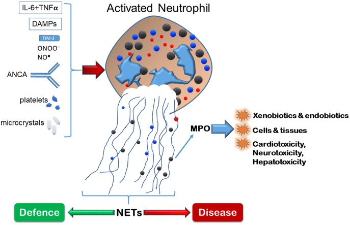
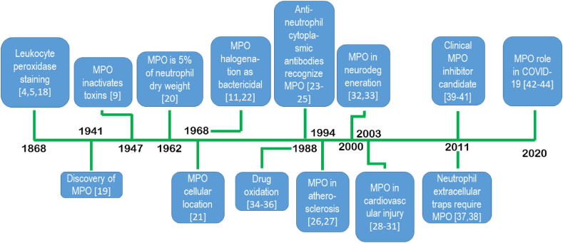
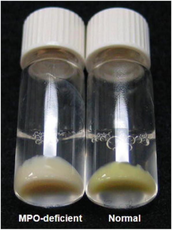
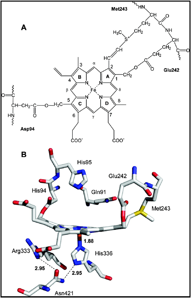
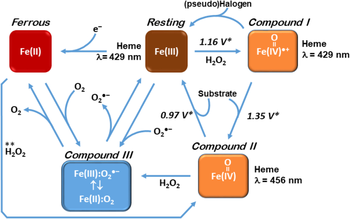
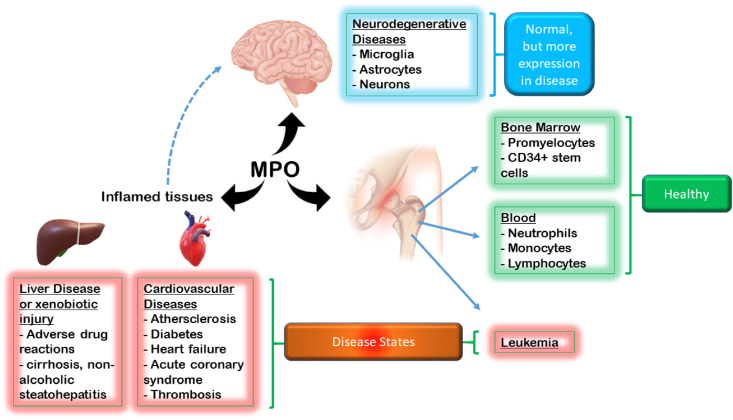
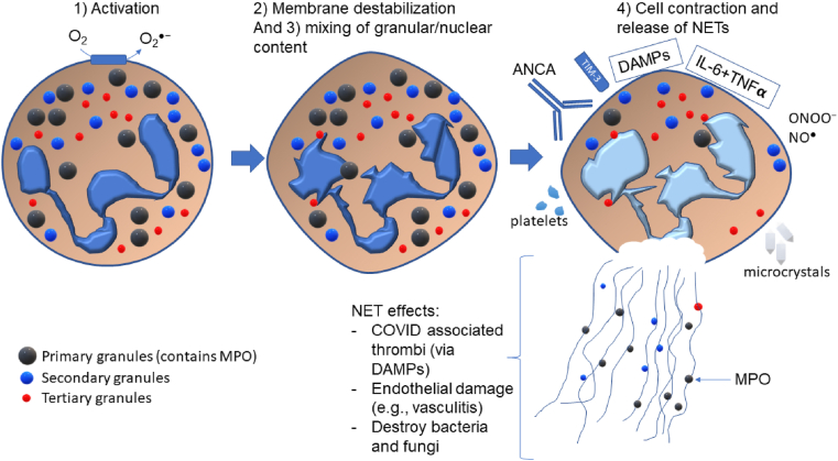
This review provides a practical guide to myeloperoxidase (MPO) and presents to the reader the diversity of its presence in biology. The review provides a historical background, from peroxidase activity to the discovery of MPO, to its role in disease and drug development. MPO is discussed in terms of its necessity, as specific individuals lack MPO expression. An underlying theme presented throughout brings up the question of the benefit and burden of MPO activity. Enzyme structure is discussed, including accurate masses and glycosylation sites. The catalytic cycle of MPO and its corresponding pathways are presented, with a discussion of the importance of the redox couples of the different states of MPO. Cell lines expressing MPO are discussed and practically summarized for the reader, and locations of MPO (primary and secondary) are provided. Useful methods of MPO detection are discussed, and how these can be used for studying disease processes are implied through the presentation of MPO as a biomarker. The presence of MPO in neutrophil extracellular traps is presented, and the activators of the former are provided. Lastly, the transition from drug metabolism to a target for drug development is where the review concludes.
Overview
The claims and statistics of this paper haven’t been checked yet.
Run the check — free, no account needed. It reads the open-access text and checks the sources it cites.
Integrity signals
Registry
Cites
Statistics
Figures
Verification
Claims the paper makes
The claims and statistics of this paper haven’t been checked yet.
Run the check — free, no account needed. It reads the open-access text and checks the sources it cites.
Claims other papers make about this one
Each is checked against this paper’s own text: does it say what the citing paper says it does?
In the citing paper
“The most common of these acids is hypochlorous acid, an important inorganic bactericidal molecule present in the innate immune system, which is effective against a wide range of micro-organisms [147].”
From The Role of Reactive Species on Innate Immunity (2022) · cites this paper as [147]
Self-check
Percentages, sums and reported differences re-computed from the paper’s own numbers.
The self-check needs the paper’s full text.
Citations
Cites
Neutrophil Extracellular Traps Kill Bacteria
Brinkmann, V., Reichard, U., Goosmann, C., Fauler, B., Uhlemann, Y., Weiss, D. S., Weinrauch, Y., & Zychlinsky, A. (2004) · Science · doi:10.1126/science.1092385 · Cited by 10,268
Novel cell death program leads to neutrophil extracellular traps
Fuchs, T. A., Abed, U., Goosmann, C., Hurwitz, R., Schulze, I., Wahn, V., Weinrauch, Y., Brinkmann, V., & Zychlinsky, A. (2007) · The Journal of Cell Biology · doi:10.1083/jcb.200606027 · Cited by 3,197
Platelet TLR4 activates neutrophil extracellular traps to ensnare bacteria in septic blood
Clark, S. R., Ma, A. C., Tavener, S. A., McDonald, B., Goodarzi, Z., Kelly, M. M., Patel, K. D., Chakrabarti, S., McAvoy, E., Sinclair, G. D., et al. (2007) · Nature Medicine · doi:10.1038/nm1565 · Cited by 2,309
Neutrophil elastase and myeloperoxidase regulate the formation of neutrophil extracellular traps
Papayannopoulos, V., Metzler, K. D., Hakkim, A., & Zychlinsky, A. (2010) · Journal of Cell Biology · doi:10.1083/jcb.201006052 · Cited by 2,103
Urban, C. F., Ermert, D., Schmid, M., Abed, U. A., Goosmann, C., Nacken, W. K. F., Brinkmann, V., Jungblut, P. R., & Zychlinsky, A. (2009) · PLoS Pathogens · doi:10.1371/journal.ppat.1000639 · Cited by 1,720
Netting neutrophils in autoimmune small-vessel vasculitis
Kessenbrock, K., Krumbholz, M., Schönermarck, U., Back, W. E., Gross, W. L., Werb, Z., Gröne, H., Brinkmann, V., & Jenne, D. E. (2009) · Nature Medicine · doi:10.1038/nm.1959 · Cited by 1,687
Middleton, E. A., He, X., Denorme, F., Campbell, R. A., Ng, D. C., Salvatore, S., Mostyka, M., Baxter-Stoltzfus, A., Borczuk, A., Loda, M. F., et al. (2020) · Blood · doi:10.1182/blood.2020007008 · Cited by 1,515
Falk, R. J., & Jennette, J. C. (1988) · New England Journal of Medicine · doi:10.1056/nejm198806233182504 · Cited by 1,435
Löwenberg, B., Downing, J. R., & Burnett, A. K. (1999) · New England Journal of Medicine · doi:10.1056/nejm199909303411407 · Cited by 1,283
Daugherty, A., Dunn, J. L., Rateri, D. L., & Heinecke, J. W. (1994) · Journal of Clinical Investigation · doi:10.1172/jci117342 · Cited by 1,269
Research works printed but not linked: 6
- Agner, 1941, Verdoperoxidase: a ferment isolated from leukocytes, Acta Physiol. Scand., 2, 1
- Mason, 1987, Free-radical metabolite formation by mammalian peroxidases, Life Sci. Res. Rep., 37, 67
- Lanza, 1998, Clinical manifestation of myeloperoxidase deficiency, J. Mol. Med., 76, 676, 10.1007/s001090050267 · DOI
- Qasim A, P.J. ANCA Positive Vasculitis. StatPearls [Internet] 2020 2021 Jan 5; Available from: https://www.ncbi.nlm.nih.gov/books/NBK554372/.
- Delgado-Rizo, 2017, Neutrophil extracellular traps and its implications in inflammation: an overview, Front. Immunol., 8
- Subrahmanyam, 1991, Hydroxylation of phenol to hydroquinone catalyzed by a human myeloperoxidase-superoxide complex: possible implications in benzene-induced myelotoxicity, Free Radic. Res. Commun., 15, 285, 10.3109/10715769109105224 · DOI
Cited by
Sorted by citations
Jomova, K., Alomar, S. Y., Alwasel, S. H., Nepovimova, E., Kuca, K., & Valko, M. (2024) · Archives of Toxicology · doi:10.1007/s00204-024-03696-4 · Cited by 1,445
Neutrophil diversity and function in health and disease
Zhang, F., Xia, Y., Su, J., Quan, F., Zhou, H., Li, Q., Feng, Q., Lin, C., Wang, D., & Jiang, Z. (2024) · Signal Transduction and Targeted Therapy · doi:10.1038/s41392-024-02049-y · Cited by 306
The Roles of Neutrophil-Derived Myeloperoxidase (MPO) in Diseases: The New Progress
Lin, W. C., Chen, H., Chen, X., & Guo, C. (2024) · Antioxidants · doi:10.3390/antiox13010132 · Cited by 290
The Role of Reactive Species on Innate Immunity
Andrés, C., Pérez de la Lastra, J., Juan, C., Plou, F., & Pérez-Lebeña, E. (2022) · Vaccines · doi:10.3390/vaccines10101735 · Cited by 142
Nanodrugs alleviate acute kidney injury: Manipulate RONS at kidney
Chen, Q., Nan, Y., Yang, Y., Xiao, Z., Liu, M., Huang, J., Xiang, Y., Long, X., Zhao, T., Wang, X., et al. (2022) · Bioactive Materials · doi:10.1016/j.bioactmat.2022.09.021 · Cited by 116
Metadata
- Authors
- Siraki, A. G.
- Affiliations
- University of Alberta (CA)
- Corresponding author
- Siraki, Arno G.
- ORCID
- Siraki, Arno G.
- Venue
- Redox Biology
- Publisher
- Elsevier BV
- Published
- 2021-10
- Volume
- 46
- Pages
- 102109
- ISSN
- 2213-2317 (ISSN-L)
- PMC
- PMC8403760
- OpenAlex
- W3195599952
- Cited by
- 150
- References
- 197
- Citations by year
- 2026: 32 · 2025: 36 · 2024: 36 · 2023: 28 · 2022: 17 · 2021: 1
- FWCI
- 2.10
- Type
- journal article
- Language
- en
- Open access
- gold
- doi.org
- Licence
- Locations
- Redox Biology (publishedVersion, cc-by-nc-nd)
- PubMed Central (publishedVersion, cc-by-nc-nd)
- PubMed (publishedVersion)
- DOAJ (DOAJ: Directory of Open Access Journals) (submittedVersion, cc-by-sa)
- Funders
- Natural Sciences and Engineering Research Council of Canada (RGPIN-2014-04878, RGPIN-2020-06305)
- Topics
- Life Sciences › Immunology and Microbiology › Immunology › Neutrophil, Myeloperoxidase and Oxidative Mechanisms
- Life Sciences › Biochemistry, Genetics and Molecular Biology › Molecular Biology › Redox biology and oxidative stress
- Life Sciences › Pharmacology, Toxicology and Pharmaceutics › Toxicology › Bioactive Compounds and Antitumor Agents
- Keywords
- myeloperoxidase, inflammation, immunity, biomarkers, drug metabolism, drug discovery, redox couples, neutrophil extracellular traps
- MeSH terms
- Pharmaceutical Preparations, Humans, Inflammation, Peroxidase, Neutrophils, Biomarkers, Drug Discovery
About these checks
Registry, retraction, Cites and statistics results are computed, not judged, with one exception: a printed reference that no index lists is read once by a model before it counts as a citation error. Claims are extracted by a model and kept only when their quote appears verbatim in the text; each cited source is then checked, with a model’s help, for whether it says what the claim relies on. Those verdicts can be wrong — read the claim’s quote and the cited source before relying on one.
Statistics and arithmetic are recomputed, not judged.
Confidence bands are shown only for fields calibrated on our annotated test set. None is calibrated yet, so no band is shown.
What counts as a claim ›How we measure confidence ›How we check this ›
Are you an author, or do you see a problem with this page? Request a review
