

The DOI matches its Crossref record.
A silk fibroin/chitosan/nanohydroxyapatite biomimetic bone scaffold combined with autologous concentrated growth factor promotes the proliferation and osteogenic differentiation of BMSCs and repair of critical bone defects
Open accessAbstract
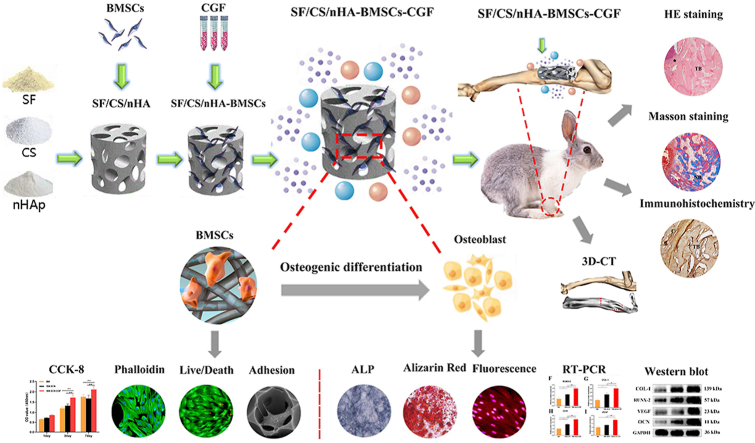
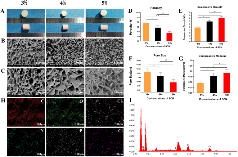
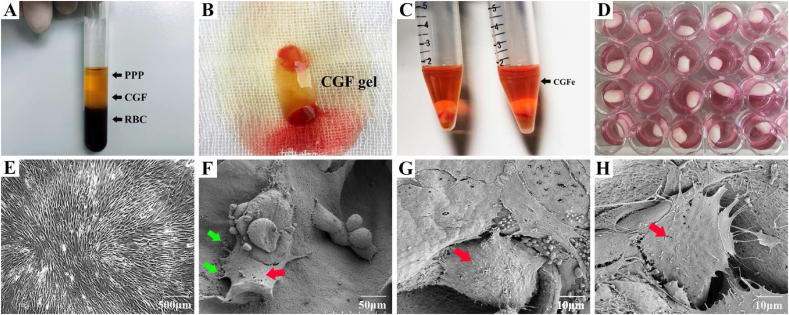
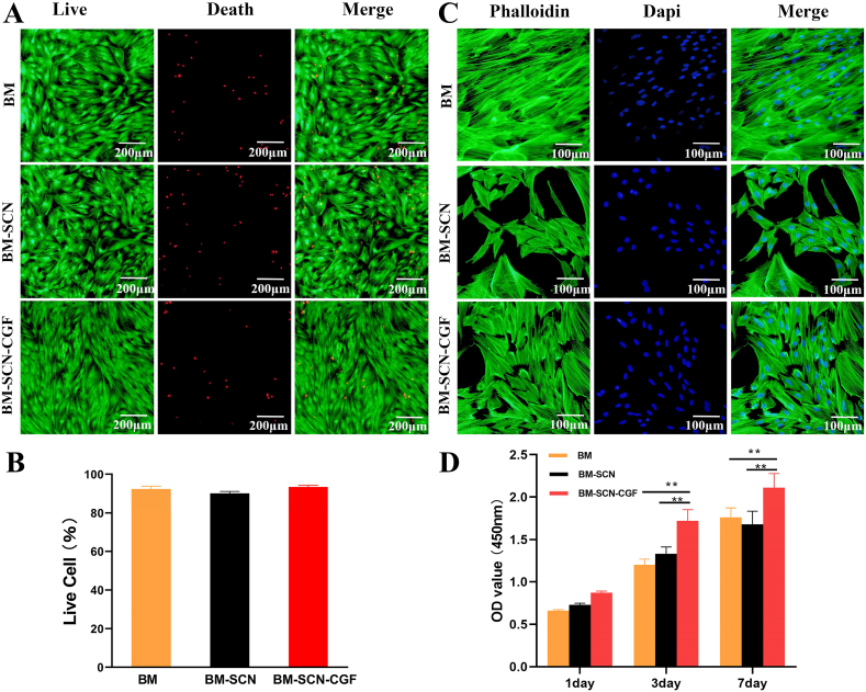
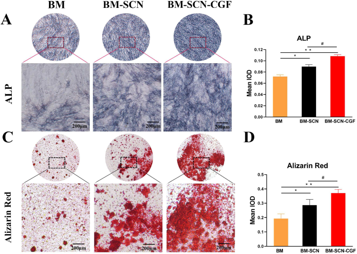
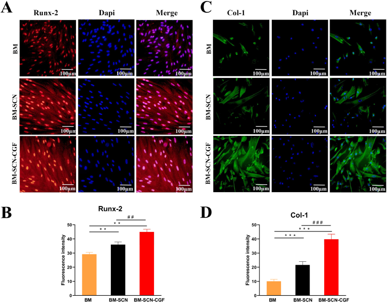
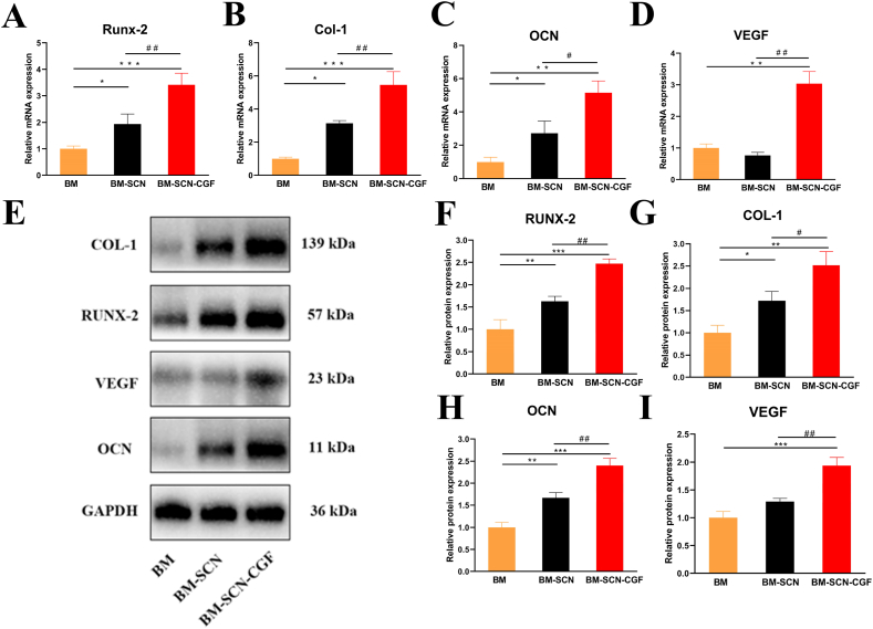
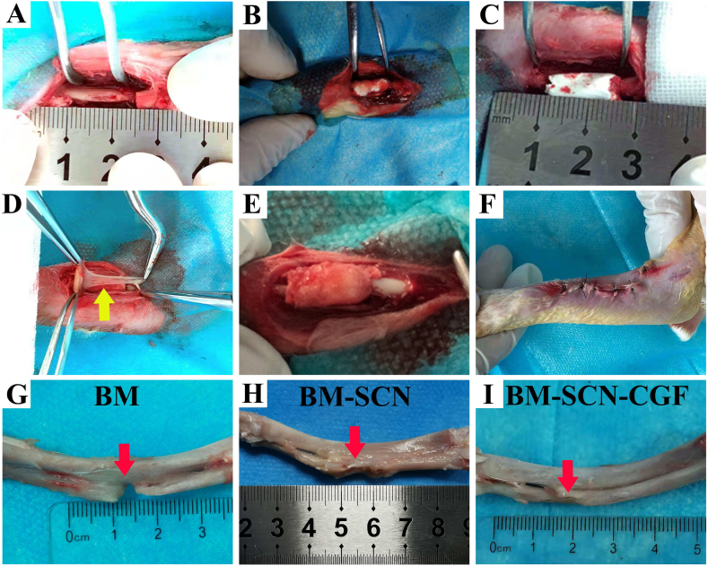
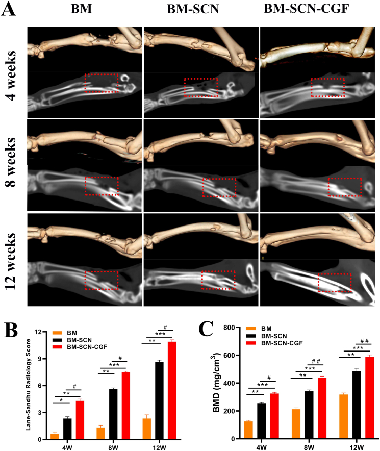
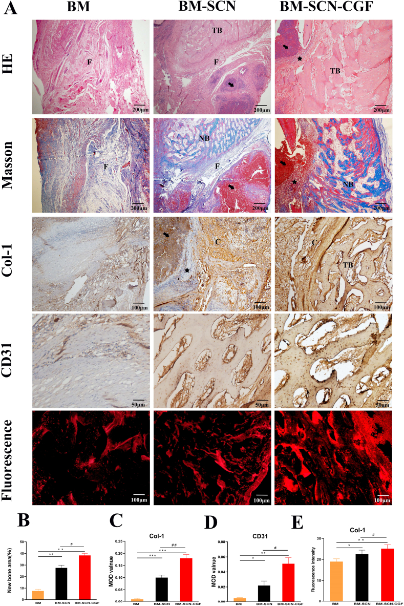
Purpose: With the goal of increasing the translational efficiency of bone tissue engineering for practical clinical applications, biomimetic composite scaffolds combined with autologous endogenous growth factors for repairing bone defects have become a current research hotspot. In this study, we prepared a silk fibroin/chitosan/nanohydroxyapatite (SF/CS/nHA) composite biomimetic scaffold and then combined it with autologous concentrated growth factor (CGF) to explore the effect of this combination on the proliferation and osteogenic differentiation of bone marrow mesenchymal stem cells (BMSCs) and the efficiency of repairing critical radial defects. Methods: Three kinds of SF/CS/nHA composite biomimetic scaffolds with mass fractions of 3%, 4%, and 5% were prepared by vacuum freeze-drying and chemical cross-linking methods, and the characteristics of the scaffolds were evaluated. In vitro, BMSCs were seeded on SF/CS/nHA scaffolds, and then CGF was added. The morphology and proliferation of BMSCs were evaluated by live-dead staining, phalloidin staining, and CCK-8 assays. ALP staining, alizarin red staining, cellular immunofluorescence, RT-PCR, and Western blotting were used to detect the osteogenic differentiation of BMSCs. In vivo, a rabbit radius critical bone defect model was constructed, and the SF/CS/nHA-BMSC scaffold cell complex combined with CGF was implanted. The effect on bone defect repair was evaluated by 3D CT scanning, HE staining, Masson staining, and immunohistochemistry. Results: The characteristics of 4% SF/CS/nHA were the most suitable for repairing bone defects. In vitro, the SF/CS/nHA combined CGF group showed better adhesion, cell morphology, proliferation, and osteogenic differentiation of BMSCs than the other groups (P < 0.05 for all). In vivo imaging examination and histological analysis demonstrated that the SF/CS/nHA scaffold combined with CGF had better efficiency in bone defect repair than the other scaffolds (P < 0.05 for all). Conclusions: A SF/CS/nHA composite biomimetic bone scaffold combined with autologous CGF promoted the proliferation and osteogenic differentiation of BMSCs in vitro and improved the repair efficiency of critical bone defects in vivo. This combination may have the potential for clinical translation due to its excellent biocompatibility.
Overview
The claims and statistics of this paper haven’t been checked yet.
Run the check — free, no account needed. It reads the open-access text and checks the sources it cites.
Integrity signals
Registry
Cites
Statistics
Figures
Verification
Claims the paper makes
The claims and statistics of this paper haven’t been checked yet.
Run the check — free, no account needed. It reads the open-access text and checks the sources it cites.
Claims other papers make about this one
Each is checked against this paper’s own text: does it say what the citing paper says it does?
The cited source says what the claim attributes to it.
In the citing paper
“The results of animal experiments show that the composite scaffold has higher efficiency in repairing bone defects [71].”
From Recent Advances in Silk Fibroin-Based Composites for Bone Repair Applications: A Review (2025) · cites this paper as [71]
The cited source says what the claim attributes to it.
In the citing paper
“Some scholars have prepared a composite bionic scaffold of silk fibroin/chitosan/nano-hydroxyapatite combined with autologous concentrated growth factor (CGF) and found that this composite scaffold is more conducive to the adhesion, proliferation, and osteogenic differentiation of bone marrow mesenchymal stem cells (BMSCs).”
From Recent Advances in Silk Fibroin-Based Composites for Bone Repair Applications: A Review (2025) · cites this paper as [71]
Self-check
Percentages, sums and reported differences re-computed from the paper’s own numbers.
The self-check needs the paper’s full text.
Citations
Cites
No references could be resolved yet.
Research works printed but not linked: 53
- Fu, 2021, An overview of polyester/hydroxyapatite composites for bone tissue repairing, J Orthop Translat, 28, 118, 10.1016/j.jot.2021.02.005 · DOI
- Song, 2022, Osteon-mimetic 3D nanofibrous scaffold enhances stem cell proliferation and osteogenic differentiation for bone regeneration, Biomater Sci, 10, 1090, 10.1039/D1BM01489G · DOI
- Wang, 2022, Bone marrow mesenchymal stem cell sheets with high expression of hBD3 and CTGF promote periodontal regeneration, Mater Sci Eng C Mater Biol Appl
- Grande, 2016, Titanium dioxide nanoparticles: a risk for human Health?, Mini Rev Med Chem, 16, 762, 10.2174/1389557516666160321114341 · DOI
- Han, 2019, Strontium-incorporated mineralized PLLA nanofibrous membranes for promoting bone defect repair, Colloids Surf B Biointerfaces, 179, 363, 10.1016/j.colsurfb.2019.04.011 · DOI
- Li, 2013, Discerning the subfibrillar structure of mineralized collagen fibrils: a model for the ultrastructure of bone, PLoS One, 8
- Song, 2021, Constructing a biomimetic nanocomposite with the in situ deposition of spherical hydroxyapatite nanoparticles to induce bone regeneration, J Mater Chem B, 9, 2469, 10.1039/D0TB02648D · DOI
- Zhao, 2021, In situ forming cellulose nanofibril-reinforced hyaluronic acid hydrogel for cartilage regeneration, Biomacromolecules, 22, 5097, 10.1021/acs.biomac.1c01063 · DOI
- Zhan, 2021, Biomineralized composite liquid crystal fiber scaffold promotes bone regeneration by enhancement of osteogenesis and angiogenesis, Front Pharmacol, 12, 10.3389/fphar.2021.736301 · DOI
- Hassanzadeh, 2021, Development and biocompatibility of the injectable collagen/nano-hydroxyapatite scaffolds as in situ forming hydrogel for the hard tissue engineering application, Artif Cell Nanomed Biotechnol, 49, 136, 10.1080/21691401.2021.1877153 · DOI
- Madhavasarma, 2020, Studies on physiochemical modifications on biologically important hydroxyapatite materials and their characterization for medical applications, Biophys Chem, 267, 10.1016/j.bpc.2020.106474 · DOI
- Guo, 2021, Photothermal regenerated fibers with enhanced toughness: silk fibroin/MoS2 nanoparticles, Polymers, 13, 3937, 10.3390/polym13223937 · DOI
- Khosropanah, 2022, Biomedical applications of silkworm (Bombyx Mori) proteins in regenerative medicine (a narrative review), J Tissue Eng Regen Med, 16, 91, 10.1002/term.3267 · DOI
- Ambaye, 2022, Preparation and applications of chitosan and cellulose composite materials, J Environ Manage, 301
- Guo, 2021, Effective immobilization of Bacillus subtilis in chitosan-sodium alginate composite carrier for ammonia removal from anaerobically digested swine wastewater, Chemosphere, 284, 10.1016/j.chemosphere.2021.131266 · DOI
- Xiao, 2019, Osteochondral repair using scaffolds with gradient pore sizes constructed with silk fibroin, chitosan, and nano-hydroxyapatite, Int J Nanomedicine, 14, 2011, 10.2147/IJN.S191627 · DOI
- Shah, 2014, Adaptive growth factor delivery from a polyelectrolyte coating promotes synergistic bone tissue repair and reconstruction, Proc Natl Acad Sci U S A, 111, 12847, 10.1073/pnas.1408035111 · DOI
- Borsani, 2020, In vitro effects of concentrated growth factors (CGF) on human SH-SY5Y neuronal cells, Eur Rev Med Pharmacol Sci, 24, 304
- Tabatabaei, 2020, In vitro and in vivo effects of concentrated growth factor on cells and tissues, J Biomed Mater Res, 108, 1338, 10.1002/jbm.a.36906 · DOI
- Rodella, 2011, Growth factors, CD34 positive cells, and fibrin network analysis in concentrated growth factors fraction, Microsc Res Tech, 74, 772, 10.1002/jemt.20968 · DOI
- Rochira, 2020, Concentrated growth factors (CGF) induce osteogenic differentiation in human bone marrow stem cells, Biology, 9, 370, 10.3390/biology9110370 · DOI
- Yamaguchi, 2020, Concentrated growth factor matrices prepared using Silica-coated plastic tubes are distinguishable from those prepared using Glass tubes in platelet distribution: application of a novel near-infrared imaging-based, quantitative technique, Front Bioeng Biotechnol, 8, 600, 10.3389/fbioe.2020.00600 · DOI
- Masuki, 2016, Growth factor and pro-inflammatory cytokine contents in platelet-rich plasma (PRP), plasma rich in growth factors (PRGF), advanced platelet-rich fibrin (A-PRF), and concentrated growth factors (CGF), Int J Implant Dent, 2, 19, 10.1186/s40729-016-0052-4 · DOI
- Aizawa, 2020, Quantitative near-infrared imaging of platelets in platelet-rich fibrin (PRF) matrices: comparative analysis of bio-PRF, leukocyte-rich PRF, advanced-PRF and concentrated growth factors, Int J Mol Sci, 21, 4426, 10.3390/ijms21124426 · DOI
- Kang, 2011, Platelet-rich fibrin is a Bioscaffold and reservoir of growth factors for tissue regeneration, Tissue Eng Part A, 17, 349, 10.1089/ten.tea.2010.0327 · DOI
- Wang, 2020, Application of a new type of natural calcined bone repair material combined with concentrated growth factors in bone regeneration in rabbit critical-sized calvarial defect, BioMed Res Int, 2020
- Fang, 2020, Clinical application of concentrated growth factor fibrin combined with bone repair materials in jaw defects, J Oral Maxillofac Surg, 78, 882, 10.1016/j.joms.2020.01.037 · DOI
- Dai, 2020, Efficacy of concentrated growth factors combined with mineralized collagen on quality of life and bone reconstruction of guided bone regeneration, Regen Biomater, 7, 313, 10.1093/rb/rbaa007 · DOI
- Chen, 2019, Modulation of proliferation and differentiation of gingiva-derived mesenchymal stem cells by concentrated growth factors: potential implications in tissue engineering for dental regeneration and repair, Int J Mol Med, 44, 37
- Honda, 2013, Bone tissue engineering with bone marrow-derived stromal cells integrated with concentrated growth factor in Rattus norvegicus calvaria defect model, J Artif Organs, 16, 305, 10.1007/s10047-013-0711-7 · DOI
- Leng, 2020, Platelet-rich plasma-enhanced osseointegration of decellularized bone matrix in critical-size radial defects in rabbits, Ann Transl Med, 8, 198, 10.21037/atm.2020.01.53 · DOI
- Liu, 2021, The effect of enhanced bone marrow in conjunction with 3D-printed PLA-HA in the repair of critical-sized bone defects in a rabbit model, Ann Transl Med, 9, 1134, 10.21037/atm-20-8198 · DOI
- Sun, 2022, The induced membrane technique: optimization of bone grafting in a rat model of segmental bone defect, Injury, 53, 1848, 10.1016/j.injury.2022.03.023 · DOI
- Kim, 2021, Effect of morphological characteristics and biomineralization of 3D-printed gelatin/hyaluronic acid/hydroxyapatite composite scaffolds on bone tissue regeneration, Int J Mol Sci, 22, 6792
- Li, 2021, CGF membrane promotes periodontal tissue regeneration mediated by hUCMSCs through upregulating TAZ and osteogenic differentiation genes, Stem Cells Int, 2021, 10.1155/2021/6644366 · DOI
- Bonazza, 2016, How the different material and shape of the blood collection tube influences the Concentrated Growth Factors production, Microsc Res Tech, 79, 1173, 10.1002/jemt.22772 · DOI
- Fang, 2022, Metformin-Incorporated gelatin/nano-hydroxyapatite scaffolds promotes bone regeneration in critical size rat alveolar bone defect model, Int J Mol Sci, 23, 558, 10.3390/ijms23010558 · DOI
- Sun, 2016, Comparison of three-dimensional printing and vacuum freeze-dried techniques for fabricating composite scaffolds, Biochem Biophys Res Commun, 477, 1085, 10.1016/j.bbrc.2016.07.050 · DOI
- Li, 2018, Silk fibroin/chitosan thin film promotes osteogenic and adipogenic differentiation of rat bone marrow-derived mesenchymal stem cells, J Biomater Appl, 32, 1164, 10.1177/0885328218757767 · DOI
- Singh, 2018, Fabrication and evaluation of non-mulberry silk fibroin fiber reinforced chitosan based porous composite scaffold for cartilage tissue engineering, Tissue Cell, 55, 83, 10.1016/j.tice.2018.10.003 · DOI
- David, 2018, Biologically anchored chitosan/gelatin-SrHAP scaffold fabricated on Titanium against chronic osteomyelitis infection, Int J Biol Macromol, 110, 206, 10.1016/j.ijbiomac.2017.11.174 · DOI
- Brennan, 2019, The effect of pore size within fibrous scaffolds fabricated using melt electrowriting on human bone marrow stem cell osteogenesis, Biomed Mater, 14, 10.1088/1748-605X/ab49f2 · DOI
- Wang, 2021, PDGF-BB induces conversion, proliferation, migration, and collagen synthesis of oral mucosal fibroblasts through PDGFR-β/PI3K/AKT signaling pathway, Cancer Biomark, 30, 407, 10.3233/CBM-201681 · DOI
- Mishima, 2021, Detection and quantification of transforming growth factor-β1 produced by murine B cells: pros and cons of different techniques, Methods Mol Biol, 2270, 113, 10.1007/978-1-0716-1237-8_7 · DOI
- Tian, 2019, Concentrated growth factor promotes dental pulp cells proliferation and mineralization and facilitates recovery of dental pulp tissue, Med Sci Monit, 25, 10016, 10.12659/MSM.919316 · DOI
- Song, 2021, Constructing a biomimetic nanocomposite with the in situ deposition of spherical hydroxyapatite nanoparticles to induce bone regeneration, J Mater Chem B, 9, 2469, 10.1039/D0TB02648D · DOI
- Chen, 2021, Curcumin modulates the crosstalk between macrophages and bone mesenchymal stem cells to ameliorate osteogenesis, Front Cell Dev Biol, 9
- Li, 2020, Synergistic anti-inflammatory and osteogenic n-HA/resveratrol/chitosan composite microspheres for osteoporotic bone regeneration, Bioact Mater, 6, 1255
- Mizokami, 2017, Osteocalcin and its endocrine functions, Biochem Pharmacol, 132, 1, 10.1016/j.bcp.2017.02.001 · DOI
- Qi, 2020, Concentrated growth factor promotes gingival regeneration through the AKT/Wnt/β-catenin and YAP signaling pathways, Artif Cell Nanomed Biotechnol, 48, 920, 10.1080/21691401.2020.1773482 · DOI
- Arıcan, 2022, Micro-ct findings of concentrated growth factors (cgf) on bone healing in masquelet's technique-an experimental study in rabbits, Arch Orthop Trauma Surg, 142, 83, 10.1007/s00402-020-03596-z · DOI
- Sadeghzadeh, 2022, PCL/Col I-based magnetic nanocomposite scaffold provides an osteoinductive environment for ADSCs in osteogenic cues-free media conditions, Stem Cell Res Ther, 13, 143, 10.1186/s13287-022-02816-0 · DOI
- Yılmaz, 2018, Effects of concentrated growth factors (CGF) on the quality of the induced membrane in Masquelet's technique - an experimental study in rabbits, Injury, 49, 1497, 10.1016/j.injury.2018.06.011 · DOI
Cited by
Sorted by citations
Recent Advances in Silk Fibroin-Based Composites for Bone Repair Applications: A Review
Zhu, S., Zhang, Q., Xu, X., Liu, Z., Cheng, G., Long, D., Cheng, L., & Dai, F. (2025) · Polymers · doi:10.3390/polym17060772 · Cited by 36
“The results of animal experiments show that the composite scaffold has higher efficiency in repairing bone defects [71].”
Metadata
- Authors
- Zhou, Y., Liu, X., She, H., Wang, R., Bai, F., & Xiang, B.
- Affiliations
- Zunyi Medical University (CN)
- Corresponding author
- Xiang, Bingyan
- Venue
- Regenerative Therapy
- Publisher
- Elsevier BV
- Published
- 2022-12
- Volume
- 21
- Pages
- 307-321
- ISSN
- 2352-3204 (ISSN-L)
- PMC
- PMC9459434
- OpenAlex
- W4294216765
- Cited by
- 49
- References
- 0
- Citations by year
- 2026: 16 · 2025: 11 · 2024: 12 · 2023: 8 · 2022: 2
- FWCI
- 2.40
- Type
- journal article
- Language
- en
- Open access
- gold
- doi.org
- Licence
- Locations
- Regenerative Therapy (publishedVersion, cc-by-nc-nd)
- PubMed Central (publishedVersion, cc-by-nc-nd)
- PubMed (publishedVersion)
- DOAJ (DOAJ: Directory of Open Access Journals) (submittedVersion, cc-by-sa)
- Funders
- Health Commission of Guizhou Province
- Topics
- Physical Sciences › Engineering › Biomedical Engineering › Bone Tissue Engineering Materials
- Physical Sciences › Materials Science › Biomaterials › Silk-based biomaterials and applications
- Health Sciences › Medicine › Surgery › Tissue Engineering and Regenerative Medicine
- Keywords
- silk fibroin, chitosan, hydroxyapatite nanoparticles, bone marrow mesenchymal stem cells, osteogenic differentiation, cell proliferation, radial bone defect, bone tissue engineering, vacuum freeze-drying, crosslinking, rabbit model, clinical translation, endogenous growth factors
About these checks
Registry, retraction, Cites and statistics results are computed, not judged, with one exception: a printed reference that no index lists is read once by a model before it counts as a citation error. Claims are extracted by a model and kept only when their quote appears verbatim in the text; each cited source is then checked, with a model’s help, for whether it says what the claim relies on. Those verdicts can be wrong — read the claim’s quote and the cited source before relying on one.
Statistics and arithmetic are recomputed, not judged.
Confidence bands are shown only for fields calibrated on our annotated test set. None is calibrated yet, so no band is shown.
What counts as a claim ›How we measure confidence ›How we check this ›
Are you an author, or do you see a problem with this page? Request a review
How to cite
Cite CoChat for the checks on this page, and the paper for its own findings.
- This page
- CoChat. (2026). Citation check: A silk fibroin/chitosan/nanohydroxyapatite biomimetic bone scaffold combined with autologous concentrated growth factor promotes the proliferation and osteogenic differentiation of BMSCs and repair of critical bone defects. https://app.cochat.ai/papers/10.1016/j.reth.2022.08.006
- The paper
- Zhou, Y., Liu, X., She, H., Wang, R., Bai, F., & Xiang, B. (2022). A silk fibroin/chitosan/nanohydroxyapatite biomimetic bone scaffold combined with autologous concentrated growth factor promotes the proliferation and osteogenic differentiation of BMSCs and repair of critical bone defects. Regenerative Therapy. https://doi.org/10.1016/j.reth.2022.08.006
