

The DOI matches its Crossref record; 93 of the 93 references it cites were found in a registry or an index.
Computational Insights into Membrane Disruption by Cell-Penetrating Peptides
Open accessPublished version of a preprint
Abstract
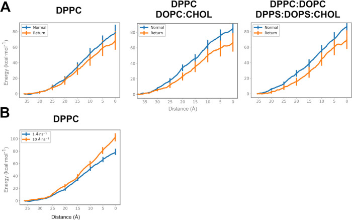
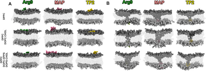
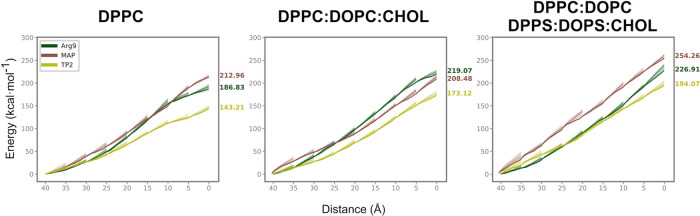
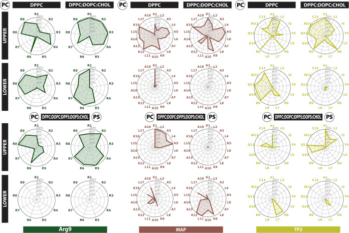
Cell-penetrating peptides (CPPs) can translocate into cells without inducing cytotoxicity. The internalization process implies several steps at different time scales ranging from microseconds to minutes. We combine adaptive Steered Molecular Dynamics (aSMD) with conventional Molecular Dynamics (cMD) to observe nonequilibrium and equilibrium states to study the early mechanisms of peptide-bilayer interaction leading to CPPs internalization. We define three membrane compositions representing bilayer sections, neutral lipids (i.e., upper leaflet), neutral lipids with cholesterol (i.e., hydrophobic core), and neutral/negatively charged lipids with cholesterol (i.e., lower leaflet) to study the energy barriers and disruption mechanisms of Arg9, MAP, and TP2, representing cationic, amphiphilic, and hydrophobic CPPs, respectively. Cholesterol and negatively charged lipids increase the energetic barriers for the peptide-bilayer crossing. TP2 interacts with the bilayer by hydrophobic insertion, while Arg9 disrupts the bilayer by forming transient or stable pores. MAP has shown both behaviors. Collectively, these findings underscore the significance of innovative computational approaches in studying membrane-disruptive peptides and, more specifically, in harnessing their potential for cell penetration.
Overview
The claims and statistics of this paper haven’t been checked yet.
Run the check — free, no account needed. It reads the open-access text and checks the sources it cites.
Integrity signals
Registry
Cites
Statistics
Figures

Verification
Claims the paper makes
The claims and statistics of this paper haven’t been checked yet.
Run the check — free, no account needed. It reads the open-access text and checks the sources it cites.
Self-check
Percentages, sums and reported differences re-computed from the paper’s own numbers.
The self-check needs the paper’s full text.
Citations
Cites
VMD: visual molecular dynamics
Humphrey, W., Dalke, A., & Schulten, K. (1996) · Journal of Molecular Graphics · doi:10.1016/0263-7855(96)00018-5 · Cited by 66,952
Highly accurate protein structure prediction with AlphaFold
Jumper, J., Evans, R., Pritzel, A., Green, T., Figurnov, M., Ronneberger, O., Tunyasuvunakool, K., Bates, R., Žídek, A., Potapenko, A., et al. (2021) · Nature · doi:10.1038/s41586-021-03819-2 · Cited by 43,764
Matplotlib: a 2D graphics environment
Hunter, J. D. (2007) · Computing in Science & Engineering · doi:10.1109/mcse.2007.55 · Cited by 39,249
A simple method for displaying the hydropathic character of a protein
Kyte, J., & Doolittle, R. F. (1982) · Journal of Molecular Biology · doi:10.1016/0022-2836(82)90515-0 · Cited by 22,843
ff14SB: Improving the Accuracy of Protein Side Chain and Backbone Parameters from ff99SB
Maier, J. A., Martinez, C., Kasavajhala, K., Wickstrom, L., Hauser, K. E., & Simmerling, C. (2015) · Journal of Chemical Theory and Computation · doi:10.1021/acs.jctc.5b00255 · Cited by 10,830
UCSF ChimeraX : Structure visualization for researchers, educators, and developers
Pettersen, E. F., Goddard, T. D., Huang, C. C., Meng, E. C., Couch, G. S., Croll, T. I., Morris, J. H., & Ferrin, T. E. (2020) · Protein Science · doi:10.1002/pro.3943 · Cited by 9,762
The Amber biomolecular simulation programs
Case, D. A., Cheatham, T. E., Darden, T., Gohlke, H., Luo, R., Merz, K. M., Onufriev, A., Simmerling, C., Wang, B., & Woods, R. J. (2005) · Journal of Computational Chemistry · doi:10.1002/jcc.20290 · Cited by 9,535
ColabFold - Making protein folding accessible to all
Mirdita, M., Schütze, K., Moriwaki, Y., Heo, L., Ovchinnikov, S., & Steinegger, M. (2021) · doi:10.1101/2021.08.15.456425 · Cited by 9,391
CHARMM‐GUI: A web‐based graphical user interface for CHARMM
Jo, S., Kim, T., Iyer, V. G., & Im, W. (2008) · Journal of Computational Chemistry · doi:10.1002/jcc.20945 · Cited by 9,284
CHARMM: The biomolecular simulation program
Brooks, B. R., Brooks, C. L., MacKerell, A. D., Nilsson, L., Petrella, R. J., Roux, B., Won, Y., Archontis, G., Bartels, C., Boresch, S., et al. (2009) · Journal of Computational Chemistry · doi:10.1002/jcc.21287 · Cited by 9,173
PTRAJ and CPPTRAJ: Software for Processing and Analysis of Molecular Dynamics Trajectory Data
Roe, D. R., & Cheatham, T. E. (2013) · Journal of Chemical Theory and Computation · doi:10.1021/ct400341p · Cited by 7,515
seaborn: statistical data visualization
Waskom, M. (2021) · Journal of Open Source Software · doi:10.21105/joss.03021 · Cited by 7,310
UCSF ChimeraX: Meeting modern challenges in visualization and analysis
Goddard, T. D., Huang, C. C., Meng, E. C., Pettersen, E. F., Couch, G. S., Morris, J. H., & Ferrin, T. E. (2018) · Protein Science · doi:10.1002/pro.3235 · Cited by 5,267
Nonequilibrium Equality for Free Energy Differences
Jarzynski, C. (1997) · Physical Review Letters · doi:10.1103/physrevlett.78.2690 · Cited by 5,231
MDAnalysis: A toolkit for the analysis of molecular dynamics simulations
Michaud‐Agrawal, N., Denning, E. J., Woolf, T. B., & Beckstein, O. (2011) · Journal of Computational Chemistry · doi:10.1002/jcc.21787 · Cited by 4,486
Lee, J., Cheng, X., Swails, J., Yeom, M. S., Eastman, P. K., Lemkul, J. A., Wei, S., Buckner, J., Jeong, J. C., Qi, Y., et al. (2015) · Journal of Chemical Theory and Computation · doi:10.1021/acs.jctc.5b00935 · Cited by 4,390
Doherty, G. J., & McMahon, H. T. (2009) · Annual Review of Biochemistry · doi:10.1146/annurev.biochem.78.081307.110540 · Cited by 3,075
CHARMM-GUI Membrane Builder toward realistic biological membrane simulations
Wu, E. L., Cheng, X., Jo, S., Rui, H., Song, K. C., Dávila-Contreras, E. M., Qi, Y., Lee, J., Monje-Galvan, V., Venable, R. M., et al. (2014) · Journal of Computational Chemistry · doi:10.1002/jcc.23702 · Cited by 2,656
MDAnalysis: A Python Package for the Rapid Analysis of Molecular Dynamics Simulations
Gowers, R. J., Linke, M., Barnoud, J., Reddy, T. J., Melo, M. N., Seyler, S. L., Domański, J., Dotson, D., Buchoux, S., Kenney, I. M., et al. (2016) · Proceedings of the Python in Science Conferences · doi:10.25080/majora-629e541a-00e · Cited by 2,228
Stothard, P. (2000) · BioTechniques · doi:10.2144/00286ir01 · Cited by 1,818
CHARMM-GUI Membrane Builder for mixed bilayers and its application to yeast membranes.
Jo, S., Lim, J. B., Klauda, J. B., & Im, W. (2009) · Biophysical Journal · doi:10.1016/j.bpj.2009.04.013 · Cited by 1,674
Futaki, S., Suzuki, T. W., Ohashi, W., Yagami, T., Tanaka, S., Ueda, K., & Sugiura, Y. (2001) · Journal of Biological Chemistry · doi:10.1074/jbc.m007540200 · Cited by 1,667
Cell Internalization of the Third Helix of the Antennapedia Homeodomain Is Receptor-independent
Derossi, D., Calvet, S., Trembleau, A., Brunissen, A., Chassaing, G., & Prochiantz, A. (1996) · Journal of Biological Chemistry · doi:10.1074/jbc.271.30.18188 · Cited by 1,073
Free energy reconstruction from nonequilibrium single-molecule pulling experiments
Hummer, G., & Szabo, A. (2001) · Proceedings of the National Academy of Sciences · doi:10.1073/pnas.071034098 · Cited by 983
Calculating potentials of mean force from steered molecular dynamics simulations
Park, S., & Schulten, K. J. (2004) · The Journal of Chemical Physics · doi:10.1063/1.1651473 · Cited by 948
Lin, Y.-Z., Yao, S., Veach, R. A., Torgerson, T. R., & Hawiger, J. (1995) · Journal of Biological Chemistry · doi:10.1074/jbc.270.24.14255 · Cited by 919
Free energy calculation from steered molecular dynamics simulations using Jarzynski’s equality
Park, S., Khalili‐Araghi, F., Tajkhorshid, E., & Schulten, K. J. (2003) · The Journal of Chemical Physics · doi:10.1063/1.1590311 · Cited by 821
Cell-penetrating peptides: classes, origin, and current landscape
Milletti, F. (2012) · Drug Discovery Today · doi:10.1016/j.drudis.2012.03.002 · Cited by 813
Lindgren, M., Hällbrink, M., Prochiantz, A., & Langel, Ü. (2000) · Trends in Pharmacological Sciences · doi:10.1016/s0165-6147(00)01447-4 · Cited by 803
Pouny, Y., Rapaport, D., Mor, A., Nicolas, P., & Shai, Y. (1992) · Biochemistry · doi:10.1021/bi00164a017 · Cited by 687
Lee, J., Patel, D. S., Ståhle, J., Park, S., Kern, N. R., Kim, S., Lee, J.-S., Cheng, X., Valvano, M. A., Holst, O., et al. (2018) · Journal of Chemical Theory and Computation · doi:10.1021/acs.jctc.8b01066 · Cited by 641
Rothbard, J. B., Garlington, S., Lin, Q., Kirschberg, T. A., Kreider, E., McGrane, P. L., Wender, P. A., & Khavari, P. A. (2000) · Nature Medicine · doi:10.1038/81359 · Cited by 632
Protein transduction: unrestricted delivery into all cells?
Schwarze, S. R., Hruska, K. A., & Dowdy, S. F. (2000) · Trends in Cell Biology · doi:10.1016/s0962-8924(00)01771-2 · Cited by 552
Herce, H. D., & Garcı́a, A. E. (2007) · Proceedings of the National Academy of Sciences · doi:10.1073/pnas.0706574105 · Cited by 488
Lipid21: Complex Lipid Membrane Simulations with AMBER
Dickson, C. J., Walker, R. C., & Gould, I. R. (2022) · Journal of Chemical Theory and Computation · doi:10.1021/acs.jctc.1c01217 · Cited by 435
CHARMM-GUI supports the Amber force fields
Lee, J., Hitzenberger, M., Rieger, M., Kern, N. R., Zacharias, M., & Im, W. (2020) · The Journal of Chemical Physics · doi:10.1063/5.0012280 · Cited by 428
Vermeer, L. S., de Groot, C. P. G. M., Réat, V., Milon, A., & Czaplicki, J. (2007) · European Biophysics Journal · doi:10.1007/s00249-007-0192-9 · Cited by 355
freud: A software suite for high throughput analysis of particle simulation data
Ramasubramani, V., Dice, B. D., Harper, E. S., Spellings, M. P., Anderson, J. A., & Glotzer, S. C. (2020) · Computer Physics Communications · doi:10.1016/j.cpc.2020.107275 · Cited by 327
Herce, H. D., Garcia, A. E., Litt, J., Kane, R. S., Martin, P., Enrique, N., Rebolledo, A., & Milesi, V. (2009) · Biophysical Journal · doi:10.1016/j.bpj.2009.05.066 · Cited by 305
Li, T., Bourgeois, J., Celli, S., Glacial, F., Sourd, A. L., Mécheri, S., Weksler, B. B., Romero, I. A., Couraud, P., Rougeon, F., et al. (2012) · The FASEB Journal · doi:10.1096/fj.11-201384 · Cited by 218
Spontaneous Membrane-Translocating Peptides by Orthogonal High-Throughput Screening
Marks, J. R., Placone, J. K., Hristova, K. A., & Wimley, W. C. (2011) · Journal of the American Chemical Society · doi:10.1021/ja2017416 · Cited by 201
Yesylevskyy, S. O., Marrink, . J., & Mark, A. E. (2009) · Biophysical Journal · doi:10.1016/j.bpj.2009.03.059 · Cited by 200
Song, W., Corey, R. A., Ansell, T. B., Cassidy, C. K., Horrell, M. R., Duncan, A. L., Stansfeld, P. J., & Sansom, M. S. P. (2022) · Journal of Chemical Theory and Computation · doi:10.1021/acs.jctc.1c00708 · Cited by 197
Mechanism of the Cell-Penetrating Peptide Transportan 10 Permeation of Lipid Bilayers
Yandek, L. E., Pokorny, A., Florén, A., Knoelke, K., Langel, Ü., & Almeida, P. F. F. (2007) · Biophysical Journal · doi:10.1529/biophysj.106.100198 · Cited by 190
Transbilayer Transport of Ions and Lipids Coupled with Mastoparan X Translocation
Matsuzaki, K., Yoneyama, S., Murase, O., & Miyajima, K. (1996) · Biochemistry · doi:10.1021/bi960342a · Cited by 166
Berlose, J., Convert, O., Derossi, D., Brunissen, A., & Chassaing, G. (1996) · European Journal of Biochemistry · doi:10.1111/j.1432-1033.1996.0372r.x · Cited by 163
Nakase, I., Konishi, Y., Ueda, M., Saji, H., & Futaki, S. (2012) · Journal of Controlled Release · doi:10.1016/j.jconrel.2012.01.016 · Cited by 157
Lee, M.-T., Hung, W.-C., Chen, F.-Y., & Huang, H. W. (2005) · Biophysical Journal · doi:10.1529/biophysj.105.068080 · Cited by 148
Cell penetrating peptides in preclinical and clinical cancer diagnosis and therapy
Tripathi, P. P., Arami, H., Banga, I. K., Gupta, J. R. P., & Gandhi, S. (2018) · Oncotarget · doi:10.18632/oncotarget.26442 · Cited by 135
Challenge to overcome current limitations of cell-penetrating peptides
Kim, G. C., Cheon, D. H., & Lee, Y. (2021) · Biochimica et Biophysica Acta (BBA) - Proteins and Proteomics · doi:10.1016/j.bbapap.2021.140604 · Cited by 132
LiPyphilic: A Python Toolkit for the Analysis of Lipid Membrane Simulations
Smith, P., & Lorenz, C. D. (2021) · Journal of Chemical Theory and Computation · doi:10.1021/acs.jctc.1c00447 · Cited by 124
Adaptive Steered Molecular Dynamics of the Long-Distance Unfolding of Neuropeptide Y
Özer, G., Valeev, E. F., Quirk, S., & Hernandez, R. (2010) · Journal of Chemical Theory and Computation · doi:10.1021/ct100320g · Cited by 123
Influence of DPH on the Structure and Dynamics of a DPPC Bilayer
Repáková, J., Holopainen, J. M., Morrow, M. R., McDonald, M. C., Čapková, P., & Vattulainen, I. (2005) · Biophysical Journal · doi:10.1529/biophysj.104.055533 · Cited by 119
Cell penetrating peptides in ocular drug delivery: State of the art
Pescina, S., Ostacolo, C., Gomez-Monterrey, I. M., Sala, M., Bertamino, A., Sonvico, F., Padula, C., Santi, P., Bianchera, A., & Nicoli, S. (2018) · Journal of Controlled Release · doi:10.1016/j.jconrel.2018.06.023 · Cited by 118
Uptake Mechanism of Cell-Penetrating Peptides
Gestin, M., Dowaidar, M., & Langel, Ü. (2017) · Advances in experimental medicine and biology · doi:10.1007/978-3-319-66095-0_11 · Cited by 114
Huang, K., & Garcı́a, A. E. (2013) · Biophysical Journal · doi:10.1016/j.bpj.2012.10.027 · Cited by 108
Transfer of Arginine into Lipid Bilayers Is Nonadditive
MacCallum, J. L., Bennett, W. F. D., & Tieleman, D. P. (2011) · Biophysical Journal · doi:10.1016/j.bpj.2011.05.038 · Cited by 101
Zhang, Y., Guo, P., Ma, Z., Lü, P., Kebebe, D., & Liu, Z. (2021) · Journal of Nanobiotechnology · doi:10.1186/s12951-021-01002-3 · Cited by 97
Özer, G., Quirk, S., & Hernandez, R. (2012) · The Journal of Chemical Physics · doi:10.1063/1.4725183 · Cited by 94
Probing a Continuous Polar Defect: A Reaction Coordinate for Pore Formation in Lipid Membranes
Hub, J. S., & Awasthi, N. (2017) · Journal of Chemical Theory and Computation · doi:10.1021/acs.jctc.7b00106 · Cited by 91
Guo, F., Fu, Q., Zhou, K., Jin, C., Wu, W.-C., Ji, X., Yan, Q., Yang, Q., Wu, D., Li, A., et al. (2020) · Journal of Nanobiotechnology · doi:10.1186/s12951-020-00595-5 · Cited by 85
Awasthi, N., & Hub, J. S. (2016) · Journal of Chemical Theory and Computation · doi:10.1021/acs.jctc.6b00369 · Cited by 80
Cellular uptake and in vivo distribution of polyhistidine peptides
Iwasaki, T., Tokuda, Y., Kotake, A., OKADA, H. M., Takeda, S., Kawano, T., & Nakayama, Y. (2015) · Journal of Controlled Release · doi:10.1016/j.jconrel.2015.05.268 · Cited by 78
Hwang, H., Schatz, G. C., & Ratner, M. A. (2006) · The Journal of Physical Chemistry B · doi:10.1021/jp0657888 · Cited by 78
Pae, J., Säälik, P., Liivamägi, L., Lubenets, D., Arukuusk, P., Langel, Ü., & Pooga, M. (2014) · Journal of Controlled Release · doi:10.1016/j.jconrel.2014.07.002 · Cited by 73
Özer, G., Quirk, S., & Hernandez, R. (2012) · Journal of Chemical Theory and Computation · doi:10.1021/ct300709u · Cited by 68
Direct Cytosolic Delivery of Polar Cargo to Cells by Spontaneous Membrane-translocating Peptides
He, J., Kauffman, W. B., Fuselier, T. T., Somanna, N. K., Voss, T. G., Hristova, K. A., & Wimley, W. C. (2013) · Journal of Biological Chemistry · doi:10.1074/jbc.m113.488312 · Cited by 67
Li, Q., Yang, J., Chen, C., Lin, X., Zhou, M., Zhou, Z., & Huang, Y. (2020) · Journal of Controlled Release · doi:10.1016/j.jconrel.2020.06.010 · Cited by 61
Biological Membrane-Penetrating Peptides: Computational Prediction and Applications
de Oliveira, E. C. L., da Costa, K. S., Taube, P. S., Lima, A. H., & Sales, C. (2022) · Frontiers in Cellular and Infection Microbiology · doi:10.3389/fcimb.2022.838259 · Cited by 60
Steered Molecular Dynamics Simulations of Na + Permeation across the Gramicidin A Channel
Liu, Z., Xu, Y., & Tang, P. S. (2006) · The Journal of Physical Chemistry B · doi:10.1021/jp060688n · Cited by 49
Pirhaghi, M., Mamashli, F., Moosavi-Movahedi, F., Arghavani, P., Amiri, A., Davaeil, B., Mohammad-Zaheri, M., Mousavi-Jarrahi, Z., Sharma, D., Langel, Ü., et al. (2024) · Molecular Pharmaceutics · doi:10.1021/acs.molpharmaceut.3c01167 · Cited by 49
Steiner, V., Schär, M., Boernsen, K. O., & Mutter, M. (1991) · Journal of Chromatography A · doi:10.1016/0021-9673(91)80023-a · Cited by 48
He, X., Lin, M., Sha, B., Feng, S., Shi, X., Qu, Z., & Xu, F. (2015) · Scientific Reports · doi:10.1038/srep12808 · Cited by 47
Effect of the Lipid Landscape on the Efficacy of Cell-Penetrating Peptides
Zákány, F., Mándity, I. M., Varga, Z., Panyi, G., Nagy, P., & Kovács, T. (2023) · Cells · doi:10.3390/cells12131700 · Cited by 47
Optimal Hydrophobicity and Reorientation of Amphiphilic Peptides Translocating through Membrane
Kabelka, I., & Vácha, R. (2018) · Biophysical Journal · doi:10.1016/j.bpj.2018.08.012 · Cited by 46
Secondary structures and cell-penetrating abilities of arginine-rich peptide foldamers
Oba, M., Nagano, Y., Kato, T., & Tanaka, M. (2019) · Scientific Reports · doi:10.1038/s41598-018-38063-8 · Cited by 45
Tian, Y., & Zhou, S. (2020) · Wiley Interdisciplinary Reviews Nanomedicine and Nanobiotechnology · doi:10.1002/wnan.1668 · Cited by 42
Cholesterol’s Condensing Effect: Unpacking a Century-Old Mystery
Regen, S. L. (2021) · JACS Au · doi:10.1021/jacsau.1c00493 · Cited by 40
Hub, J. S. (2021) · Journal of Chemical Theory and Computation · doi:10.1021/acs.jctc.0c01134 · Cited by 39
Studies on the cellular uptake of substance P and lysine-rich, KLA-derived model peptides
Oehlke, J., Lorenz, D., Wiesner, B., & Bienert, M. (2004) · Journal of Molecular Recognition · doi:10.1002/jmr.691 · Cited by 38
Recombinant peptide constructs for targeted cell penetrating peptide-mediated delivery
Zaro, J. L., Fei, L., & Shen, W. (2012) · Journal of Controlled Release · doi:10.1016/j.jconrel.2012.01.039 · Cited by 38
Yang, Y., Yang, Y., Xie, X., Cai, X., & Mei, X. (2014) · Journal of drug targeting · doi:10.3109/1061186x.2014.940589 · Cited by 38
Adaptive steered molecular dynamics of biomolecules
Zhuang, Y., Bureau, H. R., Quirk, S., & Hernandez, R. (2020) · Molecular Simulation · doi:10.1080/08927022.2020.1807542 · Cited by 34
A Second Life for MAP, a Model Amphipathic Peptide
Silva, S., Kurrikoff, K., Langel, Ü., Almeida, A. J., & Vale, N. (2022) · International Journal of Molecular Sciences · doi:10.3390/ijms23158322 · Cited by 20
Defect Formation and Peptide Permeation across Phospholipid Membranes
Elber, R. (2023) · The Journal of Physical Chemistry B · doi:10.1021/acs.jpcb.3c04895 · Cited by 19
Yue, T., Sun, M., Zhang, S., Ren, H., Ge, B., & Huang, F. L. (2016) · Physical Chemistry Chemical Physics · doi:10.1039/c6cp01133k · Cited by 17
Giménez-Dejoz, J., & Numata, K. (2021) · Nanoscale Advances · doi:10.1039/d1na00674f · Cited by 17
New dimension indices for the characterization of the solvent-accessible surface
Torrens, F., S�nchez-Mar�n, J., & Nebot‐Gil, I. (2001) · Journal of Computational Chemistry · doi:10.1002/1096-987x(20010415)22:5<501::aid-jcc1021>3.0.co;2-v · Cited by 16
Choe, S. (2023) · Scientific Reports · doi:10.1038/s41598-023-28493-4 · Cited by 16
Kenien, R., Shen, W., & Zaro, J. L. (2012) · Journal of drug targeting · doi:10.3109/1061186x.2012.719899 · Cited by 15
The relative stability of trpzip1 and its mutants determined by computation and experiment
Bureau, H. R., Quirk, S., & Hernandez, R. (2020) · RSC Advances · doi:10.1039/d0ra00920b · Cited by 14
Interaction of arginine oligomer with model membrane
Yi, D., Li, G., Gao, L., & Liang, W. P. (2007) · Biochemical and Biophysical Research Communications · doi:10.1016/j.bbrc.2007.06.015 · Cited by 10
Dual Role of Anionic Lipids in Amyloid Aggregation
Jain, M., & Matysiak, S. (2024) · The Journal of Physical Chemistry B · doi:10.1021/acs.jpcb.4c05636 · Cited by 9
Research works printed but not linked: 3
- 10.1016/S0165-6147(00)01447-4 · DOI · Paper page
- 10.1038/s41592-022-01488-1 · DOI · Paper page
- Case, D. A.; Belfon, K.; Ben-Shalom, I. Y.; Brozell, S. R.; Cerutti, D. S.; T. E. Cheatham, I. I. I.; Cruzeiro, V. W. D.; Darden, T. A.; Duke, R. E.; Giambasu, G.; Gilson, M. K.; Gohlke, H.; Goetz, A. W.; Harris, R.; Izadi, S.; Izmailov, S. A.; Kasavajhala, K.; Kovalenko, A.; Krasny, R.; Kurtzman, T.; Lee, T. S.; LeGrand, S.; Li, P.; Lin, C.; Liu, J.; Luchko, T.; Luo, R.; Man, V.; Merz, K. M.; Miao, Y.; Mikhailovskii, O.; Monard, G.; Nguyen, H.; Onufriev, A.; Pan, F.; Pantano, S.; Qi, R.; Roe, D. R.; Roitberg, A.; Sagui, C.; Schott-Verdugo, S.; Shen, J.; Simmerling, C. L.; Skrynnikov, N. R.; Smith, J.; Swails, J.; Walker, R. C.; Wang, J.; Wilson, L.; Wolf, R. M.; Wu, X.; Xiong, Y.; Xue, Y.; York, D. M.; Kollman, P. A. AMBER. 2020; Vol. 2020.
Metadata
- Authors
- Catalina-Hernandez, E., Aguilella-Arzo, M., Peralvarez-Marin, A., & Lopez-Martin, M.
- Affiliations
- Unit of Biophysics, Department of Biochemistry and Molecular Biology, Facultat de Medicina, Av. Can Domènech s/n, Universitat Autònoma de Barcelona, 08193 Cerdanyola del Vallès, Catalonia, Spain
- Laboratory of Molecular Biophysics, Department of Physics
- Venue
- Journal of Chemical Information and Modeling
- Publisher
- American Chemical Society (ACS)
- Published
- 2025-01-17
- Volume
- 65
- Issue
- 3
- Pages
- 1549-1559
- ISSN
- 1549-9596 (ISSN-L)
- 1549-960X
- PMC
- PMC11815844
- OpenAlex
- W4406508939
- Cited by
- 18
- References
- 93
- Citations by year
- 2026: 11 · 2025: 7
- FWCI
- 5.41
- Type
- journal article
- Language
- en
- Open access
- hybrid
- doi.org
- Licence
- Locations
- Journal of Chemical Information and Modeling (publishedVersion, cc-by)
- bioRxiv (Cold Spring Harbor Laboratory) (acceptedVersion, cc-by-nc-nd)
- PubMed Central (publishedVersion, cc-by)
- PubMed (publishedVersion)
- Dipòsit Digital de Documents de la UAB (Universitat Autònoma de Barcelona) (submittedVersion, cc-by)
- Funders
- Ministerio de Ciencia, Innovaci?n y Universidades (MGSD2021-10)
- Universitat Jaume I (UJI-B2022-42)
- Agencia Nacional de Promoci?n Cient?fica y Tecnol?gica (PID2020-120222GB-I00, PID2022-142795NB-I00)
- Universitat Aut?noma de Barcelona (B21P0033)
- Topics
- Life Sciences › Biochemistry, Genetics and Molecular Biology › Molecular Biology › Lipid Membrane Structure and Behavior
- Life Sciences › Biochemistry, Genetics and Molecular Biology › Molecular Biology › RNA Interference and Gene Delivery
- Life Sciences › Immunology and Microbiology › Microbiology › Antimicrobial Peptides and Activities
- Keywords
- cell-penetrating peptides, membrane damage, peptide internalization, cholesterol, negatively charged lipids, energy barrier, pore formation, hydrophobic insertion, maps, TP2, cationic peptides, peptide amphiphiles, hydrophobic peptides
- MeSH terms
- Cell Membrane, Cholesterol, Lipid Bilayers, Thermodynamics, Molecular Dynamics Simulation, Cell-Penetrating Peptides, Hydrophobic and Hydrophilic Interactions
About these checks
Registry, retraction, Cites and statistics results are computed, not judged, with one exception: a printed reference that no index lists is read once by a model before it counts as a citation error. Claims are extracted by a model and kept only when their quote appears verbatim in the text; each cited source is then checked, with a model’s help, for whether it says what the claim relies on. Those verdicts can be wrong — read the claim’s quote and the cited source before relying on one.
Statistics and arithmetic are recomputed, not judged.
Confidence bands are shown only for fields calibrated on our annotated test set. None is calibrated yet, so no band is shown.
What counts as a claim ›How we measure confidence ›How we check this ›
Are you an author, or do you see a problem with this page? Request a review
Data sources
Bibliographic data: Semantic Scholar, OpenAlex, Crossref.
Contains information from the Semantic Scholar Academic Graph Datasets, made available under the ODC Attribution License (ODC-BY 1.0).
Semantic Scholar’s data is described in: Kinney, R., et al. (2023). The Semantic Scholar Open Data Platform. arXiv:2301.10140.
