

The DOI matches its Crossref record; 41 of the 41 references it cites were found in a registry or an index.
Repurposing a Fully Reducing Polyketide Synthase toward 2-Methyl Guerbet-like Lipids
Open accessPublished version of a preprint
Abstract
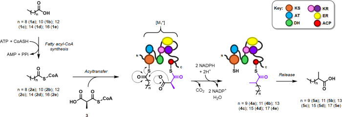
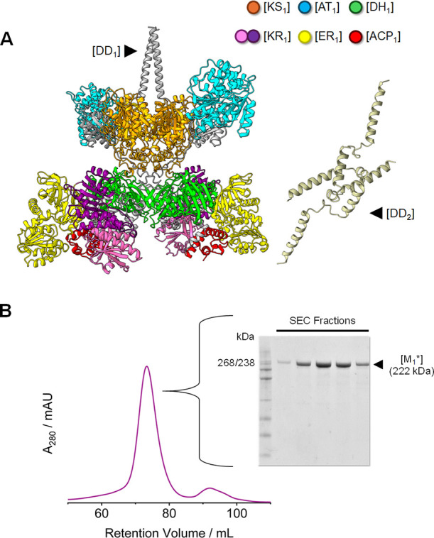
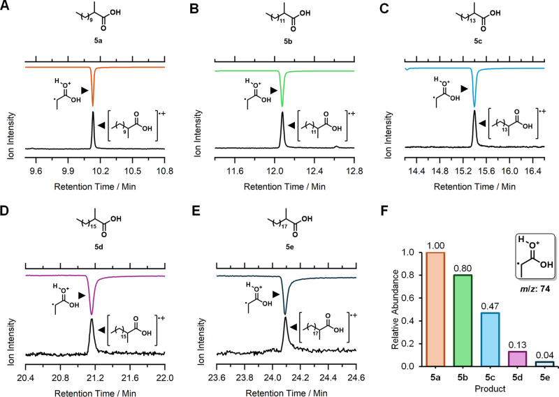
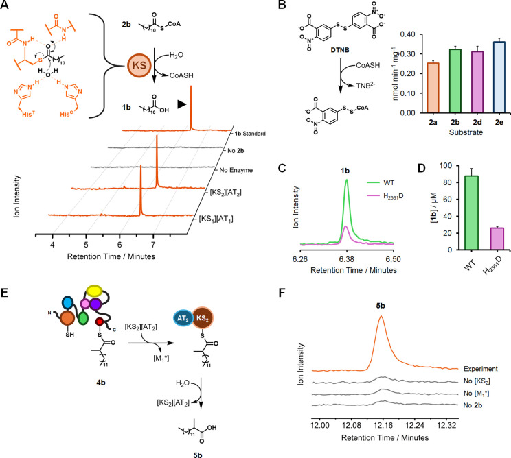
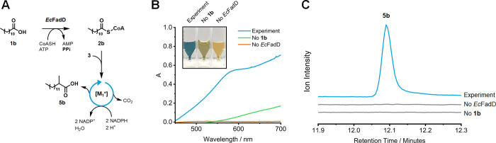
High Resolution Image Download MS PowerPoint Slide In nature, thousands of diverse and bioactive polyketides are assembled by a family of multifunctional, “assembly line” enzyme complexes called polyketide synthases (PKS). Since the late 20th century, there have been several attempts to decode, rearrange, and “reprogram” the PKS assembly line to generate valuable materials such as biofuels and platform chemicals. Here, the first module from Mycobacterium tuberculosis ( Mt ) PKS12, an unorthodox, “modularly iterative” PKS, was modified and repurposed toward the formation of 2-methyl Guerbet lipids, which have wide applications in industry. We established a robust method for the recombinant expression and purification of this modified module (named [M 1 *]), and we demonstrated its ability to catalyze the formation of several 2-methyl Guerbet-like lipids (C 13 –C 21 ). Furthermore, we studied and applied the promiscuous thioesterase activity of a neighboring β-ketoacyl synthase (KS) to release [M 1 *]-bound condensation products in a one-pot biosynthetic cascade. Finally, starting from lauric acid, we could generate our primary target compound (2-methyltetradecanoic acid) by coupling the Escherichia coli fatty acyl-CoA synthetase FadD to [M 1 *]. This work supports the biosynthetic utility of engineered PKS modules such as [M 1 *] and their ability to derive valuable Guerbet-like lipids from inexpensive fatty acids.
Overview
The claims and statistics of this paper haven’t been checked yet.
Run the check — free, no account needed. It reads the open-access text and checks the sources it cites.
Integrity signals
Registry
Cites
Statistics
Figures
Verification
Claims the paper makes
The claims and statistics of this paper haven’t been checked yet.
Run the check — free, no account needed. It reads the open-access text and checks the sources it cites.
Self-check
Percentages, sums and reported differences re-computed from the paper’s own numbers.
The self-check needs the paper’s full text.
Citations
Cites
Basic local alignment search tool
Altschul, S. F., Gish, W., Miller, W., Myers, E. W., & Lipman, D. J. (1990) · Journal of Molecular Biology · doi:10.1006/jmbi.1990.9999 · Cited by 98,669
Highly accurate protein structure prediction with AlphaFold
Jumper, J., Evans, R., Pritzel, A., Green, T., Figurnov, M., Ronneberger, O., Tunyasuvunakool, K., Bates, R., Žídek, A., Potapenko, A., et al. (2021) · Nature · doi:10.1038/s41586-021-03819-2 · Cited by 43,764
ColabFold - Making protein folding accessible to all
Mirdita, M., Schütze, K., Moriwaki, Y., Heo, L., Ovchinnikov, S., & Steinegger, M. (2021) · doi:10.1101/2021.08.15.456425 · Cited by 9,391
Biocatalysis: Enzymatic Synthesis for Industrial Applications
Wu, S., Snajdrova, R., Moore, J. C., Baldenius, K., & Bornscheuer, U. T. (2021) · Angewandte Chemie International Edition · doi:10.1002/anie.202006648 · Cited by 1,422
The Biosynthetic Logic of Polyketide Diversity
Hertweck, C. (2009) · Angewandte Chemie International Edition · doi:10.1002/anie.200806121 · Cited by 1,346
From nature to industry: Harnessing enzymes for biocatalysis
Buller, R., Lutz, S., Kazlauskas, R. J., Snajdrova, R., Moore, J. C., & Bornscheuer, U. T. (2023) · Science · doi:10.1126/science.adh8615 · Cited by 706
Quadri, L. E. N., Weinreb, P. H., Lei, M., Nakano, M. M., Zuber, P., & Walsh, C. T. (1998) · Biochemistry · doi:10.1021/bi9719861 · Cited by 693
Bell, E. L., Finnigan, W., France, S. P., Green, A. P., Hayes, M. A., Hepworth, L. J., Lovelock, S. L., Niikura, H., Osuna, S., Romero, E., et al. (2021) · Nature Reviews Methods Primers · doi:10.1038/s43586-021-00044-z · Cited by 685
Cortes, J., Haydock, S. F., Roberts, G. A., Bevitt, D. J., & Leadlay, P. F. (1990) · Nature · doi:10.1038/348176a0 · Cited by 624
Beld, J., Sonnenschein, E. C., Vickery, C. R., Noel, J. P., & Burkart, M. D. (2013) · Natural Product Reports · doi:10.1039/c3np70054b · Cited by 372
Research works printed but not linked: 2
- 10.1016/S0022-2836(05)80360-2 · DOI · Paper page
- 10.1038/s41592-022-01488-1 · DOI · Paper page
Metadata
- Authors
- Herrera, M. A., McColm, S., Craigie, L.-M., Simpson, J., Brown, F., Clarke, D. J., Carr, R., & Campopiano, D. J.
- Affiliations
- University of Edinburgh (GB)
- Ingenza (United Kingdom) (GB)
- Corresponding author
- Campopiano, Dominic J.
- Venue
- ACS Catalysis
- Publisher
- American Chemical Society (ACS)
- Published
- 2024-10-31
- Volume
- 14
- Issue
- 22
- Pages
- 16834-16842
- ISSN
- 2155-5435 (ISSN-L)
- PMC
- PMC11574752
- OpenAlex
- W4403942416
- Cited by
- 1
- References
- 41
- Citations by year
- 2025: 1
- FWCI
- 0.28
- Type
- journal article
- Language
- en
- Open access
- hybrid
- doi.org
- Licence
- Locations
- ACS Catalysis (publishedVersion, cc-by)
- ChemRxiv (acceptedVersion, cc-by-nc-nd)
- PubMed Central (publishedVersion, cc-by)
- PubMed (publishedVersion)
- Edinburgh Research Explorer (University of Edinburgh) (submittedVersion, cc-by)
- Funders
- Engineering and Physical Sciences Research Council (EP/K039717/1)
- Industrial Biotechnology Innovation Centre
- Biotechnology and Biological Sciences Research Council (BB/S506953/1)
- Topics
- Life Sciences › Immunology and Microbiology › Applied Microbiology and Biotechnology › Microbial Natural Products and Biosynthesis
- Life Sciences › Biochemistry, Genetics and Molecular Biology › Molecular Biology › Microbial Metabolic Engineering and Bioproduction
- Life Sciences › Biochemistry, Genetics and Molecular Biology › Biochemistry › Lipid metabolism and biosynthesis
- Keywords
- polyketide synthase, beta-ketoacyl synthase, lauric acid, polyketide biosynthesis, recombinant protein expression
About these checks
Registry, retraction, Cites and statistics results are computed, not judged, with one exception: a printed reference that no index lists is read once by a model before it counts as a citation error. Claims are extracted by a model and kept only when their quote appears verbatim in the text; each cited source is then checked, with a model’s help, for whether it says what the claim relies on. Those verdicts can be wrong — read the claim’s quote and the cited source before relying on one.
Statistics and arithmetic are recomputed, not judged.
Confidence bands are shown only for fields calibrated on our annotated test set. None is calibrated yet, so no band is shown.
What counts as a claim ›How we measure confidence ›How we check this ›
Are you an author, or do you see a problem with this page? Request a review
How to cite
Cite CoChat for the checks on this page, and the paper for its own findings.
- This page
- CoChat. (2026). Citation check: Repurposing a Fully Reducing Polyketide Synthase toward 2-Methyl Guerbet-like Lipids. https://app.cochat.ai/papers/10.1021/acscatal.4c04714
- The paper
- Herrera, M. A., McColm, S., Craigie, L.-M., Simpson, J., Brown, F., Clarke, D. J., Carr, R., & Campopiano, D. J. (2024). Repurposing a Fully Reducing Polyketide Synthase toward 2-Methyl Guerbet-like Lipids. ACS Catalysis. https://doi.org/10.1021/acscatal.4c04714
Data sources
Bibliographic data: Semantic Scholar, OpenAlex, Crossref.
Contains information from the Semantic Scholar Academic Graph Datasets, made available under the ODC Attribution License (ODC-BY 1.0).
Semantic Scholar’s data is described in: Kinney, R., et al. (2023). The Semantic Scholar Open Data Platform. arXiv:2301.10140.
