

The DOI matches its Crossref record; 52 of the 52 references it cites were found in a registry or an index.
Nogo-B promotes angiogenesis and improves cardiac repair after myocardial infarction via activating Notch1 signaling
Open accessAbstract
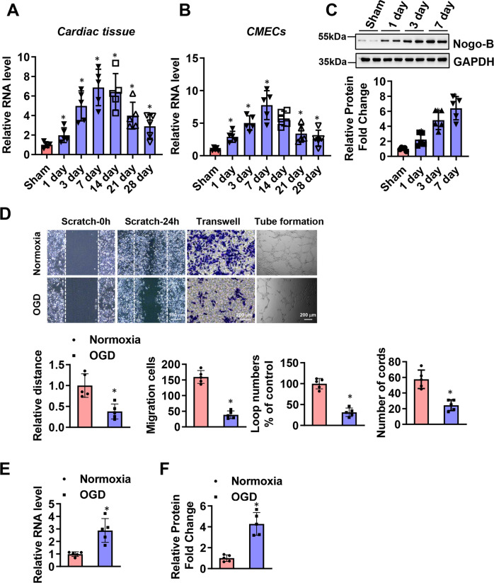
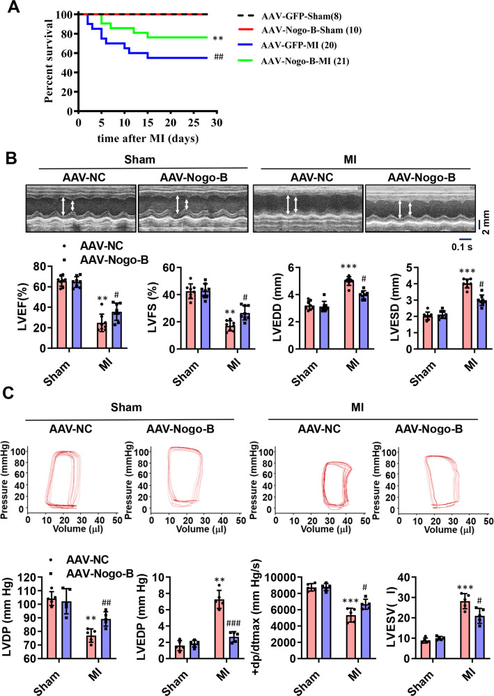
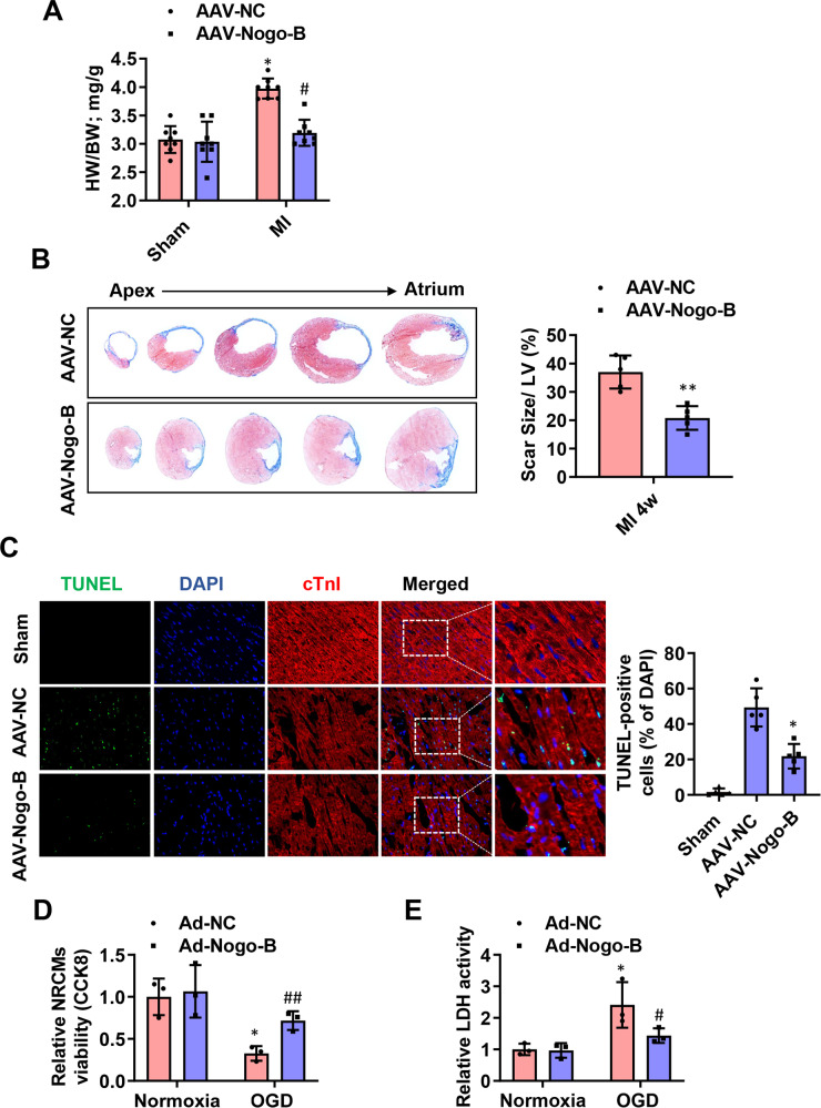
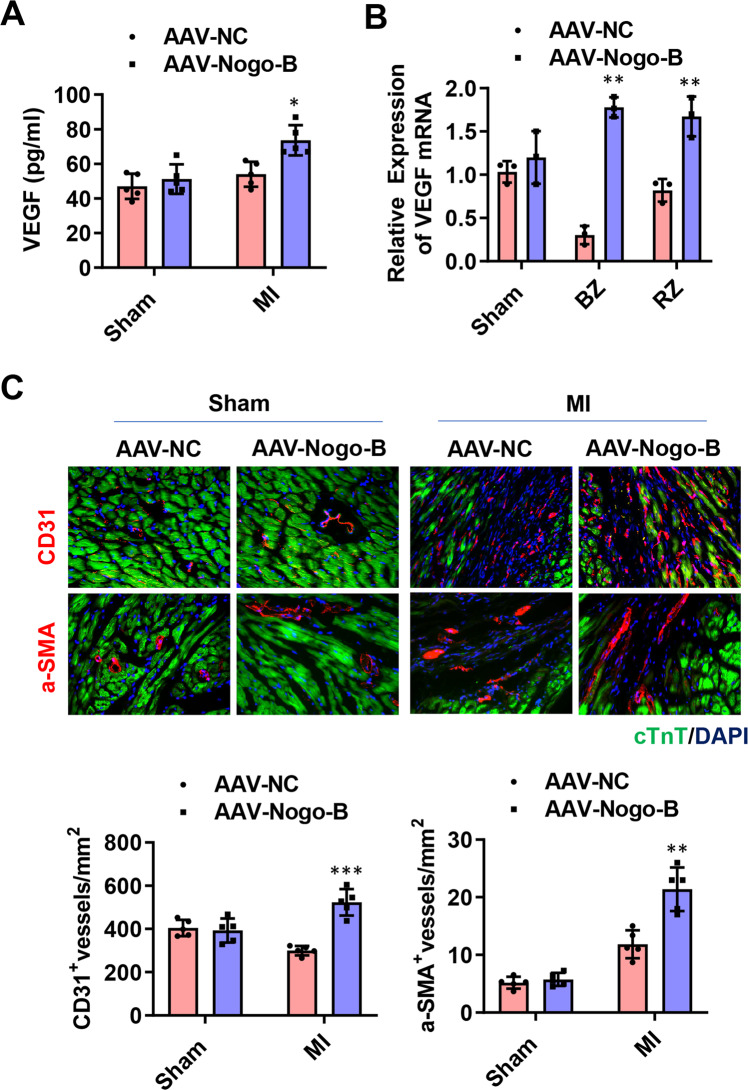
Nogo-B (Reticulon 4B) is reportedly a regulator of angiogenesis during the development and progression of cancer. However, whether Nogo-B regulates angiogenesis and post-myocardial infarction (MI) cardiac repair remains elusive. In the present study, we aimed to explore the role and underlying mechanisms of Nogo-B in cardiac repair during MI. We observed an increased expression level of Nogo-B in the heart of mouse MI models, as well as in isolated cardiac microvascular endothelial cells (CMECs). Moreover, Nogo-B was significantly upregulated in CMECs exposed to oxygen-glucose deprivation (OGD). Nogo-B overexpression in the endothelium via cardiotropic adeno-associated virus serotype 9 (AAV9) with the mouse endothelial-specific promoter Tie2 improved heart function, reduced scar size, and increased angiogenesis. RNA-seq data indicated that Notch signaling is a deregulated pathway in isolated CMECs along the border zone of the infarct with Nogo-B overexpression. Mechanistically, Nogo-B activated Notch1 signaling and upregulated Hes1 in the MI hearts. Inhibition of Notch signaling using a specific siRNA and γ-secretase inhibitor abolished the promotive effects of Nogo-B overexpression on network formation and migration of isolated cardiac microvascular endothelial cells (CMECs). Furthermore, endothelial Notch1 heterozygous deletion inhibited Nogo-B-induced cardioprotection and angiogenesis in the MI model. Collectively, this study demonstrates that Nogo-B is a positive regulator of angiogenesis by activating the Notch signaling pathway, suggesting that Nogo-B is a novel molecular target for ischemic disease.
Overview
The claims and statistics of this paper haven’t been checked yet.
Run the check — free, no account needed. It reads the open-access text and checks the sources it cites.
Integrity signals
Registry
Cites
Statistics
Figures
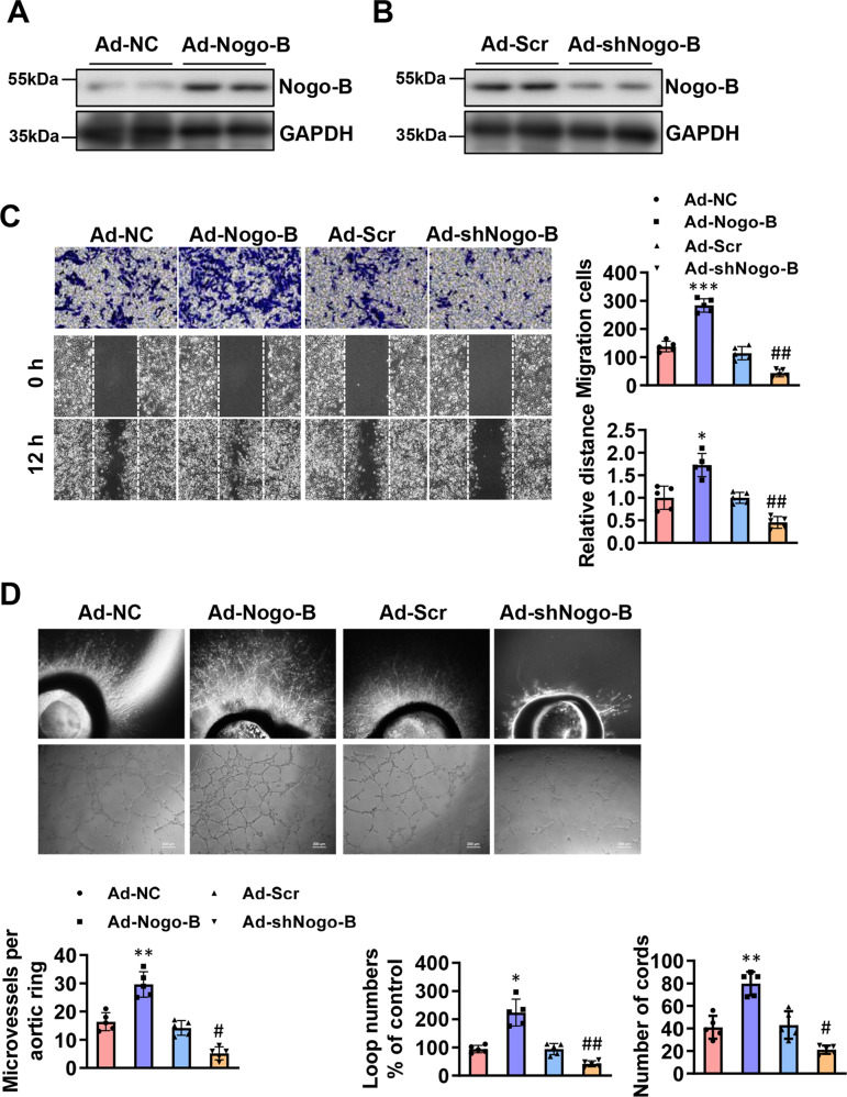
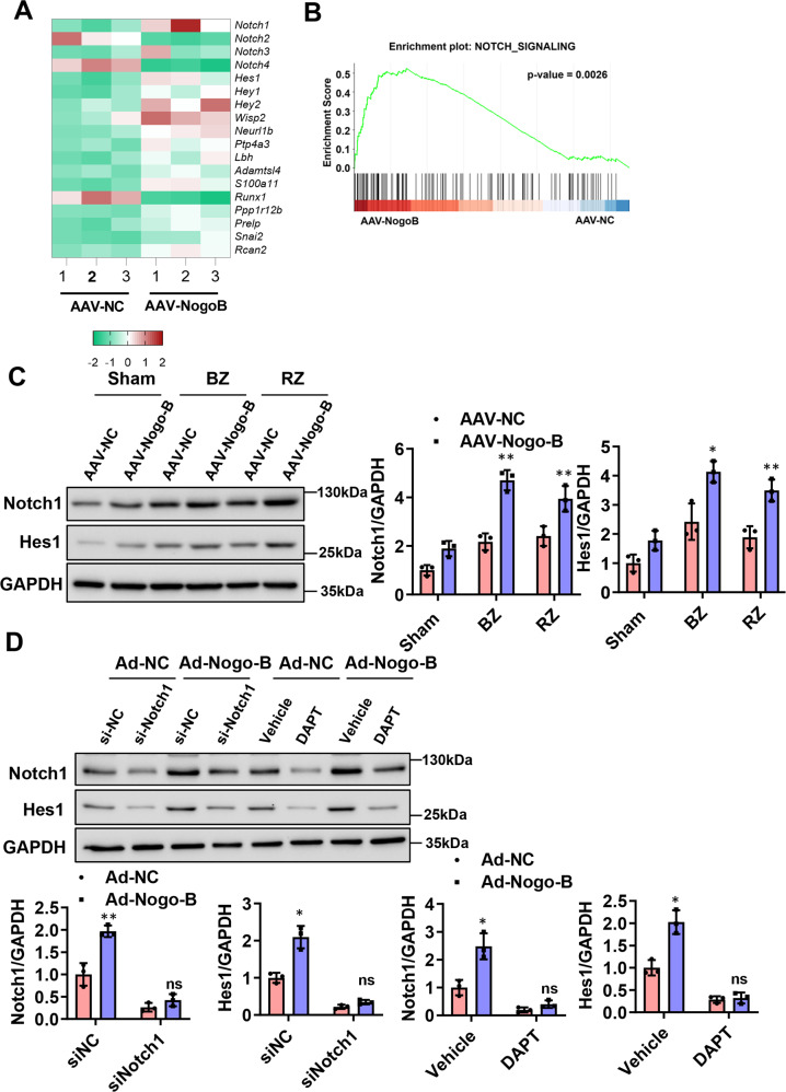
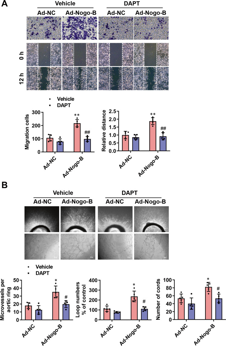
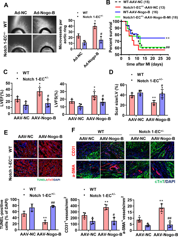
Verification
Claims the paper makes
The claims and statistics of this paper haven’t been checked yet.
Run the check — free, no account needed. It reads the open-access text and checks the sources it cites.
Claims other papers make about this one
Each is checked against this paper’s own text: does it say what the citing paper says it does?
The check wasn’t confident either way, so it gives no verdict — never disputed.
In the citing paper
“BODIPY 493/503, a dye that detects LDs consisting of neutral lipids34,36”
From INPP5D/SHIP1 regulates endo-lysosomal function and cargo-selective phagocytosis in human microglia (2025) · cites this paper as [34]
Experimental — accuracy not yet measuredThe check wasn’t confident either way, so it gives no verdict — never disputed.
In the citing paper
“the accumulation of which is considered a marker of inflammation in microglia during aging and Alzheimer’s disease32–35”
From INPP5D/SHIP1 regulates endo-lysosomal function and cargo-selective phagocytosis in human microglia (2025) · cites this paper as [34]
Experimental — accuracy not yet measured
Self-check
Percentages, sums and reported differences re-computed from the paper’s own numbers.
The self-check needs the paper’s full text.
Citations
Cites
Ahmed, A., Jorna, T., & Bhandari, S. (2016) · The Nephron journals/Nephron journals · doi:10.1159/000447068 · Cited by 11,782
The Biological Basis for Cardiac Repair After Myocardial Infarction
Prabhu, S. D., & Frangogiannis, N. G. (2016) · Circulation Research · doi:10.1161/circresaha.116.303577 · Cited by 2,340
Left Ventricular Remodeling After Myocardial Infarction
Sutton, M. G. S. J., & Sharpe, N. (2000) · Circulation · doi:10.1161/01.cir.101.25.2981 · Cited by 1,868
Khan, M., Nickoloff, E., Abramova, T. V., Johnson, J. A., Verma, S. K., Krishnamurthy, P., Mackie, A. R., Vaughan, E. E., Garikipati, V. N. S., Benedict, C., et al. (2015) · Circulation Research · doi:10.1161/circresaha.117.305990 · Cited by 754
Angiogenesis in skeletal and cardiac muscle
HUDLICKÁ, O., Brown, M. D., & Egginton, S. (1992) · Physiological Reviews · doi:10.1152/physrev.1992.72.2.369 · Cited by 617
Liu, S., Chen, J., Shi, J., Zhou, W., Wang, L., Fang, W., Zhong, Y., Chen, X., Chen, Y., Sabri, A., et al. (2020) · Basic Research in Cardiology · doi:10.1007/s00395-020-0781-7 · Cited by 281
A new role for Nogo as a regulator of vascular remodeling
Acevedo, L. M., Yu, J., Erdjument‐Bromage, H., Miao, Q., Kim, J., Fulton, D. J., Tempst, P., Strittmatter, S. M., & Sessa, W. C. (2004) · Nature Medicine · doi:10.1038/nm1020 · Cited by 244
Wu, Q., Wang, J., Tan, W. L. W., Jiang, Y. Y., Wang, S., Li, Q., Yu, X., Tan, J., Liu, S., Zhang, P., et al. (2020) · Cell Death and Disease · doi:10.1038/s41419-020-2508-y · Cited by 191
The Notch pathway controls fibrotic and regenerative repair in the adult heart
Nemir, M., Metrich, M., Plaisance, I., Lepore, M. G., Cruchet, S., Berthonneche, C., Sarre, A., Radtke, F., & Pedrazzini, T. (2012) · European Heart Journal · doi:10.1093/eurheartj/ehs269 · Cited by 154
Genome editing with CRISPR/Cas9 in postnatal mice corrects PRKAG2 cardiac syndrome
Xie, C., Zhang, Y., Song, L., Luo, J., Qi, W., Hu, J., Lu, D., Yang, Z. H., Zhang, J., Xiao, J., et al. (2016) · Cell Research · doi:10.1038/cr.2016.101 · Cited by 132
Cited by
Sorted by citations
Luo, Y., Zhang, H., Wang, Z., Jiao, J., Wang, Y., Jiang, W., Yu, T., Liu, H., Guan, L., Li, M., et al. (2025) · Regenerative Biomaterials · doi:10.1093/rb/rbaf068 · Cited by 66
Acute Myocardial Infarction: Molecular Pathogenesis, Diagnosis, and Clinical Management
Zhu, M., Li, Y., Xu, Q., Wang, W., Liu, Y., & Liu, Y. (2025) · MedComm · doi:10.1002/mco2.70418 · Cited by 49
Oligodendrocyte Maturation Alters the Cell Death Mechanisms That Cause Demyelination
Chapman, T. W., Kamen, Y., Piedra, E. T., & Hill, R. A. (2024) · The Journal of Neuroscience · doi:10.1523/jneurosci.1794-23.2024 · Cited by 33
Identifying fibrogenic cells following salivary gland obstructive injury
Altrieth, A. L., O’Keefe, K. J., Gellatly, V., Tavarez, J. R., Feminella, S. M., Moskwa, N. L., Cordi, C. V., Turrieta, J. C., Nelson, D. A., & Larsen, M. (2023) · Frontiers in Cell and Developmental Biology · doi:10.3389/fcell.2023.1190386 · Cited by 30
Chauhan, N., Koli, M. R., Ghosh, R., Majumdar, A. G., Ghosh, A., Ghanty, T. K., Mula, S., & Patro, B. S. (2024) · JACS Au · doi:10.1021/jacsau.3c00539 · Cited by 26
Metadata
- Authors
- Zheng, Y., Lin, J., Liu, D., Wan, G., Gu, X., & Ma, J.
- Affiliations
- Shanghai University of Medicine and Health Sciences (CN)
- Shanghai Jiao Tong University (CN)
- Corresponding author
- Ma, Jian
- Venue
- Cell Death & Disease
- Published
- 2022-04-05
- Volume
- 13
- Issue
- 4
- Pages
- 306-306
- ISSN
- 2041-4889 (ISSN-L)
- PMC
- PMC8983727
- OpenAlex
- W4224325360
- Cited by
- 110
- References
- 52
- Citations by year
- 2026: 20 · 2025: 53 · 2024: 21 · 2023: 14 · 2022: 1
- FWCI
- 7.27
- Type
- journal article
- Language
- en
- Open access
- gold
- doi.org
- Locations
- Cell Death and Disease (publishedVersion, cc-by)
- PubMed Central (publishedVersion, cc-by)
- PubMed (publishedVersion)
- DOAJ (DOAJ: Directory of Open Access Journals) (submittedVersion)
- Funders
- Shanghai Key Laboratory of Molecular Imaging (18DZ2260400, 81970229, 81700354)
- Ruijin Hospital
- Topics
- Life Sciences › Biochemistry, Genetics and Molecular Biology › Molecular Biology › Congenital heart defects research
- Health Sciences › Medicine › Cardiology and Cardiovascular Medicine › Cardiac Fibrosis and Remodeling
- Life Sciences › Biochemistry, Genetics and Molecular Biology › Molecular Biology › Angiogenesis and VEGF in Cancer
- Keywords
- Nogo-B, angiogenesis, myocardial infarction, cardiac repair, Notch1 signaling, Hes1, cardiac microvascular endothelial cells, oxygen-glucose deprivation, gamma-secretase inhibition, AAV9 gene delivery, ischemic heart disease, endothelial cell migration
- MeSH terms
- Animals, Endothelium, Myocardial Infarction, Neovascularization, Pathologic, Signal Transduction, Endothelial Cells, Mice
About these checks
Registry, retraction, Cites and statistics results are computed, not judged, with one exception: a printed reference that no index lists is read once by a model before it counts as a citation error. Claims are extracted by a model and kept only when their quote appears verbatim in the text; each cited source is then checked, with a model’s help, for whether it says what the claim relies on. Those verdicts can be wrong — read the claim’s quote and the cited source before relying on one.
Statistics and arithmetic are recomputed, not judged.
Confidence bands are shown only for fields calibrated on our annotated test set. None is calibrated yet, so no band is shown.
What counts as a claim ›How we measure confidence ›How we check this ›
Are you an author, or do you see a problem with this page? Request a review
