

The DOI matches its Crossref record; 52 of the 52 references it cites were found in a registry or an index.
Coherent diffractive imaging of single helium nanodroplets with a high harmonic generation source
Open accessAbstract
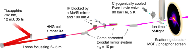
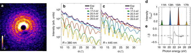
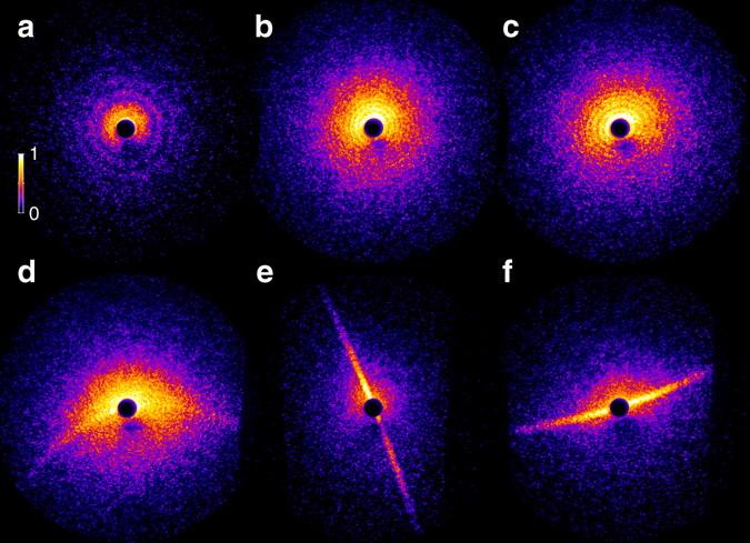
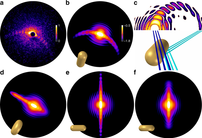
Coherent diffractive imaging of individual free nanoparticles has opened routes for the in situ analysis of their transient structural, optical, and electronic properties. So far, single-shot single-particle diffraction was assumed to be feasible only at extreme ultraviolet and X-ray free-electron lasers, restricting this research field to large-scale facilities. Here we demonstrate single-shot imaging of isolated helium nanodroplets using extreme ultraviolet pulses from a femtosecond-laser-driven high harmonic source. We obtain bright wide-angle scattering patterns, that allow us to uniquely identify hitherto unresolved prolate shapes of superfluid helium droplets. Our results mark the advent of single-shot gas-phase nanoscopy with lab-based short-wavelength pulses and pave the way to ultrafast coherent diffractive imaging with phase-controlled multicolor fields and attosecond pulses.
Overview
The claims and statistics of this paper haven’t been checked yet.
Run the check — free, no account needed. It reads the open-access text and checks the sources it cites.
Integrity signals
Registry
Cites
Statistics
Figures
Verification
Claims the paper makes
The claims and statistics of this paper haven’t been checked yet.
Run the check — free, no account needed. It reads the open-access text and checks the sources it cites.
Claims other papers make about this one
Each is checked against this paper’s own text: does it say what the citing paper says it does?
In the citing paper
“whereas as an example of (2), single-shot single-particle CDI measurements have been performed on sub-micron size helium droplets [100].”
From Roadmap of ultrafast x-ray atomic and molecular physics (2018) · cites this paper as [100]
Self-check
Percentages, sums and reported differences re-computed from the paper’s own numbers.
The self-check needs the paper’s full text.
Citations
Cites
Absorption and Scattering of Light by Small Particles
Bohren, C. F., & Huffman, D. R. (1998) · doi:10.1002/9783527618156 · Cited by 18,474
Beiträge zur Optik trüber Medien, speziell kolloidaler Metallösungen
Mie, G. (1908) · Annalen der Physik · doi:10.1002/andp.19083300302 · Cited by 11,537
Convergence Properties of the Nelder--Mead Simplex Method in Low Dimensions
Lagarias, J. C., Reeds, J. A., Wright, M. H., & Wright, P. E. (1998) · SIAM Journal on Optimization · doi:10.1137/s1052623496303470 · Cited by 7,449
Krausz, F., & Ivanov, M. (2009) · Reviews of Modern Physics · doi:10.1103/revmodphys.81.163 · Cited by 5,468
Die Röntgenkleinwinkelstreuung von dichtgepackten kolloiden Systemen
Porod, G. (1951) · Colloid & Polymer Science · doi:10.1007/bf01512792 · Cited by 1,745
Superfluid Helium Droplets: A Uniquely Cold Nanomatrix for Molecules and Molecular Complexes
Toennies, J. P., & Vilesov, A. F. (2004) · Angewandte Chemie International Edition · doi:10.1002/anie.200300611 · Cited by 1,139
Allaria, E., Appio, R., Badano, L., Barletta, W. A., Bassanese, S., Biedroń, S. G., Borga, A., Busetto, E., Castronovo, D., Cinquegrana, P., et al. (2012) · Nature Photonics · doi:10.1038/nphoton.2012.233 · Cited by 1,027
Single mimivirus particles intercepted and imaged with an X-ray laser
Seibert, M. M., Ekeberg, T., Maia, F. R. N. C., Svenda, M., Andreasson, J., Jönsson, O., Odić, D., Iwan, B., Rocker, A., Westphal, D., et al. (2011) · Nature · doi:10.1038/nature09748 · Cited by 904
Self-citationBeyond crystallography: Diffractive imaging using coherent x-ray light sources
Miao, J., Ishikawa, T., Robinson, I. K., & Murnane, M. M. (2015) · Science · doi:10.1126/science.aaa1394 · Cited by 806
Die Röntgenkleinwinkelstreuung von dichtgepackten kolloiden Systemen
Porod, G. (1952) · Colloid & Polymer Science · doi:10.1007/bf01519615 · Cited by 779
Research works printed but not linked: 1
- Gomez, L. et al. Supplemental material to: Shapes and vorticities of superfluid helium nanodroplets. Science 345, 906–909 (2014). · DOI · Paper page
Cited by
Sorted by citations
Roadmap of ultrafast x-ray atomic and molecular physics
Young, L., Ueda, K., Gühr, M., Bucksbaum, P. H., Simon, M., Mukamel, S., Rohringer, N., Prince, K. C., Masciovecchio, C., Meyer, M., et al. (2018) · Journal of Physics B: Atomic, Molecular and Optical Physics · doi:10.1088/1361-6455/aa9735 · Cited by 337
The ultrafast X-ray spectroscopic revolution in chemical dynamics
Kraus, P. M., Zürch, M. W., Cushing, S. K., Neumark, D. M., & Leone, S. R. (2018) · Nature Reviews Chemistry · doi:10.1038/s41570-018-0008-8 · Cited by 341
High-Harmonic Generation in Solids with and without Topological Edge States
Bauer, D., & Hansen, K. K. (2018) · Physical Review Letters · doi:10.1103/physrevlett.120.177401 · Cited by 198
Computational microscopy with coherent diffractive imaging and ptychography
Miao, J. (2025) · Nature · doi:10.1038/s41586-024-08278-z · Cited by 107
Femtosecond X-ray Fourier holography imaging of free-flying nanoparticles
Gorkhover, T., Ulmer, A., Ferguson, K., Bucher, M., Maia, F. R. N. C., Bielecki, J., Ekeberg, T., Hantke, M. F., Daurer, B. J., Nettelblad, C., et al. (2018) · Nature Photonics · doi:10.1038/s41566-018-0110-y · Cited by 71
Self-citation
Metadata
- Authors
- Rupp, D., Monserud, N., Langbehn, B., Sauppe, M., Zimmermann, J., Ovcharenko, Y., Möller, T., Frassetto, F., Poletto, L., Trabattoni, A., Calegari, F., Nisoli, M., Sander, K., Peltz, C., J. Vrakking, M., Fennel, T., & Rouzée, A.
- Affiliations
- Technische Universität Berlin (DE)
- Max-Born-Institute for Nonlinear Optics and Short Pulse Spectroscopy (DE)
- Istituto di Fotonica e Nanotecnologie (IT)
- Deutsches Elektronen-Synchrotron DESY (DE)
- University of Rostock (DE)
- Corresponding author
- Rupp, Daniela
- Fennel, Thomas
- Rouzée, Arnaud
- ORCID
- Venue
- Nature Communications
- Published
- 2017-09-08
- Volume
- 8
- Issue
- 1
- Pages
- 493-493
- ISSN
- 2041-1723 (ISSN-L)
- PMC
- PMC5591197
- OpenAlex
- W2951302515
- Cited by
- 89
- References
- 52
- Citations by year
- 2026: 3 · 2025: 7 · 2024: 7 · 2023: 6 · 2022: 12 · 2021: 11 · 2020: 19 · 2019: 12 · 2018: 12
- FWCI
- 5.77
- Type
- journal article
- Language
- en
- Update notices
- correction (2018-01-16)
- Open access
- gold
- doi.org
- Locations
- Nature Communications (publishedVersion, cc-by)
- Europe PMC (PubMed Central) (publishedVersion, cc-by)
- PubMed Central (publishedVersion, cc-by)
- PubMed (publishedVersion)
- arXiv (Cornell University) (submittedVersion)
- Virtual Community of Pathological Anatomy (University of Castilla La Mancha) (submittedVersion, cc-by)
- RePEc: Research Papers in Economics (submittedVersion)
- Desy Publications Database (Deutsches Elektronen-Synchrotron DESY) (submittedVersion, cc-by)
- DOAJ (DOAJ: Directory of Open Access Journals) (submittedVersion, cc-by-sa)
- Refubium (Universitätsbibliothek der Freien Universität Berlin) (submittedVersion, cc-by)
- Repository for Publications and Research Data (ETH Zurich) (submittedVersion, cc-by)
- arXiv (Cornell University)
- Universitätsbibliothek der FU Berlin Hochschulschriftenstelle u. Dokumentenserver (cc-by)
- ETH Zürich Research Collection (cc-by)
- Funders
- European Commission (637756, 641789, HORIZON2020)
- Deutsche Forschungsgemeinschaft (SFB652/3, MO 719/13, SFB 652, MO 719/13-1, 719/13, FE 1120/4-1)
- Bundesministerium für Bildung und Forschung (05K16HRB, 05K13KT2)
- FP7 Ideas: European Research Council (227355)
- Topics
- Physical Sciences › Physics and Astronomy › Atomic and Molecular Physics, and Optics › Quantum, superfluid, helium dynamics
- Physical Sciences › Physics and Astronomy › Radiation › Advanced X-ray Imaging Techniques
- Physical Sciences › Physics and Astronomy › Atomic and Molecular Physics, and Optics › Laser-Matter Interactions and Applications
- Keywords
- coherent diffraction imaging, helium nanodroplets, single-shot imaging, high harmonic generation, extreme ultraviolet pulses, femtosecond laser, superfluid helium, prolate shape, single-particle diffraction
About these checks
Registry, retraction, Cites and statistics results are computed, not judged, with one exception: a printed reference that no index lists is read once by a model before it counts as a citation error. Claims are extracted by a model and kept only when their quote appears verbatim in the text; each cited source is then checked, with a model’s help, for whether it says what the claim relies on. Those verdicts can be wrong — read the claim’s quote and the cited source before relying on one.
Statistics and arithmetic are recomputed, not judged.
Confidence bands are shown only for fields calibrated on our annotated test set. None is calibrated yet, so no band is shown.
What counts as a claim ›How we measure confidence ›How we check this ›
Are you an author, or do you see a problem with this page? Request a review
