

The DOI matches its Crossref record; 57 of the 57 references it cites were found in a registry or an index.
A positive feedback loop bi-stably activates fibroblasts
Open accessAbstract
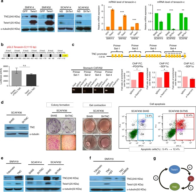
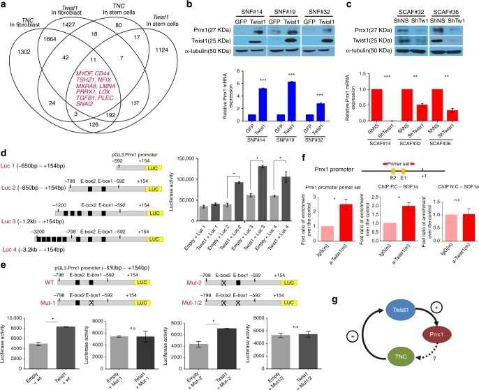
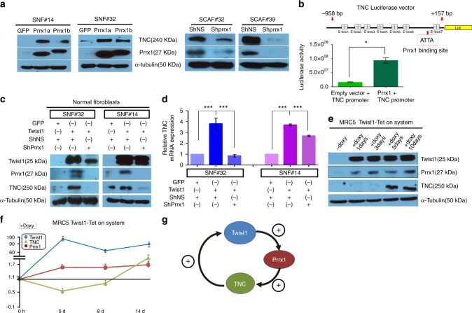
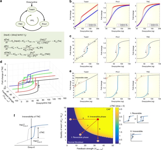
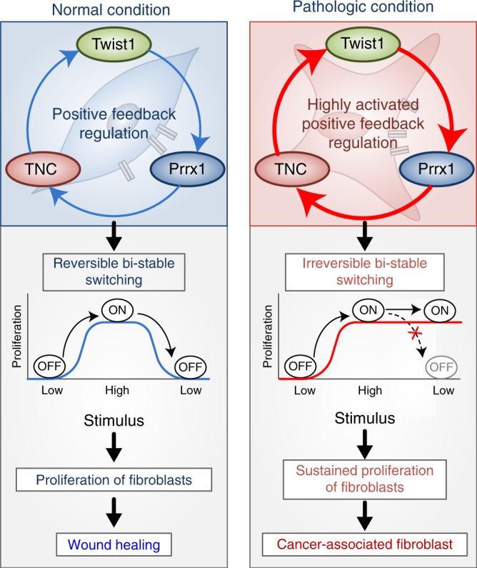
Although fibroblasts are dormant in normal tissue, they exhibit explosive activation during wound healing and perpetual activation in pathologic fibrosis and cancer stroma. The key regulatory network controlling these fibroblast dynamics is still unknown. Here, we report that Twist1, a key regulator of cancer-associated fibroblasts, directly upregulates Prrx1, which, in turn, increases the expression of Tenascin-C (TNC). TNC also increases Twist1 expression, consequently forming a Twist1-Prrx1-TNC positive feedback loop (PFL). Systems biology studies reveal that the Twist1-Prrx1-TNC PFL can function as a bistable ON/OFF switch and regulates fibroblast activation. This PFL can be irreversibly activated under pathologic conditions, leading to perpetual fibroblast activation. Sustained activation of the Twist1-Prrx1-TNC PFL reproduces fibrotic nodules similar to idiopathic pulmonary fibrosis in vivo and is implicated in fibrotic disease and cancer stroma. Considering that this PFL is specific to activated fibroblasts, Twist1-Prrx1-TNC PFL may be a fibroblast-specific therapeutic target to deprogram perpetually activated fibroblasts.
Overview
The claims and statistics of this paper haven’t been checked yet.
Run the check — free, no account needed. It reads the open-access text and checks the sources it cites.
Integrity signals
Registry
Cites
Statistics
Figures
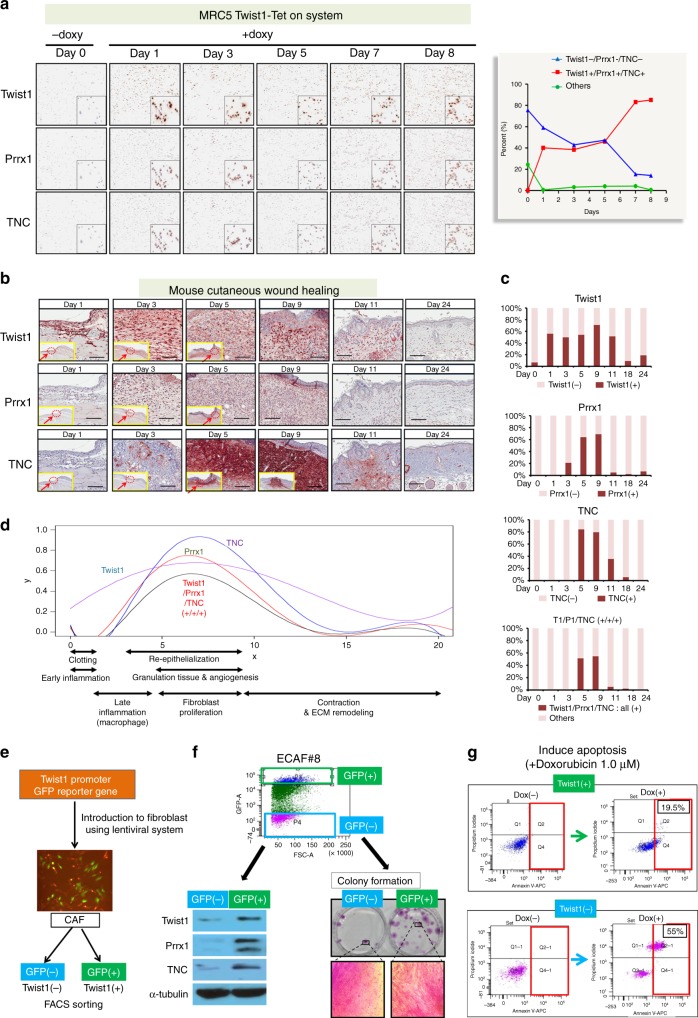
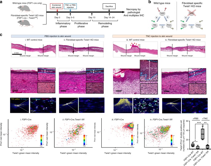
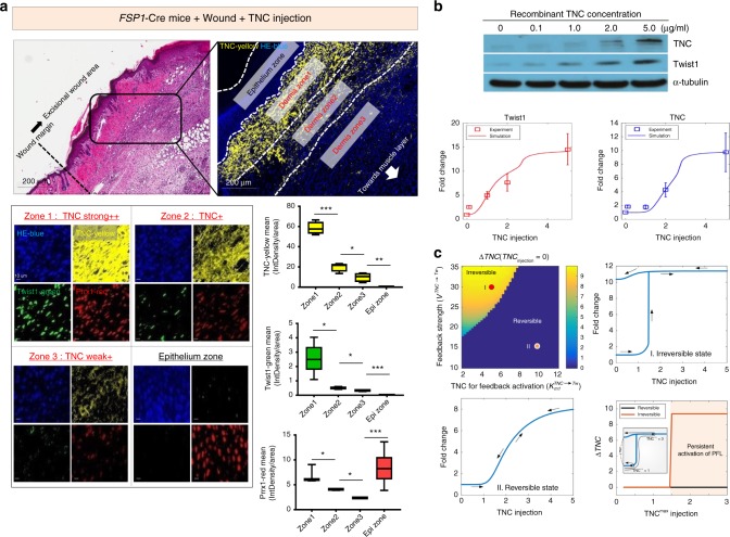
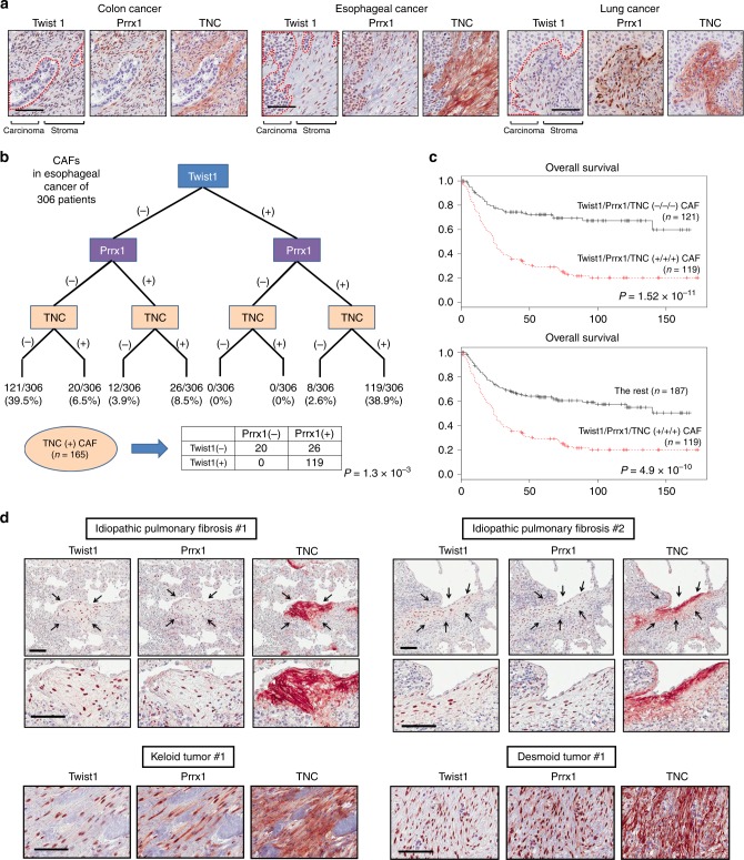
Verification
Claims the paper makes
The claims and statistics of this paper haven’t been checked yet.
Run the check — free, no account needed. It reads the open-access text and checks the sources it cites.
Claims other papers make about this one
Each is checked against this paper’s own text: does it say what the citing paper says it does?
The cited source says what the claim attributes to it.
In the citing paper
“They were both identified as key members of fibroblast transcriptional regulatory networks and involved in a positive feedback loop that leads to fibroblast activation16,17.”
From Integrative genomic analyses identify candidate causal genes for calcific aortic valve stenosis involving tissue-specific regulation (2024) · cites this paper as [17]
Self-check
Percentages, sums and reported differences re-computed from the paper’s own numbers.
The self-check needs the paper’s full text.
Citations
Cites
Kalluri, R., & Zeisberg, M. (2006) · Nature Reviews Cancer · doi:10.1038/nrc1877 · Cited by 4,538
The biology and function of fibroblasts in cancer
Kalluri, R. (2016) · Nature Reviews Cancer · doi:10.1038/nrc.2016.73 · Cited by 4,366
Myofibroblasts and mechano-regulation of connective tissue remodelling
Tomasek, J. J., Gabbiani, G., Hinz, B., Chaponnier, C., & Brown, R. A. (2002) · Nature Reviews Molecular Cell Biology · doi:10.1038/nrm809 · Cited by 4,290
The Wound Healing Process: An Overview of the Cellular and Molecular Mechanisms
Velnar, T., Bailey, T. A., & Smrkolj, V. (2009) · Journal of International Medical Research · doi:10.1177/147323000903700531 · Cited by 2,446
Desmoulière, A., Redard, M., Darby, I. A., & Gabbiani, G. (1995) · Europe PMC (PubMed Central) · Cited by 1,185
Ferrell, J. E. (2002) · Current Opinion in Cell Biology · doi:10.1016/s0955-0674(02)00314-9 · Cited by 1,121
Fibroblasts in fibrosis: novel roles and mediators
Kendall, R. T., & Feghali-Bostwick, C. A. (2014) · Frontiers in Pharmacology · doi:10.3389/fphar.2014.00123 · Cited by 1,082
Identification and characterization of a fibroblast marker: FSP1.
Strutz, F. M., Okada, H., Lo, C. W., Danoff, T. M., Carone, R. L., Tomaszewski, J. E., & Neilson, E. G. (1995) · The Journal of Cell Biology · doi:10.1083/jcb.130.2.393 · Cited by 1,075
Fibroblast heterogeneity in the cancer wound
Öhlund, D., Elyada, E., & Tuveson, D. A. (2014) · The Journal of Experimental Medicine · doi:10.1084/jem.20140692 · Cited by 982
Tsujikawa, T., Kumar, S., Borkar, R. N., Azimi, V., Thibault, G., Chang, Y. H., Balter, A. I., Kawashima, R., Choe, G. H., Sauer, D., et al. (2017) · Cell Reports · doi:10.1016/j.celrep.2017.03.037 · Cited by 560
Cited by
Sorted by citations
Fibroblast and myofibroblast activation in normal tissue repair and fibrosis
Younesi, F. S., Miller, A. E., Barker, T. H., Rossi, F. M. V., & Hinz, B. (2024) · Nature Reviews Molecular Cell Biology · doi:10.1038/s41580-024-00716-0 · Cited by 755
He, H., Suryawanshi, H., Morozov, P. S., Gay-Mimbrera, J., DUCA, E. D., Kim, H. J., Kameyama, N., Estrada, Y. D., Der, E. B., Krueger, J. G., et al. (2020) · Journal of Allergy and Clinical Immunology · doi:10.1016/j.jaci.2020.01.042 · Cited by 510
Activated fibroblasts in cancer: Perspectives and challenges
Caligiuri, G., & Tuveson, D. A. (2023) · Cancer Cell · doi:10.1016/j.ccell.2023.02.015 · Cited by 450
The interplay of fibroblasts, the extracellular matrix, and inflammation in scar formation
Moretti, L., Stalfort, J., Barker, T. H., & Abebayehu, D. (2022) · Journal of Biological Chemistry · doi:10.1016/j.jbc.2021.101530 · Cited by 393
EMT Factors and Metabolic Pathways in Cancer
Georgakopoulos‐Soares, I., Chartoumpekis, D. V., Kyriazopoulou, V. E., & Zaravinos, A. (2020) · Frontiers in Oncology · doi:10.3389/fonc.2020.00499 · Cited by 364
Huang, B., Chen, Z., Geng, L., Wang, J., Liang, H., Cao, Y., Chen, H., Huang, W., Su, M., Wang, H., et al. (2019) · Cell · doi:10.1016/j.cell.2019.10.027 · Cited by 309
The Functional Role of Extracellular Matrix Proteins in Cancer
Popova, N. V., & Jücker, M. (2022) · Cancers · doi:10.3390/cancers14010238 · Cited by 300
The Role of Myofibroblasts in Physiological and Pathological Tissue Repair
Schuster, R., Younesi, F., Ezzo, M., & Hinz, B. (2023) · Cold Spring Harbor Perspectives in Biology · doi:10.1101/cshperspect.a041231 · Cited by 232
Drivers of heterogeneity in synovial fibroblasts in rheumatoid arthritis
Smith, M. H., Gao, V. R., Periyakoil, P. K., Kochen, A., DiCarlo, E. F., Goodman, S. M., Norman, T. M., Donlin, L. T., Leslie, C. S., & Rudensky, A. Y. (2023) · Nature Immunology · doi:10.1038/s41590-023-01527-9 · Cited by 215
The inflammatory speech of fibroblasts
Schuster, R., Rockel, J. S., Kapoor, M., & Hinz, B. (2021) · Immunological Reviews · doi:10.1111/imr.12971 · Cited by 181
Sun, K., Xu, R., Ma, F., Yang, N., Li, Y., Sun, X., Jin, P., Kang, W., Jia, L., Xiong, J., et al. (2022) · Nature Communications · doi:10.1038/s41467-022-32627-z · Cited by 163
Epithelial–mesenchymal transition in tissue repair and degeneration
Youssef, K. K., & Nieto, M. A. (2024) · Nature Reviews Molecular Cell Biology · doi:10.1038/s41580-024-00733-z · Cited by 165
A myeloid–stromal niche and gp130 rescue in NOD2-driven Crohn’s disease
Nayar, S., Morrison, J. K., Giri, M., Gettler, K., Chuang, L.-S. F., Walker, L. A., Ko, H. M., Kenigsberg, E., Kugathasan, S., Mérad, M., et al. (2021) · Nature · doi:10.1038/s41586-021-03484-5 · Cited by 141
CAFs orchestrates tumor immune microenvironment—A new target in cancer therapy?
Zhang, C., Fei, Y., Wang, H., Hu, S., Liu, C., Hu, R., & Du, Q. (2023) · Frontiers in Pharmacology · doi:10.3389/fphar.2023.1113378 · Cited by 132
Lee, K.-W., Yeo, S.-Y., Gong, J.-R., Koo, O.-J., Sohn, I., Lee, W. Y., Kim, H. C., Yun, S. H., Cho, Y. B., Choi, M.-A., et al. (2022) · Nature Communications · doi:10.1038/s41467-022-30484-4 · Cited by 103
Self-citationTranscription factors in fibroblast plasticity and CAF heterogeneity
Melchionna, R., Trono, P., Carlo, A. D., Modugno, F. D., & Nisticò, P. (2023) · Journal of Experimental & Clinical Cancer Research · doi:10.1186/s13046-023-02934-4 · Cited by 92
Mesenchymal Plasticity Regulated by Prrx1 Drives Aggressive Pancreatic Cancer Biology
Feldmann, K., Maurer, H. C., Peschke, K., Teller, S., Schuck, K., Steiger, K., Engleitner, T., Öllinger, R., Nomura, A., Wirges, N., et al. (2020) · Gastroenterology · doi:10.1053/j.gastro.2020.09.010 · Cited by 88
Fibroblast Memory in Development, Homeostasis and Disease
Kirk, T., Ahmed, A., & Rognoni, E. (2021) · Cells · doi:10.3390/cells10112840 · Cited by 80
Niu, L., Cheng, B., Huang, G., Nan, K., Han, S., Ren, H., Liu, N., Li, Y., Genin, G. M., & Xu, F. (2022) · Cell Discovery · doi:10.1038/s41421-022-00427-w · Cited by 81
Li, Z., Chen, S., Cui, H., Li, X., Chen, D., Hao, W., Wang, J., Li, Z., Zheng, Z., Zhang, Z., et al. (2021) · Annals of the Rheumatic Diseases · doi:10.1136/annrheumdis-2021-220002 · Cited by 73
Determinants of renal cell carcinoma invasion and metastatic competence
Kim, K., Zhou, Q.-B., Christie, A., Stevens, C., Ma, Y., Onabolu, O., Chintalapati, S., McKenzie, T., Tcheuyap, V., Woolford, L., et al. (2021) · Nature Communications · doi:10.1038/s41467-021-25918-4 · Cited by 69
Effects of Tenascin C on the Integrity of Extracellular Matrix and Skin Aging
Choi, Y. E., Song, M. J., Hara, M., Imanaka-Yoshida, K., Lee, D. H., Chung, J. H., & Lee, S.-T. (2020) · International Journal of Molecular Sciences · doi:10.3390/ijms21228693 · Cited by 58
Single-cell analyses reveal early thymic progenitors and pre-B cells in zebrafish
Rubin, S. A., Baron, C. S., Pessoa Rodrigues, C., Duran, M., Corbin, A. F., Yang, S. P., Trapnell, C., & Zon, L. I. (2022) · Journal of Experimental Medicine · doi:10.1084/jem.20220038 · Cited by 56
Chaotic activation of developmental signalling pathways drives idiopathic pulmonary fibrosis
Froidure, A., Marchal‐Duval, E., Homps-Legrand, M., Ghanem, M., Justet, A., Crestani, B., & Mailleux, A. (2020) · European Respiratory Review · doi:10.1183/16000617.0140-2019 · Cited by 61
Khan, A. Q., Hasan, A., Mir, S. S., Rashid, K., Shahab Uddin, M. S., & Steinhoff, M. S. (2024) · Seminars in Cancer Biology · doi:10.1016/j.semcancer.2024.03.002 · Cited by 60
Tenascin-C in Heart Diseases—The Role of Inflammation
Imanaka‐Yoshida, K. (2021) · International Journal of Molecular Sciences · doi:10.3390/ijms22115828 · Cited by 54
Tenascin-C: A Key Regulator in Angiogenesis during Wound Healing
Wang, Y., Wang, G., & Liu, H. (2022) · Biomolecules · doi:10.3390/biom12111689 · Cited by 52
Ke, B.-J., Abdurahiman, S., Biscu, F., Zanella, G., Dragoni, G., Santhosh, S., Simone, V. D., Zouzaf, A., van Baarle, L., Stakenborg, M., et al. (2024) · Journal of Clinical Investigation · doi:10.1172/jci173835 · Cited by 56
Emerging Role of Extracellular Vesicles and Cellular Communication in Metastasis
Forder, A., Hsing, C.-Y., Trejo, J., & Garnis, C. (2021) · Cells · doi:10.3390/cells10123429 · Cited by 53
Sunami, Y., Boeker, V., & Kleeff, J. H. (2021) · Cancers · doi:10.3390/cancers13040697 · Cited by 56
Imodoye, S. O., Adedokun, K. A., Muhammed, A. O., Bello, I. O., Muhibi, M. A., Oduola, T., & Oyenike, M. A. (2021) · Frontiers in Oncology · doi:10.3389/fonc.2021.762817 · Cited by 50
Thériault, S., Li, Z., Abner, E., Luan, J., Manikpurage, H. D., Houessou, U., Zamani, P., Briend, M., Estonian Biobank Research Team, Boudreau, D. K., et al. (2024) · Nature Communications · doi:10.1038/s41467-024-46639-4 · Cited by 49
Trained Immunity for Personalized Cancer Immunotherapy: Current Knowledge and Future Opportunities
Lérias, J. R., de Sousa, E., Paraschoudi, G., Martins, J. D., Condeço, C., Figueiredo, N., Carvalho, C. T., Dodoo, E., Maia, A., Castillo-Martín, M., et al. (2020) · Frontiers in Microbiology · doi:10.3389/fmicb.2019.02924 · Cited by 45
Wilson, R. B., Archid, R., & Reymond, M. A. (2020) · International Journal of Molecular Sciences · doi:10.3390/ijms21114158 · Cited by 46
The Prrx1 limb enhancer marks an adult subpopulation of injury-responsive dermal fibroblasts
Currie, J. D., Grosser, L., Murawala, P., Schuez, M., Michel, M., Tanaka, E. M., & Sandoval‐Guzmán, T. (2019) · Biology Open · doi:10.1242/bio.043711 · Cited by 44
Albacete-Albacete, L., Sánchez‐Álvarez, M., & del Pozo, M. Á. (2021) · Frontiers in Immunology · doi:10.3389/fimmu.2021.671485 · Cited by 43
Zhang, W., Wang, J., Liu, C., Li, Y., Sun, C., Wu, J., & Wu, Q. (2023) · Journal of Translational Medicine · doi:10.1186/s12967-023-04714-2 · Cited by 42
Freag, M. S., Mohammed, M. T., Kulkarni, A., Emam, H. E., Maremanda, K. P., & Elzoghby, A. O. (2024) · Science Advances · doi:10.1126/sciadv.adk3074 · Cited by 37
Mechanobiology across timescales
Cheng, B., Li, M., Lin, M., Guo, H., & Xu, F. (2025) · Nature Reviews Physics · doi:10.1038/s42254-025-00874-w · Cited by 38
Cancer-associated fibroblasts: Mediators of head and neck tumor microenvironment remodeling
Raudenská, M., Balvan, J., Hánělová, K., Bugajová, M., & Masařík, M. (2023) · Biochimica et Biophysica Acta (BBA) - Reviews on Cancer · doi:10.1016/j.bbcan.2023.188940 · Cited by 37
Peng, K., Sant, D., Andersen, N., Silvera, R., Camarena, V., Piñero, G., Graham, R. M., Khan, A., Xu, X., Wang, G., et al. (2020) · Scientific Reports · doi:10.1038/s41598-020-74128-3 · Cited by 35
Nonequilibrium polarity-induced chemotaxis: Emergent Galilean symmetry and exact scaling exponents
Mahdisoltani, S., Zinati, R. B. A., Duclut, C., Gambassi, A., & Golestanian, R. (2021) · Physical Review Research · doi:10.1103/physrevresearch.3.013100 · Cited by 35
Valenzi, E., Bahudhanapati, H., Tan, J., Tabib, T., Sullivan, D. I., Nouraie, M. S., Sembrat, J., Fan, L., Chen, K., Liu, S., et al. (2023) · European Respiratory Journal · doi:10.1183/13993003.00474-2022 · Cited by 34
Dankel, S. N., Grytten, E., Bjune, J.-I., Nielsen, H. J., Dietrich, A., Blüher, M., Sagen, J. V., & Mellgren, G. (2020) · Scientific Reports · doi:10.1038/s41598-020-77406-2 · Cited by 33
Yang, J. H., Gong, L., Liu, Q., Zhao, H., Wang, Z., Li, X., Tian, W., & Zhou, Q. (2023) · Frontiers in Immunology · doi:10.3389/fimmu.2023.1142273 · Cited by 35
Menche, C., Schuhwerk, H., Armstark, I., Gupta, P., Fuchs, K., van Roey, R., Mosa, M. H., Hartebrodt, A., Hajjaj, Y., Ezquerra, A. C., et al. (2024) · EMBO Reports · doi:10.1038/s44319-024-00186-7 · Cited by 31
Николаевна, Д. И., Shirmanova, M. V., Ignatova, N. I., Dudenkova, V. V., Lukina, M. M., Вадимовна, З. Е., Safina, D. R., Kostrov, S. V., Didych, D. A., Kuzmich, A. I., et al. (2020) · International Journal of Molecular Sciences · doi:10.3390/ijms21218119 · Cited by 31
Biology of Tenascin C and its Role in Physiology and Pathology
Abedsaeidi, M., Hojjati, F., Tavassoli, A., & Sahebkar, A. (2023) · Current Medicinal Chemistry · doi:10.2174/0929867330666230404124229 · Cited by 28
Siddiqui, S. H., Subramaniyan, S. A., Kang, D., Park, J., Khan, M., Choi, H. W., & Shim, K. S. (2020) · Cell Stress and Chaperones · doi:10.1007/s12192-020-01140-x · Cited by 27
Obesity Inhibits Angiogenesis Through TWIST1-SLIT2 Signaling
Hunyenyiwa, T., Hendee, K., Matus, K., Kyi, P., Mammoto, T., & Mammoto, A. (2021) · Frontiers in Cell and Developmental Biology · doi:10.3389/fcell.2021.693410 · Cited by 30
Fibroblasts from Distinct Pancreatic Pathologies Exhibit Disease-Specific Properties
Barrera, L. N., Evans, A., Lane, B. S., Brumskill, S., Oldfield, F. E., Campbell, F., Andrews, T., Lu, Z., Pérez-Mancera, P. A., Liloglou, T., et al. (2020) · Cancer Research · doi:10.1158/0008-5472.can-19-3534 · Cited by 28
iPSC-derived mesenchymal cells that support alveolar organoid development
Tamai, K., Sakai, K., Yamaki, H., Moriguchi, K., Igura, K., Maehana, S., Suezawa, T., Takehara, K., Hagiwara, M., Hirai, T., et al. (2022) · Cell Reports Methods · doi:10.1016/j.crmeth.2022.100314 · Cited by 27
Lee, S., Lee, C., Hwang, C. Y., Kim, D., Han, Y.-H., Hong, S. N., Kim, S., & Cho, K. (2020) · Molecular Cancer Research · doi:10.1158/1541-7786.mcr-19-0450 · Cited by 27
Self-citationRintz, E., Gaffke, L., Podlacha, M., Brokowska, J., Cyske, Z., Węgrzyn, G., & Pierzynowska, K. (2020) · International Journal of Molecular Sciences · doi:10.3390/ijms21093194 · Cited by 27
Fibroblasts in cancer dormancy: foe or friend?
Dai, L., Mao, L., Zhang, W., Tang, Y.-J., Tang, Y., & Liang, X. (2021) · Cancer Cell International · doi:10.1186/s12935-021-01883-2 · Cited by 27
Kim, H. J., Shim, J., Park, J., Shin, H., Shim, J. S., Jang, K., Park, W., Lee, K.-H., Kwon, E. J., Jang, H., et al. (2021) · Communications Biology · doi:10.1038/s42003-021-02223-w · Cited by 29
Self-citationPhysiology and Pathophysiology of Wound Healing in Diabetes
Pastar, I., Balukoff, N. C., Sawaya, A. P., Vecin, N. M., & Tomic‐Canic, M. (2024) · Contemporary diabetes · doi:10.1007/978-3-031-55715-6_7 · Cited by 27
Son, S. W., Chau, G. C., Kim, S., & Um, S. H. (2019) · Cells · doi:10.3390/cells8101137 · Cited by 24
Self-citationDecoding sequence determinants of gene expression in diverse cellular and disease states
Lal, A., Karollus, A., Gunsalus, L. M., Garfield, D., Nair, S., Tseng, A. M., Gordon, M. G., Blischak, J., Geijn, B. V. D., Bhangale, T., et al. (2026) · Nature Methods · doi:10.1038/s41592-026-03102-0 · Cited by 34
Strobel, B., Klein, H., Leparc, G., Stierstorfer, B., Gantner, F., & Kreuz, S. (2022) · Scientific Reports · doi:10.1038/s41598-022-16344-7 · Cited by 24
Chowkwale, M., Lindsey, M. L., & Saucerman, J. J. (2022) · The Journal of Physiology · doi:10.1113/jp283346 · Cited by 24
The Hidden Control Architecture of Complex Brain Networks
Lee, B., Kang, U., Chang, H., & Cho, K. (2019) · iScience · doi:10.1016/j.isci.2019.02.017 · Cited by 24
Self-citationEmbryo-derive TNF promotes decidualization via fibroblast activation
Chen, S., Shi, W., Lin, Y.-Q., Yang, Z., Wang, Y., Li, Y., Liu, A.-X., Liu, A.-X., Hu, Y., & Yang, Z. (2023) · eLife · doi:10.7554/elife.82970 · Cited by 22
[Retracted] Fibroblasts in Scar Formation: Biology and Clinical Translation
Qian, H., Shan, Y., Gong, R., Lin, D., Zhang, M., Wang, C., & Wang, L. (2022) · Oxidative Medicine and Cellular Longevity · doi:10.1155/2022/4586569 · Cited by 20
RetractedHarihar, S., & Welch, D. R. (2023) · Cancer and Metastasis Reviews · doi:10.1007/s10555-023-10090-6 · Cited by 19
Marchal‐Duval, E., Homps-Legrand, M., Froidure, A., Jaillet, M., Ghanem, M., Deneuville, L., Justet, A., Maurac, A., Vadel, A., Fortas, E., et al. (2023) · eLife · doi:10.7554/elife.79840 · Cited by 20
Transcription factor networks in cellular quiescence
Mitra, M., Batista, S., & Coller, H. A. (2025) · Nature Cell Biology · doi:10.1038/s41556-024-01582-w · Cited by 16
Exploring cellular changes in ruptured human quadriceps tendons at single‐cell resolution
Mimpen, J. Y., Baldwin, M. J., Paul, C., Ramos‐Mucci, L., Kurjan, A., Cohen, C., Sharma, S., Florquin, M. S. N. C., Hulley, P. A., McMaster, J., et al. (2025) · The Journal of Physiology · doi:10.1113/jp287812 · Cited by 16
Yu, H., Tiao, M., Huang, S., Sheu, J. J., Tain, Y., Sheen, J., Lin, I.-C. F., Tsai, C., Huang, L., Hsu, C., et al. (2025) · Ecotoxicology and Environmental Safety · doi:10.1016/j.ecoenv.2025.118397 · Cited by 15
Twist1 signaling in age-dependent decline in angiogenesis and lung regeneration
Hendee, K., Hunyenyiwa, T., Matus, K., Toledo, M., Mammoto, A., & Mammoto, T. (2021) · Aging · doi:10.18632/aging.202875 · Cited by 14
Traveling wave of inflammatory response to regulate the expansion or shrinkage of skin erythema
Sudo, M., & Fujimoto, K. (2022) · PLoS ONE · doi:10.1371/journal.pone.0263049 · Cited by 13
The paired‐related homeobox protein 1 promotes cardiac fibrosis via the Twist1‐Prrx1‐tenascin‐C loop
Wang, F.-Y., Zhao, H., Yin, L., Zhang, W., Tang, Y., Wang, X., & Huang, C. (2022) · Cell Biology International · doi:10.1002/cbin.11944 · Cited by 13
Moschini, G., Mohanan, A. G., Niewczas, I. S., Taylor, D. E., Jaeger, P. K., Turiel, G., Hussien, A. A., Wunderli, S. L., Baumberger, O., Wolleb, M., et al. (2026) · Science Translational Medicine · doi:10.1126/scitranslmed.adt1228 · Cited by 12
Chung, H. C., Cho, E. J., Lee, H., Kim, W., Oh, J., Kim, S., Lee, D., & Sung, C. O. (2021) · Clinical and Translational Medicine · doi:10.1002/ctm2.487 · Cited by 12
Self-citationLee, E., Yeo, S.-Y., Lee, K., Lee, J. A., Kim, K. K., & Kim, S. (2020) · Scientific Reports · doi:10.1038/s41598-020-63996-4 · Cited by 11
Self-citationThe reactive stroma response regulates the immune landscape in prostate cancer
Thomas, R., Jerome, J. M., Krieger, K. L., Ashraf, N., & Rowley, D. R. (2024) · Journal of Translational Genetics and Genomics · doi:10.20517/jtgg.2024.15 · Cited by 11
Brücher, B. L. D. M., & Jamall, I. S. (2019) · 4open · doi:10.1051/fopen/2018996 · Cited by 10
The Importance of Cancer-Associated Fibroblasts in the Pathogenesis of Head and Neck Cancers
Raudenská, M., Svobodová, M., Gumulec, J., Falk, M., & Masařík, M. (2020) · Klinicka onkologie · doi:10.14735/amko202039 · Cited by 10
Bell, J. A., Davies, E. R., Brereton, C. J., Vukmirovic, M., Roberts, J., Lunn, K., Wickens, L., Conforti, F., Ridley, R. A., Ceccato, J., et al. (2024) · Cell Reports Medicine · doi:10.1016/j.xcrm.2024.101695 · Cited by 12
Secchia, S., Beilinson, V., Chen, X., Yang, Z. F., Wayman, J. A., Dhaliwal, J., Jurickova, I., Angerman, E., Denson, L. A., Miraldi, E. R., et al. (2025) · Cell Reports · doi:10.1016/j.celrep.2025.115896 · Cited by 14
Fujita, M., Sasada, M., Eguchi, M., Iyoda, T., Okuyama, S., Osawa, T., Tsuzuranuki, K., Sakamoto, M., Hagihara, Y., Matsumura, M., et al. (2021) · PubMed Central · Cited by 9
Turano, M., Vicidomini, R., Cammarota, F., D’Agostino, V., Duraturo, F., Izzo, P., & Rosa, M. D. (2023) · Biomedicines · doi:10.3390/biomedicines11051428 · Cited by 9
Ku, J., Jeong, E., Gong, J., Cho, K., Sung, C. O., & Kim, S. (2025) · Molecules and Cells · doi:10.1016/j.mocell.2025.100263 · Cited by 8
Self-citationEpitranscriptomics as a Candidate Universal Modulator of Dormancy Transitions
Ahi, E. P. (2026) · Ecology and Evolution · doi:10.1002/ece3.73007 · Cited by 7
Canalizing kernel for cell fate determination
Kim, N., Lee, J., Kim, J., Kim, Y., & Cho, K. (2024) · Briefings in Bioinformatics · doi:10.1093/bib/bbae406 · Cited by 6
Self-citationSiddiqui, S., Subramaniyan, S., Park, J., Kang, D., Khan, M., Belal, S., Lee, S. C., & Shim, K. (2022) · Journal of Applied Animal Research · doi:10.1080/09712119.2022.2060986 · Cited by 7
Regulation of cell behavior by extracellular proteins
Bradshaw, A. D. (2020) · Elsevier eBooks · doi:10.1016/b978-0-12-818422-6.00013-7 · Cited by 8
TWISTed fibroblasts: New drivers of intestinal fibrosis in Crohn's disease
Lovisa, S., & Vetrano, S. (2024) · Heliyon · doi:10.1016/j.heliyon.2024.e40604 · Cited by 6
Validation of ZMYND8 as a new treatment target in hepatocellular carcinoma
Choi, S., Lee, K., Koh, H. H., Park, S., Yeo, S.-Y., Joh, J., Choi, M. S., Kim, S., Park, C.-K., & Ha, S. Y. (2021) · Journal of Cancer Research and Clinical Oncology · doi:10.1007/s00432-021-03768-3 · Cited by 6
Self-citationYamamoto, S., Kashimoto, R., Furukawa, S., Sakamoto, H., & Satoh, A. (2021) · Journal of Experimental Zoology Part B Molecular and Developmental Evolution · doi:10.1002/jez.b.23093 · Cited by 4
McDaniels, J. M., Shetty, A. C., Rousselle, T. V., Bardhi, E., Maluf, D. G., & Mas, V. R. (2022) · Frontiers in Transplantation · doi:10.3389/frtra.2022.988238 · Cited by 4
Nonequilibrium phenomena in driven and active Coulomb field theories
Mahdisoltani, S., & Golestanian, R. (2022) · Physica A Statistical Mechanics and its Applications · doi:10.1016/j.physa.2022.127947 · Cited by 4
Bocchi, V. D., To, K., Weber, L., Zumbo, P., Kim, L., Shida, I. Y., Yang, D., Storm, P., Fiorenzano, A., Sozzi, E., et al. (2026) · bioRxiv (Cold Spring Harbor Laboratory) · doi:10.64898/2026.06.19.733041 · Cited by 4
Liu, J., Zhang, Y., Liu, K., Shi, J., Yan, M., Xu, J., Liu, Y., & Zhang, S. (2025) · Cancer Cell International · doi:10.1186/s12935-025-04120-2 · Cited by 3
Zeng, Y., Zhang, W., Chen, J., Luo, S., Wu, Y., Hu, C., Cai, Y., Yang, L., & Zhan, J. P. (2026) · ACS Nano · doi:10.1021/acsnano.5c17134 · Cited by 3
Koudouna, E., Young, R. D., Quantock, A. J., & Ralphs, J. R. (2023) · International Journal of Molecular Sciences · doi:10.3390/ijms24043555 · Cited by 3
Mechanical network motifs as targets for mechanomedicine
Lu, M., Zhao, X., Xu, F., Yin, C., Guo, H., & Cheng, B. (2024) · Drug Discovery Today · doi:10.1016/j.drudis.2024.104145 · Cited by 3
Juan, L. S., de Pedro, I., Rodríguez-Luna, A., Villalba, M., Guerrero, A., González, S., & Gandarillas, A. (2021) · Cosmetics · doi:10.3390/cosmetics9010002 · Cited by 3
Plant pathogenic nematode exosomes remodel vector tracheae to enhance pathogen transmission
Chang, Y., Zhou, J., Ye, F., Zhang, H., Sun, J., & Zhao, L. (2026) · Proceedings of the National Academy of Sciences · doi:10.1073/pnas.2600386123 · Cited by 2
Mirshahidi, S., Yuan, I. J., Chen, Z., Simental, A., Lee, S. C., Filho, P. A. A., Murry, T., Zeng, F., Duerksen-Hughes, P. J., Wang, C., et al. (2024) · Cancers · doi:10.3390/cancers16152730 · Cited by 2
Metadata
- Authors
- Yeo, S.-Y., Lee, K.-W., Shin, D., An, S., Cho, K.-H., & Kim, S.-H.
- Affiliations
- Sungkyunkwan University (KR)
- Korea Advanced Institute of Science and Technology (KR)
- Samsung Medical Center (KR)
- Corresponding author
- Cho, Kwang-Hyun
- Kim, Seok-Hyung
- Venue
- Nature Communications
- Published
- 2018-08-01
- Volume
- 9
- Issue
- 1
- Pages
- 3016-3016
- ISSN
- 2041-1723 (ISSN-L)
- PMC
- PMC6070563
- OpenAlex
- W2883191015
- Cited by
- 115
- References
- 57
- Citations by year
- 2026: 11 · 2025: 8 · 2024: 18 · 2023: 16 · 2022: 17 · 2021: 21 · 2020: 18 · 2019: 5 · 2018: 1
- FWCI
- 4.00
- Type
- journal article
- Language
- en
- Open access
- gold
- doi.org
- Locations
- Nature Communications (publishedVersion, cc-by)
- Europe PMC (PubMed Central) (publishedVersion, cc-by)
- PubMed Central (publishedVersion, cc-by)
- PubMed (publishedVersion)
- RePEc: Research Papers in Economics (submittedVersion)
- DOAJ (DOAJ: Directory of Open Access Journals) (submittedVersion)
- DOAJ (DOAJ: Directory of Open Access Journals) (submittedVersion, cc-by-sa)
- Funders
- National Research Foundation (2015R1A2A1A15054021)
- Ministry of Health, Welfare and Family Affairs | Korea National Institute of Health (HI14C2517)
- National Research Foundation of Korea (2017R1A2A1A17069642, 2015M3A9A7067220, 2013M3A9A7046303)
- Topics
- Life Sciences › Biochemistry, Genetics and Molecular Biology › Molecular Biology › TGF-β signaling in diseases
- Health Sciences › Medicine › Pulmonary and Respiratory Medicine › Interstitial Lung Diseases and Idiopathic Pulmonary Fibrosis
- Health Sciences › Medicine › Surgery › Wound Healing and Treatments
- Keywords
- fibroblast activation, positive feedback loop, TWIST1, PRRX1, tenascin-C, bistability, cancer-associated fibroblasts, fibrosis, idiopathic pulmonary fibrosis, tumor stroma, wound healing, gene regulatory networks
- MeSH terms
- Cancer-Associated Fibroblasts, Animals, Cell Line, Feedback, Fibroblasts, Fibrosis, Humans, Mice, Inbred BALB C, Nuclear Proteins, Homeodomain Proteins, Tenascin, Systems Biology, Twist-Related Protein 1
About these checks
Registry, retraction, Cites and statistics results are computed, not judged, with one exception: a printed reference that no index lists is read once by a model before it counts as a citation error. Claims are extracted by a model and kept only when their quote appears verbatim in the text; each cited source is then checked, with a model’s help, for whether it says what the claim relies on. Those verdicts can be wrong — read the claim’s quote and the cited source before relying on one.
Statistics and arithmetic are recomputed, not judged.
Confidence bands are shown only for fields calibrated on our annotated test set. None is calibrated yet, so no band is shown.
What counts as a claim ›How we measure confidence ›How we check this ›
Are you an author, or do you see a problem with this page? Request a review
