

The DOI matches its Crossref record; 3 of the 3 references it cites were found in a registry or an index.
Indoleacetate decarboxylase is a glycyl radical enzyme catalysing the formation of malodorant skatole
Abstract
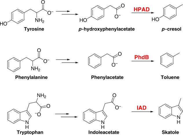
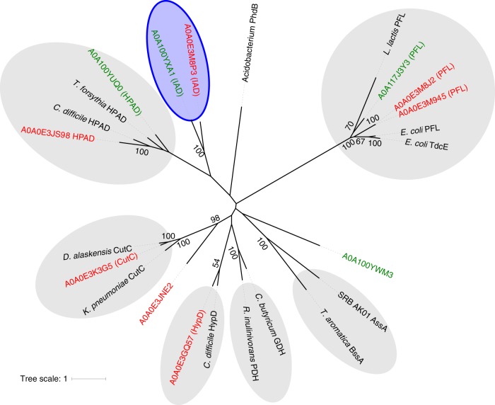
Skatole is a malodorous compound that contributes to the characteristic smell of animal faeces. Although skatole has long been known to originate from bacterial tryptophan fermentation, the enzyme catalysing its formation has so far remained elusive. Here we report the use of comparative genomics for the discovery of indoleacetate decarboxylase, an O 2-sensitive glycyl radical enzyme catalysing the decarboxylation of indoleacetate to form skatole as the terminal step of tryptophan fermentation in certain anaerobic bacteria. We describe its biochemical characterization and compare it to other glycyl radical decarboxylases. Indoleacetate decarboxylase may serve as a genetic marker for the identification of skatole-producing environmental and human-associated bacteria, with impacts on human health and the livestock industry.
Overview
The claims and statistics of this paper haven’t been checked yet.
Run the check — free, no account needed. It reads the open-access text and checks the sources it cites.
Integrity signals
Registry
Cites
Statistics
Figures
Verification
Claims the paper makes
The claims and statistics of this paper haven’t been checked yet.
Run the check — free, no account needed. It reads the open-access text and checks the sources it cites.
Claims other papers make about this one
Each is checked against this paper’s own text: does it say what the citing paper says it does?
In the citing paper
“but reduced corrinoids have been demonstrated to have defluorination activity with polyfluorinated compounds (Ochoa‐Herrera et al., 2016; Liu et al., 2018, 2018,2018, 2018; Sun et al., 2021).”
From Nothing lasts forever: understanding microbial biodegradation of polyfluorinated compounds and perfluorinated alkyl substances (2022) · cites this paper as Liu et al., 2018
Self-check
Percentages, sums and reported differences re-computed from the paper’s own numbers.
The self-check needs the paper’s full text.
Citations
Cites
A gut bacterial pathway metabolizes aromatic amino acids into nine circulating metabolites
Dodd, D., Spitzer, M. H., Van Treuren, W., Merrill, B. D., Hryckowian, A. J., Higginbottom, S. K., Le, A., Cowan, T. M., Nolan, G. P., Fischbach, M. A., et al. (2017) · Nature · doi:10.1038/nature24661 · Cited by 1,425
The end products of the metabolism of aromatic amino acids by Clostridia
Elsden, S. R., Hilton, M. G., & Waller, J. M. (1976) · Archives of Microbiology · doi:10.1007/bf00425340 · Cited by 396
Olagbemiro, T. O., Birkett, M. A., (Luntz), A. J. M., & Pickett, J. A. (2004) · Journal of Chemical Ecology · doi:10.1023/b:joec.0000028461.86243.19 · Cited by 53
Research works printed but not linked: 47
- Yokoyama, M. T. & Carlson, J. R. Microbial metabolites of tryptophan in the intestinal tract with special reference to skatole. Am. J. Clin. Nutr. 32, 173–178 (1979). · DOI
- Persico, A. M. & Napolioni, V. Urinary p-cresol in autism spectrum disorder. Neurotoxicol. Teratol. 36, 82–90 (2013). · DOI · Paper page
- Selmer, T. & Andrei, P. I. p-Hydroxyphenylacetate decarboxylase from Clostridium difficile. A novel glycyl radical enzyme catalysing the formation of p-cresol. Eur. J. Biochem. 268, 1363–1372 (2001). · DOI · Paper page
- Zargar, K. et al. In vitro characterization of phenylacetate decarboxylase, a novel enzyme catalyzing toluene biosynthesis in an anaerobic microbial community. Sci. Rep. 6, 31362 (2016). · DOI
- Beller, H. R. et al. Discovery of enzymes for toluene synthesis from anoxic microbial communities. Nat. Chem. Biol. 14, 451–457 (2018). · DOI
- Whitehead, T. R., Price, N. P., Drake, H. L. & Cotta, M. A. Catabolic pathway for the production of skatole and indoleacetic acid by the acetogen Clostridium drakei, Clostridium scatologenes, and swine manure. Appl. Environ. Microbiol. 74, 1950–1953 (2008). · DOI
- Schiffman, S. S., Bennett, J. L. & Raymer, J. H. Quantification of odors and odorants from swine operations in North Carolina. Agric. For. Meteorol. 108, 213–240 (2001). · DOI
- Deslandes, Bt, Gariépy, C. & Houde, A. Review of microbiological and biochemical effects of skatole on animal production. Livest. Prod. Sci. 71, 193–200 (2001). · DOI
- Lundström, K. et al. Skatole, androstenone and taint in boars fed two different diets. Livest. Prod. Sci. 18, 55–67 (1988). · DOI
- Linden, A., Desmecht, D., Vandenput, S., Van de Weerdt, M. L. & Lekeux, P. Effect of serotonergic blockade on calf pulmonary function after the intravenous administration of 3-methylindole. J. Comp. Pathol. 114, 361–371 (1996). · DOI
- Yost, G. S. Mechanisms of 3-methylindole pneumotoxicity. Chem. Res. Toxicol. 2, 273–279 (1989). · DOI
- Bray, T. M. & Kirkland, J. B. The metabolic basis of 3-methylindole-induced pneumotoxicity. Pharmacol. Ther. 46, 105–118 (1990). · DOI
- Weems, J. M. et al. 3-Methylindole is mutagenic and a possible pulmonary carcinogen. Toxicol. Sci. 112, 59–67 (2009). · DOI
- Rasmussen, M. K., Balaguer, P., Ekstrand, B., Daujat-Chavanieu, M. & Gerbal-Chaloin, S. Skatole (3-methylindole) is a partial aryl hydrocarbon receptor agonist and induces CYP1A1/2 and CYP1B1 expression in primary human hepatocytes. PLoS ONE 11, e0154629 (2016). · DOI
- Hughes, D. T., Pelletier, J., Luetje, C. W. & Leal, W. S. Odorant receptor from the southern house mosquito narrowly tuned to the oviposition attractant skatole. J. Chem. Ecol. 36, 797–800 (2010). · DOI
- Yu, L., Blaser, M., Andrei, P. I., Pierik, A. J. & Selmer, T. 4-Hydroxyphenylacetate decarboxylases: properties of a novel subclass of glycyl radical enzyme systems. Biochemistry 45, 9584–9592 (2006). · DOI
- Levin, B. J. et al. A prominent glycyl radical enzyme in human gut microbiomes metabolizes trans-4-hydroxy-l-proline. Science 355, eaai8386 (2017). · DOI
- Backman, L. R. F., Funk, M. A., Dawson, C. D. & Drennan, C. L. New tricks for the glycyl radical enzyme family. Crit. Rev. Biochem. Mol. Biol. 52, 674–695 (2017). · DOI
- Shisler, K. A. & Broderick, J. B. Glycyl radical activating enzymes: structure, mechanism, and substrate interactions. Arch. Biochem. Biophys. 546, 64–71 (2014). · DOI
- Honeyfield, D. C. & Carlson, J. R. Assay for the enzymatic conversion of indoleacetic acid to 3-methylindole in a ruminal Lactobacillus species. Appl. Environ. Microbiol. 56, 724–729 (1990). · DOI
- Martins, B. M. et al. Structural basis for a Kolbe-type decarboxylation catalyzed by a glycyl radical enzyme. J. Am. Chem. Soc. 133, 14666–14674 (2011). · DOI
- Feliks, M., Martins, B. M. & Ullmann, G. M. Catalytic mechanism of the glycyl radical enzyme 4-hydroxyphenylacetate decarboxylase from continuum electrostatic and QC/MM calculations. J. Am. Chem. Soc. 135, 14574–14585 (2013). · DOI
- Li, X., Jensen, R. L., Hojberg, O., Canibe, N. & Jensen, B. B. Olsenella scatoligenes sp. nov., a 3-methylindole- (skatole) and 4-methylphenol- (p-cresol) producing bacterium isolated from pig faeces. Int. J. Syst. Evol. Microbiol. 65, 1227–1233 (2015). · DOI
- Finn, R. D. et al. InterPro in 2017-beyond protein family and domain annotations. Nucleic Acids Res. 45, D190–D199 (2017). · DOI
- Wei, Y. et al. The class III ribonucleotide reductase from Neisseria bacilliformis can utilize thioredoxin as a reductant. Proc. Natl Acad. Sci. USA 111, E3756–E3765 (2014). · DOI
- Wei, Y. et al. A ferredoxin disulfide reductase delivers electrons to the Methanosarcina barkeri class III ribonucleotide reductase. Biochemistry 54, 7019–7028 (2015). · DOI
- Sofia, H. J., Chen, G., Hetzler, B. G., Reyes-Spindola, J. F. & Miller, N. E. Radical SAM, a novel protein superfamily linking unresolved steps in familiar biosynthetic pathways with radical mechanisms: functional characterization using new analysis and information visualization methods. Nucleic Acids Res. 29, 1097–1106 (2001). · DOI
- Selvaraj, B., Buckel, W., Golding, B. T., Ullmann, G. M. & Martins, B. M. Structure and function of 4-hydroxyphenylacetate decarboxylase and its cognate activating enzyme. J. Mol. Microbiol. Biotechnol. 26, 76–91 (2016). · DOI
- Krebs, C., Henshaw, T. F., Cheek, J., Huynh, B. H. & Broderick, J. B. Conversion of 3Fe-4S to 4Fe-4S clusters in native pyruvate formate-lyase activating enzyme: Mössbauer characterization and implications for mechanism. J. Am. Chem. Soc. 122, 12497–12506 (2000). · DOI
- Atkinson, H. J., Morris, J. H., Ferrin, T. E. & Babbitt, P. C. Using sequence similarity networks for visualization of relationships across diverse protein superfamilies. PLoS ONE 4, e4345 (2009). · DOI
- Gerlt, J. A. et al. Enzyme Function Initiative-Enzyme Similarity Tool (EFI-EST): a web tool for generating protein sequence similarity networks. Biochim. Biophys. Acta 1854, 1019–1037 (2015). · DOI
- Shannon, P. et al. Cytoscape: a software environment for integrated models of biomolecular interaction networks. Genome Res. 13, 2498–2504 (2003). · DOI · Paper page
- Sievers, F. et al. Fast, scalable generation of high-quality protein multiple sequence alignments using Clustal Omega. Mol. Syst. Biol. 7, 539 (2011). · DOI
- Olsen, I., Johnson, J. L., Moore, L. V. & Moore, W. E. Lactobacillus uli sp. nov. and Lactobacillus rimae sp. nov. from the human gingival crevice and emended descriptions of lactobacillus minutus and Streptococcus parvulus. Int. J. Syst. Bacteriol. 41, 261–266 (1991). · DOI
- Sakamoto, M., Iino, T. & Ohkuma, M. Faecalimonas umbilicata gen. nov., sp. nov., isolated from human faeces, and reclassification of Eubacterium contortum, Eubacterium fissicatena and Clostridium oroticum as Faecalicatena contorta gen. nov., comb. nov., Faecalicatena fissicatena comb. nov. and Faecalicatena orotica comb. nov. Int. J. Syst. Evol. Microbiol. 67, 1219–1227 (2017). · DOI
- Tonzetich, J. Production and origin of oral malodor: a review of mechanisms and methods of analysis. J. Periodontol. 48, 13–20 (1977). · DOI
- Van Petegem, F., Clark, K. A., Chatelain, F. C. & Minor, D. L. Jr. Structure of a complex between a voltage-gated calcium channel beta-subunit and an alpha-subunit domain. Nature 429, 671–675 (2004). · DOI
- Fish, W. W. Rapid colorimetric micromethod for the quantitation of complexed iron in biological samples. Methods Enzymol. 158, 357–364 (1988). · DOI
- Siegel, L. M. A direct microdetermination for sulfide. Anal. Biochem. 11, 126–132 (1965). · DOI
- Zhang, Y. et al. Dre2, a conserved eukaryotic Fe/S cluster protein, functions in cytosolic Fe/S protein biogenesis. Mol. Cell Biol. 28, 5569–5582 (2008). · DOI
- Shen, G. et al. SufR coordinates two [4Fe-4S]2+, 1+clusters and functions as a transcriptional repressor of the sufBCDS operon and an autoregulator of sufR in cyanobacteria. J. Biol. Chem. 282, 31909–31919 (2007). · DOI
- Duboc-Toia, C. et al. Very high-field EPR study of glycyl radical enzymes. J. Am. Chem. Soc. 125, 38–39 (2003). · DOI
- Guindon, S. & Gascuel, O. A simple, fast, and accurate algorithm to estimate large phylogenies by maximum likelihood. Syst. Biol. 52, 696–704 (2003). · DOI
- Le, S. Q. & Gascuel, O. An improved general amino acid replacement matrix. Mol. Biol. Evol. 25, 1307–1320 (2008). · DOI · Paper page
- Eyre-Walker, A. & Keightley, P. D. The distribution of fitness effects of new mutations. Nat. Rev. Genet. 8, 610–618 (2007). · DOI
- Felsenstein, J. Cases in which parsimony or compatibility methods will be positively misleading. Syst. Zool. 27, 401–410 (1978). · DOI · Paper page
- Letunic, I. & Bork, P. Interactive tree of life (iTOL) v3: an online tool for the display and annotation of phylogenetic and other trees. Nucleic Acids Res. 44, W242–W245 (2016). · DOI
Cited by
Sorted by citations
Wackett, L. P. (2022) · Microbial Biotechnology · doi:10.1111/1751-7915.13928 · Cited by 165
Metadata
- Authors
- Liu, D., Wei, Y., Liu, X., Zhou, Y., Jiang, L., Yin, J., Wang, F., Hu, Y., Nanjaraj Urs, A. N., Liu, Y., Ang, E. L., Zhao, S., Zhao, H., & Zhang, Y.
- Venue
- Nature Communications
- Published
- 2018-10-11
- Volume
- 9
- Issue
- 1
- ISSN
- 2041-1723
- PMC
- PMC6181972
- Cited by
- 63
- References
- 3
- Language
- en
- Funders
- National Natural Science Foundation of China (31570060)
About these checks
Registry, retraction, Cites and statistics results are computed, not judged, with one exception: a printed reference that no index lists is read once by a model before it counts as a citation error. Claims are extracted by a model and kept only when their quote appears verbatim in the text; each cited source is then checked, with a model’s help, for whether it says what the claim relies on. Those verdicts can be wrong — read the claim’s quote and the cited source before relying on one.
Statistics and arithmetic are recomputed, not judged.
Confidence bands are shown only for fields calibrated on our annotated test set. None is calibrated yet, so no band is shown.
What counts as a claim ›How we measure confidence ›How we check this ›
Are you an author, or do you see a problem with this page? Request a review
