

The DOI matches its Crossref record; 57 of the 59 references it cites were found in a registry or an index.
APOE ε2 is associated with increased tau pathology in primary tauopathy
Open accessAbstract
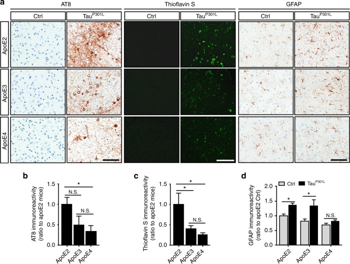
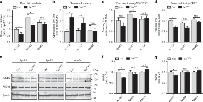
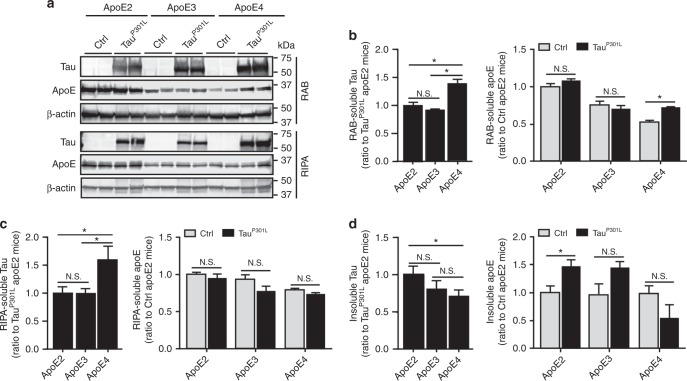
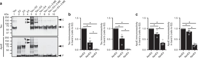
Apolipoprotein E ( APOE ) ε4 allele is the strongest genetic risk factor for late-onset Alzheimer’s disease mainly by modulating amyloid-β pathology. APOE ε4 is also shown to exacerbate neurodegeneration and neuroinflammation in a tau transgenic mouse model. To further evaluate the association of APOE genotype with the presence and severity of tau pathology, we express human tau via an adeno-associated virus gene delivery approach in human APOE targeted replacement mice. We find increased hyperphosphorylated tau species, tau aggregates, and behavioral abnormalities in mice expressing APOE ε2/ε2. We also show that in humans, the APOE ε2 allele is associated with increased tau pathology in the brains of progressive supranuclear palsy (PSP) cases. Finally, we identify an association between the APOE ε2/ε2 genotype and risk of tauopathies using two series of pathologically-confirmed cases of PSP and corticobasal degeneration. Our data together suggest APOE ε2 status may influence the risk and progression of tauopathy.
Overview
The claims and statistics of this paper haven’t been checked yet.
Run the check — free, no account needed. It reads the open-access text and checks the sources it cites.
Integrity signals
Registry
Cites
Statistics
Figures
Verification
Claims the paper makes
The claims and statistics of this paper haven’t been checked yet.
Run the check — free, no account needed. It reads the open-access text and checks the sources it cites.
Claims other papers make about this one
Each is checked against this paper’s own text: does it say what the citing paper says it does?
The source backs part of the claim, not all of it — or backs it for a different population, condition or comparison.
In the citing paper
“This is especially critical after a recent study in the preclinical literature demonstrated APOE2 expression via viral delivery exacerbated pathology in tauopathy mice (Zhao et al., 2018).”
From Alzheimer Disease: An Update on Pathobiology and Treatment Strategies (2019) · cites this paper as Zhao et al., 2018
Experimental — accuracy not yet measuredThe source backs part of the claim, not all of it — or backs it for a different population, condition or comparison.
In the citing paper
“One study found that the APOEε2 allele is associated with increased odds of neuropathologically confirmed primary tauopathy (PSP and corticobasal degeneration)66.”
From Multifaceted roles of APOE in Alzheimer disease (2024) · cites this paper as [66]
Experimental — accuracy not yet measuredThe cited source says what the claim attributes to it.
In the citing paper
“Furthermore, the APOE ε2 allele was associated with increased tau pathology in the brains of human progressive supranuclear palsy, a primary tauopathy.”
From Deletion of neuronal Idol ameliorates Alzheimer's disease–related pathologies via APOE receptors (2025) · cites this paper as [71]
The cited source says what the claim attributes to it.
In the citing paper
“For example, APOE2 mice exhibited more tau pathology, compared to APOE3 and APOE4 mice, and behavioral abnormalities in a tauopathy model.”
From Deletion of neuronal Idol ameliorates Alzheimer's disease–related pathologies via APOE receptors (2025) · cites this paper as [71]
The cited source says what the claim attributes to it.
In the citing paper
“ApoE can also affect tauopathy and tau-mediated neurodegeneration (Brecht et al., 2004; Shi and Holtzman, 2018; Zhao et al., 2018).”
From Selective removal of astrocytic APOE4 strongly protects against tau-mediated neurodegeneration and decreases synaptic phagocytosis by microglia (2021) · cites this paper as Zhao et al., 2018
In the citing paper
“Studies have shown that hyperphosphorylated tau species, tau aggregates and behavioral abnormalities were observed in APOE ε2/ε2 mice [471].”
From Molecular and cellular mechanisms underlying the pathogenesis of Alzheimer’s disease (2020) · cites this paper as [471]
In the citing paper
“In addition, APOE ε2 is also associated with increased pathological tau levels in the presence of amyloid [471, 472].”
From Molecular and cellular mechanisms underlying the pathogenesis of Alzheimer’s disease (2020) · cites this paper as [471]
In the citing paper
“A recent study failed to replicate the neurotoxic effect of ApoE4 in tauopathy mice.”
From Alzheimer Disease: An Update on Pathobiology and Treatment Strategies (2019) · cites this paper as Zhao et al., 2018
Self-check
Percentages, sums and reported differences re-computed from the paper’s own numbers.
The self-check needs the paper’s full text.
Citations
Cites
Neuropathological stageing of Alzheimer-related changes.
Braak, H., & Braak, E. (1991) · Acta Neuropathologica · doi:10.1007/bf00308809 · Cited by 16,123
Phases of Aβ-deposition in the human brain and its relevance for the development of AD
Thal, D. R., Rüb, U., Orantes, M., & Braak, H. (2002) · Neurology · doi:10.1212/wnl.58.12.1791 · Cited by 3,490
Apolipoprotein E and Alzheimer disease: risk, mechanisms and therapy
Liu, C.-C., Kanekiyo, T., Xu, H., & Bu, G. (2013) · Nature Reviews Neurology · doi:10.1038/nrneurol.2012.263 · Cited by 3,459
Self-citationBlennow, K., de Leon, M. J., & Zetterberg, H. (2006) · The Lancet · doi:10.1016/s0140-6736(06)69113-7 · Cited by 3,134
Tau in physiology and pathology
Wang, Y., & Mandelkow, E. (2016) · Nature Reviews Neuroscience · doi:10.1038/nrn.2015.1 · Cited by 2,251
Primary age-related tauopathy (PART): a common pathology associated with human aging
Crary, J. F., Trojanowski, J. Q., Schneider, J. A., Abisambra, J. F., Abner, E. L., Alafuzoff, I., Arnold, S. E., Attems, J., Beach, T. G., Bigio, E. H., et al. (2014) · Acta Neuropathologica · doi:10.1007/s00401-014-1349-0 · Cited by 1,467
Self-citationApoE4 markedly exacerbates tau-mediated neurodegeneration in a mouse model of tauopathy
Initiative, A. D. N., Shi, Y., Yamada, K., Liddelow, S. A., Smith, S. T., Zhao, L., Luo, W., Tsai, R. M., Spina, S., Grinberg, L. T., et al. (2017) · Nature · doi:10.1038/nature24016 · Cited by 1,220
Apolipoprotein E and its receptors in Alzheimer's disease: pathways, pathogenesis and therapy
Bu, G. (2009) · Nature Reviews Neuroscience · doi:10.1038/nrn2620 · Cited by 1,168
Self-citationHauw, J., Daniel, S. E., Dickson, D. W., Horoupian, D. S., Jellinger, K. A., Lantos, P. L., McKee, A. C., Tabaton, M., & Litvan, I. (1994) · Neurology · doi:10.1212/wnl.44.11.2015 · Cited by 854
Self-citationParkinson's Disease and Parkinsonism: Neuropathology
Dickson, D. W. (2012) · Cold Spring Harbor Perspectives in Medicine · doi:10.1101/cshperspect.a009258 · Cited by 847
Self-citation
Research works printed but not linked: 1
- Dickson, D. W., Hauw, J.-J., Agid, Y. and Litvan, I. in Neurodegeneration: The Molecular Pathology of Dementia and Movement Disorders 2nd edn (eds Dickon, D. W. & Weller, R. O.) Ch. 15 (Wiley-Blackwell, Chichester, 2011).
Cited by
Sorted by citations
Items 101–142 of 142
Ikeda, M., Okamoto, K., Suzuki, K., Takai, E., Kasahara, H., Furuta, N., Furuta, M., Tashiro, Y., Shimizu, C., TAKATAMA, S., et al. (2021) · Frontiers in Neurology · doi:10.3389/fneur.2021.645625 · Cited by 3
The extracellular chaperone Clusterin enhances Tau aggregate seeding in a cellular modelPreprint
Yuste‐Checa, P., Trinkaus, V. A., Riera-Tur, I., Imamoglu, R., Schaller, T., Wang, H., Dudanova, I., Hipp, M. S., Bracher, A., & Hartl, F. U. (2021) · bioRxiv (Cold Spring Harbor Laboratory) · doi:10.1101/2021.07.16.452659 · Cited by 4
ApoE expression across the CNS: Who, What, Where, When, and How (much)?
Nolt, G. L., Lucido, C. C., Smith, L. M., & Johnson, L. A. (2026) · Molecular Neurodegeneration Advances · doi:10.1186/s44477-026-00028-x · Cited by 2
Deletion of neuronal Idol ameliorates Alzheimer's disease–related pathologies via APOE receptors
Karahan, H., Hartigan, K., Al‐Amin, M. M., John, S. K., McCord, B., Wijeratne, H. R. S., Acri, D. J., Smith, D. C., Dabin, L. C., Cordero, H. M. R., et al. (2025) · Alzheimer's & Dementia · doi:10.1002/alz.70949 · Cited by 2
“71 Furthermore, the APOE ε 2 allele was associated with increased tau pathology in the brains of human progressive supranuclear palsy, a primary tauopathy.”
A Toxicogenic Interaction between Intracellular Amyloid-β and Apolipoprotein-E
Dey, A., Verma, A., Bhaskar, U., Sarkar, B., Kallianpur, M., Vishvakarma, V., Das, A. K., Garai, K., Mukherjee, O., Ishii, K., et al. (2024) · ACS Chemical Neuroscience · doi:10.1021/acschemneuro.4c00048 · Cited by 3
Mediation Analysis with Multiple Exposures and Multiple Mediators
Zhao, Y. (2024) · Statistics in Medicine · doi:10.1002/sim.10215 · Cited by 2
Sasaki, R., Morimoto, S., Ozawa, F., Okano, H., Yoshida, M., Ishiura, H., Tsuji, S., Kuzuhara, S., & Kokubo, Y. (2022) · Neurology · doi:10.1212/wnl.0000000000201156 · Cited by 2
“Researchers showed increased hyperphosphorylated tau in mice expressing APOE e2/e2, and the e2 allele was associated with increased tau pathology in the brains of progressive supranuclear palsy cases.(6) They suggested that APOE e2 influences the risk and progression of tauopathy.”
BackgroundCommentary: LRP1 Is a Master Regulator of Tau Uptake and Spread
Fearon, C., & Lynch, T. (2020) · Frontiers in Neurology · doi:10.3389/fneur.2020.557509 · Cited by 2
“Conversely, Aβ42 amyloid deposition is not a prominent feature in the primary tauopathies (such as PSP, CBD and FTD).”
BackgroundInfluentialNetwork mechanism analysis between hippocampal development and Alzheimer's disease
Xu, S., Liu, Z.-T., Yang, X.-Y., Huang-Fu, X., Yue, B., & Tang, X.-H. (2025) · New medicine. · doi:10.61958/nmfu1373 · Cited by 1
Chen, K., Chen, Y., Ren, Y., Wang, Z., Huang, Y., Pan, M., Li, Z., Casey, M. C., Qiao, W., Shi, J., et al. (2026) · Nature Communications · doi:10.1038/s41467-026-78407-x
Self-citationWhat are the Prevalence and Risk Factors of Alzheimer’s Disease in China and the United States?
Xu, Y. (2020) · E3S Web of Conferences · doi:10.1051/e3sconf/202018503031
ROLE OF PATHOGENIC PROTEINS AND CHRONIC INFLAMMATION IN THE OCCURRENCE OF ALZHEIMER'S DISEASE
Denysenko, A., Haikova, O. O., & Moskalenko, R. (2020) · Eastern Ukrainian Medical Journal · doi:10.21272/eumj.2020;8(4):475-490
Aghaei, F., & Jahromi, S. R. (2023) · Animal Gene · doi:10.1016/j.angen.2023.200152
Han, X., Rosenberg, G. M., Kisling, V. M., Zhang, T., Lee, C., Ravi, A., Melnik, M., Bilousova, T., Spina, S., Nana, A. L., et al. (2026) · Nature Communications · doi:10.1038/s41467-026-73007-1
“C1 both enriched in process of cell population proliferation, developmental growth and lipid biosynthetic process (Supplementary Data 6).”
InfluentialUnveiling the Nexus Between the APOE Gene and Anxiety/Depression: A Comprehensive Review
Xu, C. D., Gamez, D., Saveron, J. M., Guajardo, E., Su, B., & Wang, K. (2024) · Springer eBooks · doi:10.1007/978-3-031-32035-4_9-1
Unveiling the Nexus Between the APOE Gene and Anxiety/Depression: A Comprehensive Review
Xu, C. D., Gamez, D., Saveron, J. M., Guajardo, E., Su, B., & Wang, K. (2025) · Springer eBooks · doi:10.1007/978-3-031-73368-0_9
Di, C., Tang, T., Wu, H., Gan, J., Shi, Z., Yi, T., Chen, R., Liu, S., & Ji, Y. (2025) · Journal of Alzheimer s Disease · doi:10.1177/13872877251393713
Wang, H., Chang, T. S., Dombroski, B. A., Cheng, P.-L., Patil, V., Valiente‐Banuet, L., Farrell, K. W., Mclean, C., Molina‐Porcel, L., Rajput, A., et al. (2024) · Figshare · doi:10.6084/m9.figshare.26777941
Self-citationWilliams, T., Bathe, T., Vo, Q., Sacilotto, P., McFarland, K., Ruiz, A. J., Hery, G. P., Sullivan, P. M., Borchelt, D. R., Prokop, S., et al. (2023) · Figshare · doi:10.6084/m9.figshare.23543276
Williams, T., Bathe, T., Vo, Q., Sacilotto, P., McFarland, K., Ruiz, A. J., Hery, G. P., Sullivan, P. M., Borchelt, D. R., Prokop, S., et al. (2023) · Figshare · doi:10.6084/m9.figshare.23543279
Wang, H., Chang, T. S., Dombroski, B. A., Cheng, P.-L., Patil, V., Valiente‐Banuet, L., Farrell, K. W., Mclean, C., Molina‐Porcel, L., Rajput, A., et al. (2024) · Figshare · doi:10.6084/m9.figshare.26777944
Self-citationWilliams, T., Bathe, T., Vo, Q., Sacilotto, P., McFarland, K., Ruiz, A. J., Hery, G. P., Sullivan, P. M., Borchelt, D. R., Prokop, S., et al. (2023) · Figshare · doi:10.6084/m9.figshare.23543273
Williams, T., Bathe, T., Vo, Q., Sacilotto, P., McFarland, K., Ruiz, A. J., Hery, G. P., Sullivan, P. M., Borchelt, D. R., Prokop, S., et al. (2023) · Figshare · doi:10.6084/m9.figshare.23543264
Additional file 12 of Impact of APOE genotype on prion-type propagation of tauopathy
Williams, T., Ruiz, A. J., Ruiz, A. M., Vo, Q., Tsering, W., Xu, G., McFarland, K., Giasson, B. I., Sullivan, P. M., Borchelt, D. R., et al. (2022) · Figshare · doi:10.6084/m9.figshare.19619142
Williams, T., Bathe, T., Vo, Q., Sacilotto, P., McFarland, K., Ruiz, A. J., Hery, G. P., Sullivan, P. M., Borchelt, D. R., Prokop, S., et al. (2023) · Figshare · doi:10.6084/m9.figshare.23543267
Additional file 1 of Clusterin ameliorates tau pathology in vivo by inhibiting fibril formation
Wojtas, A., Carlomagno, Y., Sens, J. P., Kang, S. S., Jensen, T. D., Kurti, A., Baker, K. E., Berry, T. J., Phillips, V. R., Castanedes, M. C., et al. (2020) · Figshare · doi:10.6084/m9.figshare.13316710
Self-citationWilliams, T., Bathe, T., Vo, Q., Sacilotto, P., McFarland, K. N., Ruiz, A. J., Hery, G. P., Sullivan, P. M., Borchelt, D. R., Prokop, S., et al. (2023) · Figshare · doi:10.6084/m9.figshare.23543261
Valentino, R. R., Tamvaka, N., Heckman, M. G., Johnson, P. W., Soto‐Beasley, A. I., Walton, R. L., Koga, S., Uitti, R. J., Wszolek, Z. K., Dickson, D. W., et al. (2020) · Figshare · doi:10.6084/m9.figshare.12972312
Self-citationAPOE genotype differentially modulates prion pathology in a mouse model
Lizińczyk, A. M., Pankiewicz, J. E., Cullina, W. L., Franco, L. A., Sullivan, P. M., & Sadowski, M. J. (2026) · Acta Neuropathologica Communications · doi:10.1186/s40478-025-02207-5
APOE, Biomarkers, and Proteogenomics in Alzheimer’s Disease
Pereira, J. D., Tosatti, J. A. G., & Gomes, K. B. (2026) · Springer eBooks · doi:10.1007/978-3-031-52641-1_66-1
APOE Gene Editing as a Therapeutic Target Toward Precision Medicine in AD
Teter, B., Campagna, J. J., Spilman, P. R., & John, V. (2025) · Springer eBooks · doi:10.1007/978-3-031-52641-1_73-1 · Cited by 1
A critical appraisal of the link between apolipoprotein E and Tau
Das, S., Hyman, B. T., & Serrano‐Pozo, A. (2026) · Current Opinion in Neurology · doi:10.1097/wco.0000000000001459
APOE, Biomarkers, and Proteogenomics in Alzheimer’s Disease
Pereira, J. D., Tosatti, J. A. G., & Gomes, K. B. (2026) · Springer eBooks · doi:10.1007/978-3-031-96089-5_66
APOE Gene Editing as a Therapeutic Target Toward Precision Medicine in AD
Teter, B., Campagna, J. J., Spilman, P., & John, V. (2026) · Springer eBooks · doi:10.1007/978-3-031-96089-5_73
Apoprotein E: Structural insights functionality and genetic variability
Tewari, S., & Ranade, S. D. (2026) · Elsevier eBooks · doi:10.1016/b978-0-443-40563-1.00008-9
Hariharan, P., Bagheri, M., Asamoah, E., Voiculescu, I., Singh, P., Machipisa, T., Pottinger, T. D., & Opekun, A. (2026) · BMJ Neurology Open · doi:10.1136/bmjno-2026-001584
“Our observations highlight the role of APO ( ε ) genotyping in CABG patients for IC risk assessment.”
Savignac, C., St‐Onge, F., Villeneuve, S., Badhwar, A., Taliun, S. A. G., Farhan, S., Geddes, M. R., Medina, Y. I., Poirier, J., Spreng, R. N., et al. (2026) · Cell Reports Medicine · doi:10.1016/j.xcrm.2026.102943
Czech, J., Weyland, C. S., Neumann, M., Drzezga, A. E., & Schulz, J. B. (2026) · Springer eBooks · doi:10.1007/978-3-032-13040-2_6
Liampas, I., Polyviou, A., Demiri, S., Malataras, G., Oikonomakis, V., Siokas, V., Kargiotis, O., Kefalopoulou, Z. M., Chroni, E., & Dardiotis, E. (2026) · International Journal of Molecular Sciences · doi:10.3390/ijms27177752
“Among the included studies, 31 involved participants with FTD [20–50], 8 with PSP [37,51–57], 5 with CBS or pathologically confirmed corticobasal degeneration (CBD) [37,52,54,55,58], and 3 with FTD-MND [24,41,59], with some studies including more than one phenotype.”
InfluentialGroaning in Progressive Supranuclear Palsy: Insights From a Large Asian Cohort
Lim, T. Q., Tan, L. Y., Tee, Y. X., Lim, D. W. P., Chen, K. K. N., Lim, K. S., Anuar, A. N. K., Beh, Y. Y., Yap, K. H., Lim, Y. Z., et al. (2026) · Movement Disorders Clinical Practice · doi:10.1002/mdc3.70785
Genetic correlates of corticobasal degeneration: a systematic review and meta-analysis
Thompson, J., & Johnson, P. K. (2026) · Journal of Neurology · doi:10.1007/s00415-026-14218-w
Metadata
- Authors
- Zhao, N., Liu, C.-C., Van Ingelgom, A. J., Linares, C., Kurti, A., Knight, J. A., Heckman, M. G., Diehl, N. N., Shinohara, M., Martens, Y. A., Attrebi, O. N., Petrucelli, L., Fryer, J. D., Wszolek, Z. K., Graff-Radford, N. R., Caselli, R. J., Sanchez-Contreras, M. Y., Rademakers, R., Murray, M. E., Koga, S., Dickson, D. W., Ross, O. A., & Bu, G.
- Affiliations
- Mayo Clinic (US)
- Mayo Clinic in Florida (US)
- Corresponding author
- Liu, Chia-Chen
- Bu, Guojun
- ORCID
- Venue
- Nature Communications
- Published
- 2018-10-22
- Volume
- 9
- Issue
- 1
- Pages
- 4388-4388
- ISSN
- 2041-1723 (ISSN-L)
- PMC
- PMC6197187
- OpenAlex
- W2897709706
- Cited by
- 139
- References
- 59
- Citations by year
- 2026: 14 · 2025: 11 · 2024: 13 · 2023: 15 · 2022: 20 · 2021: 20 · 2020: 30 · 2019: 15 · 2018: 1
- FWCI
- 11.35
- Type
- journal article
- Language
- en
- Open access
- gold
- doi.org
- Locations
- Nature Communications (publishedVersion, cc-by)
- Europe PMC (PubMed Central) (publishedVersion, other-oa)
- PubMed Central (publishedVersion, cc-by)
- PubMed (publishedVersion)
- RePEc: Research Papers in Economics (submittedVersion)
- DOAJ (DOAJ: Directory of Open Access Journals) (submittedVersion, cc-by-sa)
- DOAJ (DOAJ: Directory of Open Access Journals) (submittedVersion)
- Rare & Special e-Zone (The Hong Kong University of Science and Technology) (submittedVersion)
- Funders
- U.S. Department of Health & Human Services | NIH | National Institute on Aging (R01AG046205, R37AG027924, RF1AG051504, P50AG016574)
- Topics
- Life Sciences › Neuroscience › Cellular and Molecular Neuroscience › Alzheimer's disease research and treatments
- Health Sciences › Medicine › Psychiatry and Mental health › Dementia and Cognitive Impairment Research
- Health Sciences › Medicine › Clinical Neurology › Parkinson's Disease Mechanisms and Treatments
- Keywords
- APOE ε2, apolipoprotein E, tau pathology, primary tauopathies, progressive supranuclear palsy, corticobasal degeneration, tau hyperphosphorylation, tau aggregates, AAV gene delivery, genetic risk, neurodegeneration, behavioral abnormalities
- MeSH terms
- Alleles, Alzheimer Disease, Animals, Humans, Supranuclear Palsy, Progressive, Disease Progression, Tauopathies, Mice, Apolipoprotein E4, Apolipoprotein E2
About these checks
Registry, retraction, Cites and statistics results are computed, not judged, with one exception: a printed reference that no index lists is read once by a model before it counts as a citation error. Claims are extracted by a model and kept only when their quote appears verbatim in the text; each cited source is then checked, with a model’s help, for whether it says what the claim relies on. Those verdicts can be wrong — read the claim’s quote and the cited source before relying on one.
Statistics and arithmetic are recomputed, not judged.
Confidence bands are shown only for fields calibrated on our annotated test set. None is calibrated yet, so no band is shown.
What counts as a claim ›How we measure confidence ›How we check this ›
Are you an author, or do you see a problem with this page? Request a review
