Cited by 12Plant biochemistry and biosynthesisPlant tissue culture and regenerationPlant Gene Expression Analysis

The DOI matches its Crossref record; 1 of the 1 references it cites were found in a registry or an index.

Nicotine biosynthesis is completed by cryptic activating glucosylation

Open access

Published version of a preprint

Abstract

Nicotine is a neuroactive alkaloid produced by tobacco ( Nicotiana tabacum ) as a defence against herbivory, and an addictive stimulant that has been used by humans for millennia. Despite its significance, the core steps of its biosynthesis have remained elusive. Here, we demonstrate the in vitro reconstruction of a four-enzyme stereoselective biocatalytic cascade that forms ( S )-nicotine from nicotinic acid and N -methylpyrrolinium. This cascade includes two glucose-processing enzymes that participate in a cryptic activating glucosylation step. We also reconstruct this pathway in planta and present high resolution X-ray structures of the key carbon-carbon bond forming reductase-oxidase pair bound to their substrate and product, respectively. This work establishes the complete biosynthetic pathway to nicotine, providing new gene targets for controlling alkaloid production in Nicotiana and unlocking enzymatic routes to pyridine alkaloids.

Overview

The claims and statistics of this paper haven’t been checked yet.

Run the check — free, no account needed. It reads the open-access text and checks the sources it cites.

A few free checks a day · papers already verified open instantly · results are public

Integrity signals

Registry

Crossref · 10.1038/s41467-026-72705-0

Cites

1 of 1 references verified0 citation errors · 0 not checkable

Statistics

Figures

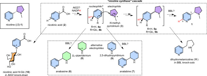
Fig. 1 Proposed biosynthesis of nicotine. Hypothesized biosynthetic route to (S)-nicotine ((S)-1), showing origins of the pyridine (blue) and N-methylpyrrolidine (purple) rings. The Mannich-like scaffold formation is proposed to proceed via a multi-step ‘nicotine synthase’ cascade16. Compounds that accumulate in Nicotiana silencing/knock-out experiments are depicted. The red hydrogens follow the fate of the hydride transferred via enzymatic reduction of 2.
Fig. 1 Proposed biosynthesis of nicotine. Hypothesized biosynthetic route to (S)-nicotine ((S)-1), showing origins of the pyridine (blue) and N-methylpyrrolidine (purple) rings. The Mannich-like scaffold formation is proposed to proceed via a multi-step ‘nicotine synthase’ cascade16. Compounds that accumulate in Nicotiana silencing/knock-out experiments are depicted. The red hydrogens follow the fate of the hydride transferred via enzymatic reduction of 2.CC BY · PubMed Central
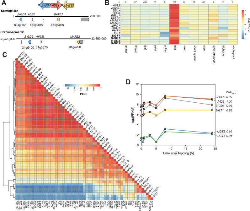
Fig. 2 Identification of biosynthetic gene clusters and gene candidates. A Homeologous biosynthetic gene clusters identified in the N. tabacum 2017 genome assembly37 consisting of β-glucosidase (β-GD, blue), reductase (A622, red) and transporter (MATE, yellow) genes. Scaffold/chromosome number is described above the linear genome strand, with numbers at either end representing nucleotide position. Scaffold 884 is unplaced in this assembly but in the Sierro et al. 2024 assembly it is part of chromosome 1675. The gene model between A622 and MATE1 in scaffold 884 (884g0040) encodes a protein that contains a domain with homology to a reverse transcriptase domain (PANTHER ID PTHR33116). B Heatmap of expression of selected nicotine genes, including β-GD and UGT candidates across tissue types. Average expression calculated in each tissue type and values normalized per gene. Number of samples in each category above each column. C Clustered heatmap showing all-by-all correlations (PCC) of nicotine biosynthesis and related genes across 824 samples. TPM values obtained from PEO40. D Root expression levels of selected nicotine biosynthesis genes and UGT candidates following topping treatment, and their correlation (PCC) with A622 responses. Data obtained from Qin et al. 202041. Details of genes available in Supplementary Data 1.
Fig. 2 Identification of biosynthetic gene clusters and gene candidates. A Homeologous biosynthetic gene clusters identified in the N. tabacum 2017 genome assembly37 consisting of β-glucosidase (β-GD, blue), reductase (A622, red) and transporter (MATE, yellow) genes. Scaffold/chromosome number is described above the linear genome strand, with numbers at either end representing nucleotide position. Scaffold 884 is unplaced in this assembly but in the Sierro et al. 2024 assembly it is part of chromosome 1675. The gene model between A622 and MATE1 in scaffold 884 (884g0040) encodes a protein that contains a domain with homology to a reverse transcriptase domain (PANTHER ID PTHR33116). B Heatmap of expression of selected nicotine genes, including β-GD and UGT candidates across tissue types. Average expression calculated in each tissue type and values normalized per gene. Number of samples in each category above each column. C Clustered heatmap showing all-by-all correlations (PCC) of nicotine biosynthesis and related genes across 824 samples. TPM values obtained from PEO40. D Root expression levels of selected nicotine biosynthesis genes and UGT candidates following topping treatment, and their correlation (PCC) with A622 responses. Data obtained from Qin et al. 202041. Details of genes available in Supplementary Data 1.CC BY · PubMed Central
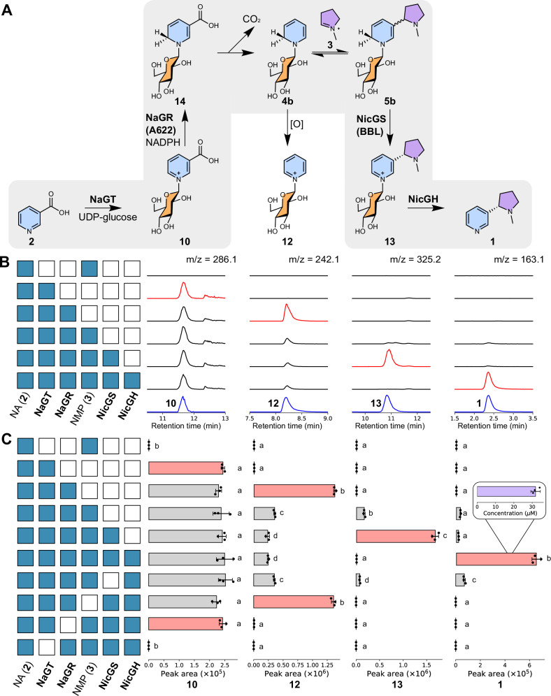
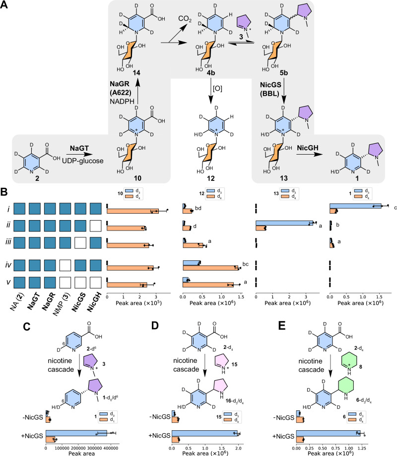
Fig. 3 In vitro reconstitution of nicotine biosynthesis. A Proposed chemical transformations in nicotine biosynthesis reconstitution. B Products of in vitro cascades. Each row corresponds to an in vitro reaction, with the matrix showing presence/absence (blue/white) of reaction components, alongside UDP-Glc and NADPH. Each column shows extracted ion chromatograms (EICs, m/z ± 0.15). Blue chromatograms are chemically verified standards, scaled for clarity. Red chromatograms highlight key products. MS2 data is available in Supplementary Fig. 3. C Quantification of in vitro cascades. Each row of bars corresponds to an in vitro reaction, with the matrix showing presence/absence (blue/white) of reaction components. Each column of bars shows the peak area of EICs (m/z ± 0.15) corresponding to the masses of the chemical depicted above (left-to-right: 10, 12, 13, 1). Compound identity was validated by standard retention time, and MS2 spectra (Supplementary Fig. 3). Red bars highlight key products. The letters above bars are significant groupings of peak area, determined per chemical (p < 0.05, Tukey post-hoc test, see Supplementary Data 5). The concentration of nicotine produced (from 1 mM of 2) was determined via an external standard curve; error bars show standard error (inset). Bars show mean EIC peak area (n = 3); points are individual replicates; error bars show standard deviation. Source data are provided as a Source Data file.
Fig. 3 In vitro reconstitution of nicotine biosynthesis. A Proposed chemical transformations in nicotine biosynthesis reconstitution. B Products of in vitro cascades. Each row corresponds to an in vitro reaction, with the matrix showing presence/absence (blue/white) of reaction components, alongside UDP-Glc and NADPH. Each column shows extracted ion chromatograms (EICs, m/z ± 0.15). Blue chromatograms are chemically verified standards, scaled for clarity. Red chromatograms highlight key products. MS2 data is available in Supplementary Fig. 3. C Quantification of in vitro cascades. Each row of bars corresponds to an in vitro reaction, with the matrix showing presence/absence (blue/white) of reaction components. Each column of bars shows the peak area of EICs (m/z ± 0.15) corresponding to the masses of the chemical depicted above (left-to-right: 10, 12, 13, 1). Compound identity was validated by standard retention time, and MS2 spectra (Supplementary Fig. 3). Red bars highlight key products. The letters above bars are significant groupings of peak area, determined per chemical (p < 0.05, Tukey post-hoc test, see Supplementary Data 5). The concentration of nicotine produced (from 1 mM of 2) was determined via an external standard curve; error bars show standard error (inset). Bars show mean EIC peak area (n = 3); points are individual replicates; error bars show standard deviation. Source data are provided as a Source Data file.CC BY · PubMed Central
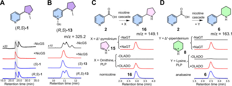
Fig. 4 Stereoselectivity and substrate scope of nicotine biosynthetic cascade. A Stereoselective formation of (S)-nicotine (1). Nicotine formation was achieved through one-pot enzymatic cascade (2, 3, NaGT, NaGR, NicGH) with and without NicGS detected by chiral HPLC (260 nm). Signal intensity of peaks and standards (blue) was scaled for comparison. B Induction of stereoselectivity by NicGS. EIC (m/z 325.2 ± 0.15) showing (R, S)-nicotine glucoside (13) formation from 2 with NaGT, NaGR and NMP, with or without NicGS. Chemically verified standards are blue. Signal intensity was scaled for comparison. C Formation of nornicotine (16) and D anabasine (6) via the nicotine biosynthetic cascade (2, NaGT, NaGR, NicGS, NicGH) with 15 or 8, respectively, either synthesized or formed enzymatically by OLADO.
Fig. 4 Stereoselectivity and substrate scope of nicotine biosynthetic cascade. A Stereoselective formation of (S)-nicotine (1). Nicotine formation was achieved through one-pot enzymatic cascade (2, 3, NaGT, NaGR, NicGH) with and without NicGS detected by chiral HPLC (260 nm). Signal intensity of peaks and standards (blue) was scaled for comparison. B Induction of stereoselectivity by NicGS. EIC (m/z 325.2 ± 0.15) showing (R, S)-nicotine glucoside (13) formation from 2 with NaGT, NaGR and NMP, with or without NicGS. Chemically verified standards are blue. Signal intensity was scaled for comparison. C Formation of nornicotine (16) and D anabasine (6) via the nicotine biosynthetic cascade (2, NaGT, NaGR, NicGS, NicGH) with 15 or 8, respectively, either synthesized or formed enzymatically by OLADO.CC BY · PubMed Central
Fig. 5 In vitro enzymatic cascade with labelled nicotinic acid. A Schematic of reaction cascade with nicotinic acid-(ring-d4) (2-d4). B Selected in vitro cascades with 2-d4 substrate. Bar charts show EIC peak areas of product (from L-to-R: 10, 12, 13, 1) isotopologues (all m/z ± 0.15 except for d3-12 which was m/z ± 0.5 due to peak shape distortion). Reactions performed in triplicate, bars show mean peak area, points show individual measurements, and error bars show standard deviation. Each row corresponds to an in vitro reaction, with the matrix showing presence/absence (blue/white) of reaction components. Letters above bars are significant groupings of d3/d4 isotopologue ratios, determined per chemical across all reaction combinations (p < 0.05, Tukey’s HSD test, Supplementary Data 6, see Supplementary Fig. 8 for all reactions). C–E Conversion of nicotinic acid (2) isotopologues by the nicotine biosynthetic cascade (NaGT, NaGR, NicGH, NADPH and UDP-Glc) with and without NicGS. Bar charts show peak areas of products from EICs. Reactions were performed in triplicate (points); bars show mean peak area; error bars show standard deviation. Source data are provided as a Source Data file.
Fig. 5 In vitro enzymatic cascade with labelled nicotinic acid. A Schematic of reaction cascade with nicotinic acid-(ring-d4) (2-d4). B Selected in vitro cascades with 2-d4 substrate. Bar charts show EIC peak areas of product (from L-to-R: 10, 12, 13, 1) isotopologues (all m/z ± 0.15 except for d3-12 which was m/z ± 0.5 due to peak shape distortion). Reactions performed in triplicate, bars show mean peak area, points show individual measurements, and error bars show standard deviation. Each row corresponds to an in vitro reaction, with the matrix showing presence/absence (blue/white) of reaction components. Letters above bars are significant groupings of d3/d4 isotopologue ratios, determined per chemical across all reaction combinations (p < 0.05, Tukey’s HSD test, Supplementary Data 6, see Supplementary Fig. 8 for all reactions). C–E Conversion of nicotinic acid (2) isotopologues by the nicotine biosynthetic cascade (NaGT, NaGR, NicGH, NADPH and UDP-Glc) with and without NicGS. Bar charts show peak areas of products from EICs. Reactions were performed in triplicate (points); bars show mean peak area; error bars show standard deviation. Source data are provided as a Source Data file.CC BY · PubMed Central
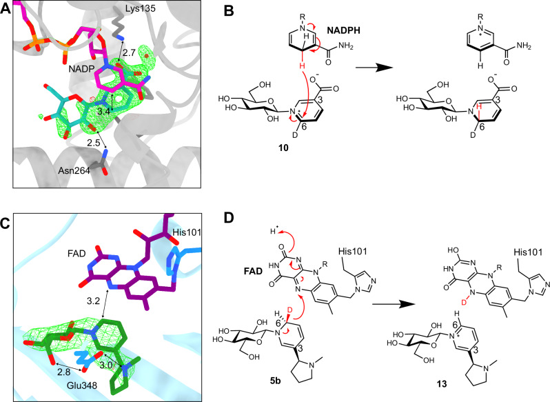
Fig. 6 X-ray crystal structures of NaGR (A622) and NicGS (BBLa). A The crystal structure of NaGR bound to NADP+ and nicotinic acid N-glucoside (green net shows unrefined ligand density Fo-Fc at 3 σ). B Proposed mechanism of NaGR: NADPH reduces nicotinic acid N-glucoside-d6 on the re-face, adding a hydride in the pro-R position. C The crystal structure of NicGS with covalently bound FAD and ligand (S)-nicotine glucoside (green net shows unrefined ligand density Fo-Fc at 3 σ). D Proposed mechanism of NicGS, abstracting the pro-S hydride from dihydronicotine glucoside-d6 to yield nicotine glucoside.
Fig. 6 X-ray crystal structures of NaGR (A622) and NicGS (BBLa). A The crystal structure of NaGR bound to NADP+ and nicotinic acid N-glucoside (green net shows unrefined ligand density Fo-Fc at 3 σ). B Proposed mechanism of NaGR: NADPH reduces nicotinic acid N-glucoside-d6 on the re-face, adding a hydride in the pro-R position. C The crystal structure of NicGS with covalently bound FAD and ligand (S)-nicotine glucoside (green net shows unrefined ligand density Fo-Fc at 3 σ). D Proposed mechanism of NicGS, abstracting the pro-S hydride from dihydronicotine glucoside-d6 to yield nicotine glucoside.CC BY · PubMed Central
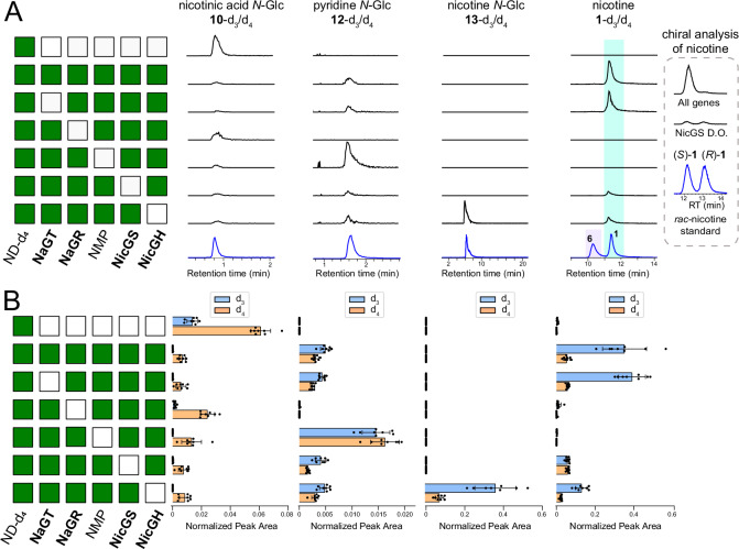
Fig. 7 In planta reconstitution of nicotine biosynthesis. Leaves of N. benthamiana were agroinfiltrated with a combination of nicotine biosynthesis genes (day 0), fed with nicotinamide-d4 (ND-d4) at day 4, and harvested for metabolite analysis at day 6. The gene combination producing the N-methylpyrrolinium (ODC, PMT and MPO) is abbreviated as NMP. A Accumulation of the labelled compounds is indicated at the top (d3 and d4 versions together). Each row corresponds to the gene combination shown in the matrix on the left (green/white indicates presence/absence). Traces are representative extracted ion chromatograms (EICs, m/z ± 0.005) corresponding to the labelled compounds (black) in comparison to chemically synthesized or commercial standards (blue) (see Supplementary Fig. 12 for MS2 spectra). Signal intensities are normalized to the largest peak on each column; standards were rescaled to match the largest peak. The nicotine and anabasine standards were analysed as a mixed sample. The insert on the right shows the chiral analysis of nicotine in the presence of all genes and in the NicGS drop-out (D.O.). B Individual quantification of d3 and d4 isotopologues corresponding to the compounds and gene combinations shown in (A). Bars show mean EIC peak area (n = 8); points are individual replicates; error bars show standard deviation (see statistics in Supplementary Data 11). Source data are provided as a Source Data file.
Fig. 7 In planta reconstitution of nicotine biosynthesis. Leaves of N. benthamiana were agroinfiltrated with a combination of nicotine biosynthesis genes (day 0), fed with nicotinamide-d4 (ND-d4) at day 4, and harvested for metabolite analysis at day 6. The gene combination producing the N-methylpyrrolinium (ODC, PMT and MPO) is abbreviated as NMP. A Accumulation of the labelled compounds is indicated at the top (d3 and d4 versions together). Each row corresponds to the gene combination shown in the matrix on the left (green/white indicates presence/absence). Traces are representative extracted ion chromatograms (EICs, m/z ± 0.005) corresponding to the labelled compounds (black) in comparison to chemically synthesized or commercial standards (blue) (see Supplementary Fig. 12 for MS2 spectra). Signal intensities are normalized to the largest peak on each column; standards were rescaled to match the largest peak. The nicotine and anabasine standards were analysed as a mixed sample. The insert on the right shows the chiral analysis of nicotine in the presence of all genes and in the NicGS drop-out (D.O.). B Individual quantification of d3 and d4 isotopologues corresponding to the compounds and gene combinations shown in (A). Bars show mean EIC peak area (n = 8); points are individual replicates; error bars show standard deviation (see statistics in Supplementary Data 11). Source data are provided as a Source Data file.CC BY · PubMed Central
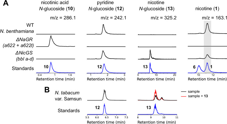
Fig. 8 Identification of pathway intermediates and shunt products in Nicotiana roots. A Metabolite analysis of roots from N. benthamiana wild type, ΔNaGR29 and ΔNicGS31 lines (black) in comparison to standards (blue) (see MS2 verification in Supplementary Fig. 13). Traces are representative extracted ion chromatograms (EICs, m/z ± 0.005) and were normalized to the largest peak in each column; standards were re-scaled to match the largest peak. B Metabolite analysis of roots from N. tabacum var Samsun (black), compared to standards (blue) (see MS2 verification in Supplementary Fig. 14). The red trace is root extract sample spiked with 10 μM nicotine N-glucoside (13) to verify peak identity. Traces are representative EICs (m/z ± 005); standards were re-scaled to match the corresponding peaks in the N. tabacum samples.
Fig. 8 Identification of pathway intermediates and shunt products in Nicotiana roots. A Metabolite analysis of roots from N. benthamiana wild type, ΔNaGR29 and ΔNicGS31 lines (black) in comparison to standards (blue) (see MS2 verification in Supplementary Fig. 13). Traces are representative extracted ion chromatograms (EICs, m/z ± 0.005) and were normalized to the largest peak in each column; standards were re-scaled to match the largest peak. B Metabolite analysis of roots from N. tabacum var Samsun (black), compared to standards (blue) (see MS2 verification in Supplementary Fig. 14). The red trace is root extract sample spiked with 10 μM nicotine N-glucoside (13) to verify peak identity. Traces are representative EICs (m/z ± 005); standards were re-scaled to match the corresponding peaks in the N. tabacum samples.CC BY · PubMed Central
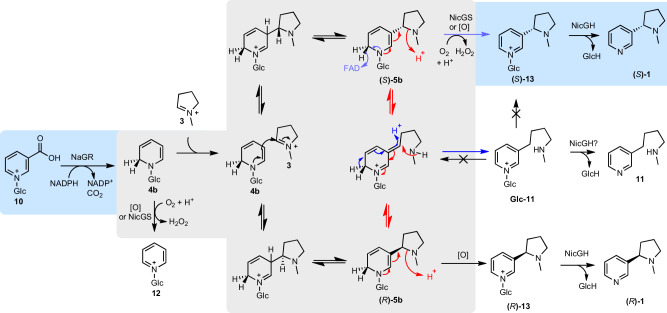
Fig. 9 Putative activities of NicGS (BBL). Possible mechanistic routes towards (S)−1 from 10. The blue background shows key NaGR and NicGH reactions and major products. The grey background shows possible reactions that are either non-enzymatically or catalysed by NicGS. Only compounds with blue or white background have been directly detected. To induce the stereoselective outcome, NicGS may operate through: dynamic kinetic resolution (catalysing just purple arrows and allowing other intermediates to non-enzymatically equilibrate), epimerisation of 5b via a ring opened intermediate (catalysing red and purple arrows), catalysis of the stereoselective Mannich reaction between 3 and 10 prior to oxidation, reversal of the non-desired Mannich reaction (opening (R)-5b into 3 and 10). Blue arrows show the predicted mechanism of Glc−11 formation.
Fig. 9 Putative activities of NicGS (BBL). Possible mechanistic routes towards (S)−1 from 10. The blue background shows key NaGR and NicGH reactions and major products. The grey background shows possible reactions that are either non-enzymatically or catalysed by NicGS. Only compounds with blue or white background have been directly detected. To induce the stereoselective outcome, NicGS may operate through: dynamic kinetic resolution (catalysing just purple arrows and allowing other intermediates to non-enzymatically equilibrate), epimerisation of 5b via a ring opened intermediate (catalysing red and purple arrows), catalysis of the stereoselective Mannich reaction between 3 and 10 prior to oxidation, reversal of the non-desired Mannich reaction (opening (R)-5b into 3 and 10). Blue arrows show the predicted mechanism of Glc−11 formation.CC BY · PubMed Central

Verification

Claims the paper makes

The claims and statistics of this paper haven’t been checked yet.

Run the check — free, no account needed. It reads the open-access text and checks the sources it cites.

A few free checks a day · papers already verified open instantly · results are public

Self-check

Percentages, sums and reported differences re-computed from the paper’s own numbers.

The self-check needs the paper’s full text.

Citations

Cites

  • Highly accurate protein structure prediction with AlphaFold

    Jumper, J., Evans, R., Pritzel, A., Green, T., Figurnov, M., Ronneberger, O., Tunyasuvunakool, K., Bates, R., Žídek, A., Potapenko, A., et al. (2021) · Nature · doi:10.1038/s41586-021-03819-2 · Cited by 43,764 · cited 1×

    “The structures were solved by molecular replacement using MOLREP 68 with the AlphaFold structures AF-P52579-F1-v4andAF-F1T160-F1-v4usedasthemodelsforNaGR and NicGS, respectively (https://alphafold.ebi.ac.uk/) 69 .”

Research works printed but not linked: 74

  1. Duke, D. et al. Earliest evidence for human use of tobacco in the Pleistocene Americas. Nat. Hum. Behav. 6, 183–192 (2022). · DOI
  2. Wipfli, H. & Samet, J. M. One hundred years in the making: The global tobacco epidemic. Annu. Rev. Public Health 37, 149–166 (2016). · DOI
  3. Langley, J. N. On the reaction of cells and of nerve-endings to certain poisons, chiefly as regards the reaction of striated muscle to nicotine and to curari. J. Physiol. 33, 374–413 (1905). · DOI
  4. Baldwin, I. T. An ecologically motivated analysis of plant-herbivore interactions in native tobacco. Plant Physiol. 127, 1449–1458 (2001). · DOI
  5. Millar, N. S. & Denholm, I. Nicotinic acetylcholine receptors: targets for commercially important insecticides. Invert. Neurosci. 7, 53–66 (2007). · DOI
  6. Kessler, D. et al. Unpredictability of nectar nicotine promotes outcrossing by hummingbirds in Nicotiana attenuata. Plant J. 71, 529–538 (2012). · DOI
  7. Shitan, N., Hayashida, M. & Yazaki, K. Translocation and accumulation of nicotine via distinct spatio-temporal regulation of nicotine transporters in Nicotiana tabacum. Plant Signal Behav. 10, e1035852 (2015). · DOI
  8. Shoji, T., Kajikawa, M. & Hashimoto, T. Clustered transcription factor genes regulate nicotine biosynthesis in tobacco. Plant Cell 22, 3390–3409 (2010). · DOI
  9. Elser, D. et al. Evolutionary metabolomics of specialized metabolism diversification in the genus Nicotiana highlights N-acylnornicotine innovations. Sci. Adv. 9, eade8984 (2023). · DOI
  10. Benvenuto, E. et al. Plant molecular farming in the wake of the closure of Medicago Inc. Nat. Biotechnol. 41, 893–894 (2023). · DOI
  11. Golubova, D., Tansley, C., Su, H. & Patron, N. J. Engineering Nicotiana benthamiana as a platform for natural product biosynthesis. Curr. Opin. Plant Biol. 81, 102611 (2024). · DOI
  12. Lichman, B. R. The scaffold-forming steps of plant alkaloid biosynthesis. Nat. Prod. Rep. 38, 103–129 (2021). · DOI
  13. Dawson, R. F. et al. Biosynthesis of the pyridine ring of nicotine. J. Am. Chem. Soc. 78, 2645–2646 (1956). · DOI
  14. Leete, E. Biosynthesis of the Nicotiana alkaloids. XI. Investigation of tautomerism in N-methyl-delta-pyrrolinium chloride and its incorporation into nicotine. J. Am. Chem. Soc. 89, 7081–7084 (1967). · DOI
  15. Dawson, R. F. Biosynthesis of the Nicotiana alkaloids. Am. Sci. 48, 321–340 (1960).
  16. Brent Friesen, J. & Leete, E. Nicotine synthase—an enzyme from Nicotiana species which catalyzes the formation of (S)-nicotine from nicotinic acid and 1-methyl-δ’pyrrolinium chloride. Tetrahedron Lett. 31, 6295–6298 (1990). · DOI
  17. Yang, K. S., Gholson, R. K. & Waller, G. R. Studies on nicotine biosynthesis. J. Am. Chem. Soc. 87, 4184–4188 (1965). · DOI
  18. Scott, T. A. & Glynn, J. P. The incorporation of [2,3,7-14C] nicotinic acid into nicotine by Nicotiana tabacum. Phytochemistry 6, 505–510 (1967). · DOI
  19. Leete, E. The incorporation of [5,6-13C2]Nicotinic acid into the tobacco alkaloids examined by the use of 13C nuclear magnetic resonance. Bioorg. Chem. 6, 273–286 (1977). · DOI
  20. Dawson, R. F., Christman, D. R., D’Adamo, A., Solt, M. L. & Wolf, A. P. The biosynthesis of nicotine from isotopically labeled nicotinic acids. J. Am. Chem. Soc. 82, 2628–2633 (1960). · DOI
  21. Leete, E. & Liu, Y.-Y. Metabolism of [2-3H]- and [6-3H]-nicotinic acid in intact Nicotiana tabacum plants. Phytochemistry 12, 593–596 (1973). · DOI
  22. Leete, E. Stereochemistry of the reduction of nicotinic acid when it serves as a precursor of anatabine. J. Chem. Soc. Chem. Commun. 610 (1978) https://doi.org/10.1039/c39780000610. · DOI
  23. Cai, B., Jack, A. M., Lewis, R. S., Dewey, R. E. & Bush, L. P. (R)-nicotine biosynthesis, metabolism and translocation in tobacco as determined by nicotine demethylase mutants. Phytochemistry 95, 188–196 (2013). · DOI
  24. Leete, E. Biosynthesis of anatabine and anabasine in Nicotiana glutinosa. J. Chem. Soc. Chem. Commun. 9–10. https://doi.org/10.1039/C39750000009 (1975). · DOI
  25. Hibi, N., Higashiguchi, S., Hashimoto, T. & Yamada, Y. Gene expression in tobacco low-nicotine mutants. Plant Cell 6, 723–735 (1994).
  26. Kajikawa, M., Hirai, N. & Hashimoto, T. A PIP-family protein is required for biosynthesis of tobacco alkaloids. Plant Mol. Biol. 69, 287–298 (2009). · DOI
  27. Deboer, K. D., Lye, J. C., Aitken, C. D., Su, A. K.-K. & Hamill, J. D. The A622 gene in Nicotiana glauca (tree tobacco): evidence for a functional role in pyridine alkaloid synthesis. Plant Mol. Biol. 69, 299–312 (2009). · DOI
  28. Burner, N., Kernodle, S. P., Steede, T. & Lewis, R. S. Editing of A622 genes results in ultra-low nicotine whole tobacco plants at the expense of dramatically reduced growth and development. Mol. Breed. 42, 20 (2022). · DOI
  29. Negin, B., Wang, F., Fischer, H. D. & Jander, G. Acylsugars, nicotine and a protease inhibitor provide variable protection for Nicotiana benthamiana in a natural setting. Plant Cell Environ. 48, 1073–1087 (2025). · DOI
  30. Kajikawa, M., Shoji, T., Kato, A. & Hashimoto, T. Vacuole-localized berberine bridge enzyme-like proteins are required for a late step of nicotine biosynthesis in tobacco. Plant Physiol. 155, 2010–2022 (2011). · DOI
  31. Vollheyde, K. et al. An improved Nicotiana benthamiana bioproduction chassis provides novel insights into nicotine biosynthesis. N. Phytol. 240, 302–317 (2023). · DOI
  32. Shoji, T. et al. Expression patterns of two tobacco isoflavone reductase-like genes and their possible roles in secondary metabolism in tobacco. Plant Mol. Biol. 50, 427–440 (2002). · DOI
  33. Daniel, B. et al. The family of berberine bridge enzyme-like enzymes: A treasure-trove of oxidative reactions. Arch. Biochem Biophys. 632, 88–103 (2017). · DOI
  34. Allen, Z. et al. BBL enzymes exhibit enantiospecific preferences in the biosynthesis of pyridine alkaloids in Nicotiana tabacum L. Phytochemistry 232, 114363 (2025). · DOI
  35. Posselt, W. & Reimann, L. Chemische Untersuchung des Tabaks und Darstellung eines eigenthümlich wirksamen Prinzips dieser Pflanze. Mag. f.ür. die neuesten Erfahrungen, Entdeckungen und Berichtigungen im. Geb. der Pharm. 6, 138–161 (1828).
  36. Smit, S. J. & Lichman, B. R. Plant biosynthetic gene clusters in the context of metabolic evolution. Nat. Prod. Rep. 39, 1465–1482 (2022). · DOI
  37. Edwards, K. D. et al. A reference genome for Nicotiana tabacum enables map-based cloning of homeologous loci implicated in nitrogen utilization efficiency. BMC Genomics 18, 448 (2017). · DOI
  38. Shoji, T. et al. Multidrug and toxic compound extrusion-type transporters implicated in vacuolar sequestration of nicotine in tobacco roots. Plant Physiol. 149, 708–718 (2009). · DOI
  39. Kajikawa, M. et al. Genomic insights into the evolution of the nicotine biosynthesis pathway in tobacco. Plant Physiol. 174, 999–1011 (2017). · DOI
  40. Koh, E., Goh, W., Julca, I., Villanueva, E. & Mutwil, M. PEO: Plant Expression Omnibus - a comparative transcriptomic database for 103 Archaeplastida. Plant J. 117, 1592–1603 (2024). · DOI
  41. Qin, Y. et al. Transcriptome analysis reveals key genes involved in the regulation of nicotine biosynthesis at early time points after topping in tobacco (Nicotiana tabacum L.). BMC Plant Biol. 20, 30 (2020). · DOI
  42. Wood, C. X. et al. Parallel evolution of plant alkaloid biosynthesis from bacterial-like decarboxylases. N. Phytol. 249, 2954–2973 (2026). · DOI
  43. Wang, X. et al. Crystal structure of isoflavone reductase from alfalfa (Medicago sativa L.). J. Mol. Biol. 358, 1341–1352 (2006). · DOI
  44. Winkler, A. et al. A concerted mechanism for berberine bridge enzyme. Nat. Chem. Biol. 4, 739–741 (2008). · DOI
  45. Carlson, E. D., Rajniak, J. & Sattely, E. S. Multiplicity of the Agrobacterium Infection of Nicotiana benthamiana for Transient DNA Delivery. ACS Synth. Biol. 12, 2329–2338 (2023). · DOI
  46. Dawson, R. F. The localization of the nicotine synthetic mechanism in the tobacco plant. Science 94, 396–397 (1941). · DOI
  47. Xu, B., Sheehan, M. J. & Timko, M. P. Differential induction of ornithine decarboxylase (ODC) gene family members in transgenic tobacco (Nicotiana tabacum L. cv. Bright Yellow 2) cell suspensions by methyl-jasmonate treatment. Plant Growth Regul. 44, 101–116 (2004). · DOI
  48. Junker, A., Fischer, J., Sichhart, Y., Brandt, W. & Dräger, B. Evolution of the key alkaloid enzyme putrescine N-methyltransferase from spermidine synthase. Front Plant Sci. 4, 260 (2013). · DOI
  49. Naconsie, M., Kato, K., Shoji, T. & Hashimoto, T. Molecular evolution of N-methylputrescine oxidase in tobacco. Plant Cell Physiol. 55, 436–444 (2014). · DOI
  50. Jeong, J.-H. et al. Impact of CRISPR/Cas9-induced mutations in nicotine biosynthesis core genes A622 and BBL on tobacco: reduction in nicotine content and developmental abnormalities. Curr. Plant Biol. 38, 100343 (2024). · DOI
  51. Liu, J. et al. Systematic genomic characterization of β-glucosidase genes in tobacco reveals NtBGLU50 as a multifunctional regulator of glycoside metabolism and defense-associated pathways. Ind. Crops Prod. 240, 122643 (2026). · DOI
  52. Osbourn, A. E. Preformed antimicrobial compounds and plant defense against fungal attack. Plant Cell 8, 1821 (1996). · DOI
  53. von Rad, U., Hüttl, R., Lottspeich, F., Gierl, A. & Frey, M. Two glucosyltransferases are involved in detoxification of benzoxazinoids in maize. Plant J. 28, 633–642 (2001). · DOI
  54. Cicek, M. & Esen, A. Structure and expression of a dhurrinase (beta-glucosidase) from sorghum. Plant Physiol. 116, 1469–1478 (1998). · DOI
  55. Hou, B. ingkai, Lim Gillian, E. ng-K. iat, Higgins Dianna, S. & Bowles, J. N-Glucosylation of cytokinins by glycosyltransferases of Arabidopsis thaliana. J. Biol. Chem. 279, 27822–47832 (2004). · DOI
  56. Bontpart, T., Cheynier, V., Ageorges, A. & Terrier, N. BAHD or SCPL acyltransferase? What a dilemma for acylation in the world of plant phenolic compounds. N. Phytol. 208, 695–707 (2015). · DOI
  57. Regenbrecht, J. & Strack, D. Distribution of 1-sinapoylglucose: choline sinapoyltransferase activity in the Brassicaceae. Phytochemistry 24, 407–410 (1985). · DOI
  58. Michalczuk, L. & Bandurski, R. S. UDP-glucose: indoleacetic acid glucosyl transferase and indoleacetyl-glucose: myo-inositol indoleacetyl transferase. Biochem. Biophys. Res. Commun. 93, 588–592 (1980). · DOI
  59. Qiu, F. et al. Functional genomics analysis reveals two novel genes required for littorine biosynthesis. N. Phytol. 225, 1906–1914 (2020). · DOI
  60. Jozwiak, A. et al. A cellulose synthase-like protein governs the biosynthesis of Solanum alkaloids. Science 386, eadq5721 (2024). · DOI
  61. Boccia, M. et al. A scaffold protein manages the biosynthesis of steroidal defense metabolites in plants. Science 386, 1366–1372 (2024). · DOI
  62. Zheng, X. et al. Biosynthesis of the pyrrolidine protein synthesis inhibitor anisomycin involves a novel gene ensemble and cryptic biosynthetic steps. Proc. Natl. Acad. Sci. USA 114, 4135–4140 (2017). · DOI
  63. Chang, L. et al. Complete biosynthesis of nicotine. Cell. https://doi.org/10.1016/j.cell.2026.03.034 (2026). · DOI
  64. Fernandez-Pozo, N. et al. The Sol Genomics Network (SGN)-from genotype to phenotype to breeding. Nucleic Acids Res. 43, D1036–41 (2015). · DOI
  65. Kabsch, W. XDS. Acta Crystallogr D. Biol. Crystallogr. 66, 125–132 (2010). · DOI · Paper page
  66. Evans, P. Scaling and assessment of data quality. Acta Crystallogr D. Biol. Crystallogr 62, 72–82 (2006). · DOI
  67. Winter, G. Xia2: An expert system for macromolecular crystallography data reduction. J. Appl. Crystallogr. 43, 186–190 (2010). · DOI
  68. Vagin, A. & Teplyakov, A. Molecular replacement with MOLREP. Acta Crystallogr D. Biol. Crystallogr. 66, 22–25 (2010). · DOI
  69. Emsley, P. & Cowtan, K. Coot: model-building tools for molecular graphics. Acta Crystallogr D. Biol. Crystallogr. 60, 2126–2132 (2004). · DOI · Paper page
  70. Murshudov, G. N., Vagin, A. A. & Dodson, E. J. Refinement of macromolecular structures by the maximum-likelihood method. Acta Crystallogr D. Biol. Crystallogr. 53, 240–255 (1997). · DOI
  71. Peyret, H., Brown, J. K. M. & Lomonossoff, G. P. Improving plant transient expression through the rational design of synthetic 5’ and 3’ untranslated regions. Plant Methods 15, 108 (2019). · DOI
  72. Chuang, L. & Franke, J. Rapid combinatorial coexpression of biosynthetic genes by transient expression in the plant host Nicotiana benthamiana. Methods Mol. Biol. 2489, 395–420 (2022). · DOI
  73. Florentine, S. K. et al. Influence of selected environmental factors on seed germination and seedling survival of the arid zone invasive species tobacco bush (Nicotiana glauca R. Graham). Rangel. J. 38, 417 (2016). · DOI
  74. Sierro, N., Auberson, M., Dulize, R. & Ivanov, N. V. Chromosome-level genome assemblies of Nicotiana tabacum, Nicotiana sylvestris, and Nicotiana tomentosiformis. Sci. Data 11, 135 (2024). · DOI

Metadata

Authors
Schwabe, B. T. W., Angstman, I. M., Vollheyde, K., Ingold, Z., Li, J., Stankevich, K. S., Spicer, C. D., Fascione, M. A., Grogan, G., Geu-Flores, F., & Lichman, B. R.
Affiliations
  • University of York (GB)
  • University of Copenhagen (DK)
Corresponding author
Lichman, Benjamin R.
Venue
Nature Communications
Published
2026-05-18
Volume
17
Issue
1
ISSN
2041-1723 (ISSN-L)
OpenAlex
W7161541149
Cited by
12
References
1
Citations by year
2026: 12
FWCI
24.63
Type
journal article
Language
en
Open access
Locations
  • Nature Communications (publishedVersion, cc-by)
  • bioRxiv (Cold Spring Harbor Laboratory) (acceptedVersion, cc-by-nc-nd)
  • PubMed (publishedVersion)
  • PubMed Central (publishedVersion, cc-by)
  • Research at the University of Copenhagen (University of Copenhagen) (publishedVersion, cc-by)
  • White Rose Research Online (University of Leeds, The University of Sheffield, University of York) (acceptedVersion)
  • DOAJ (DOAJ: Directory of Open Access Journals) (submittedVersion, cc-by-sa)
Funders
  • Research Councils UK (MR/S01862X/1, MR/X010260/1)
  • RCUK | Biotechnology and Biological Sciences Research Council (BB/T007222/1)
  • Det Frie Forskningsråd (5281-00267B)
Topics
  • Life Sciences › Biochemistry, Genetics and Molecular Biology › Molecular Biology › Plant biochemistry and biosynthesis
  • Life Sciences › Agricultural and Biological Sciences › Plant Science › Plant tissue culture and regeneration
  • Life Sciences › Biochemistry, Genetics and Molecular Biology › Molecular Biology › Plant Gene Expression Analysis
Keywords
nicotine biosynthesis, tobacco, alkaloid biosynthesis, nicotinic acid, C–C bond formation, pyridine alkaloids, X-ray crystal structure
MeSH terms
Alkaloids, Glucose, Glycosylation, Nicotine, Plant Proteins, Nicotiana, Crystallography, X-Ray

About these checks

Registry, retraction, Cites and statistics results are computed, not judged, with one exception: a printed reference that no index lists is read once by a model before it counts as a citation error. Claims are extracted by a model and kept only when their quote appears verbatim in the text; each cited source is then checked, with a model’s help, for whether it says what the claim relies on. Those verdicts can be wrong — read the claim’s quote and the cited source before relying on one.

Statistics and arithmetic are recomputed, not judged.

Confidence bands are shown only for fields calibrated on our annotated test set. None is calibrated yet, so no band is shown.

What counts as a claim ›How we measure confidence ›How we check this ›

Are you an author, or do you see a problem with this page? Request a review