

The DOI matches its Crossref record; 3 of the 3 references it cites were found in a registry or an index.
Computational design of highly signalling-active membrane receptors through solvent-mediated allosteric networks
Open accessPublished version of a preprint
Abstract
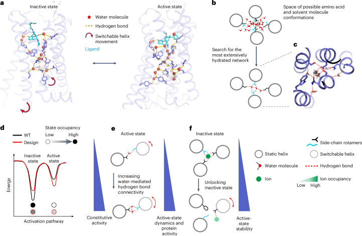
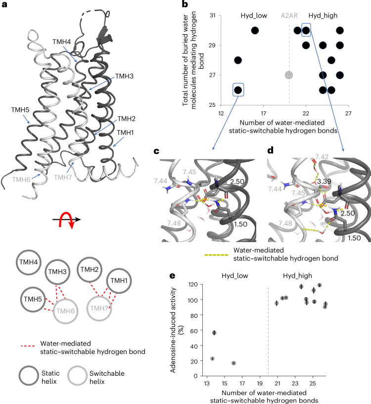
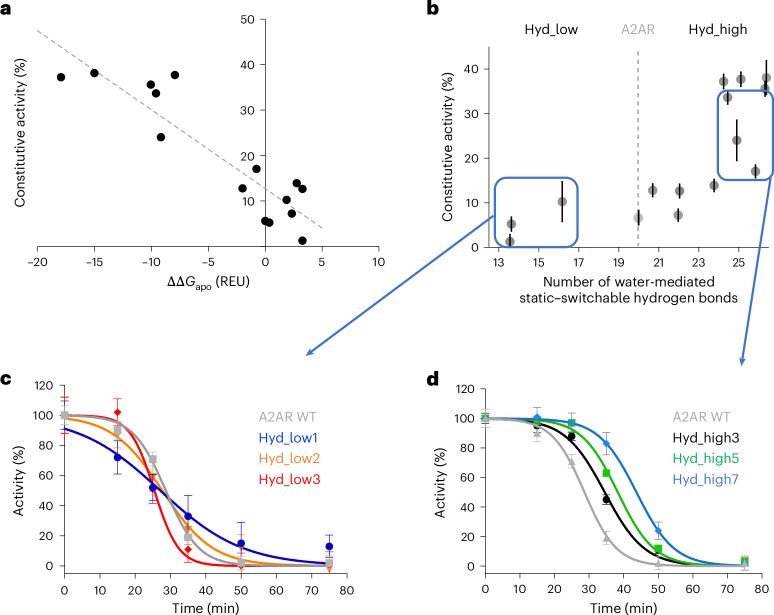
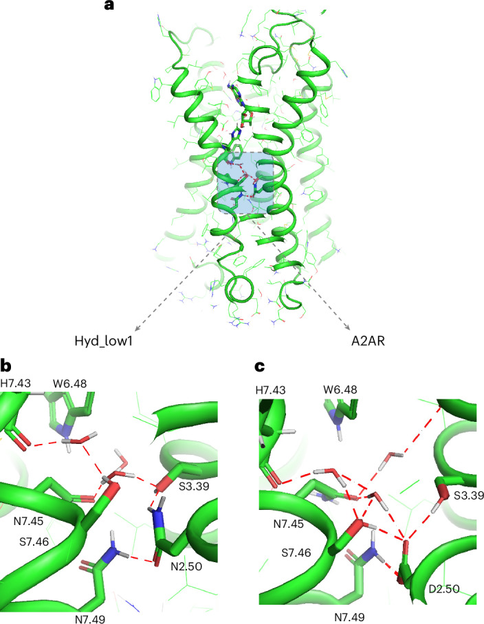
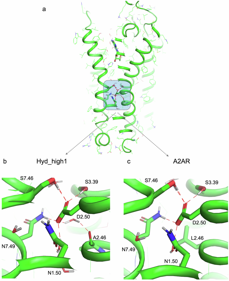
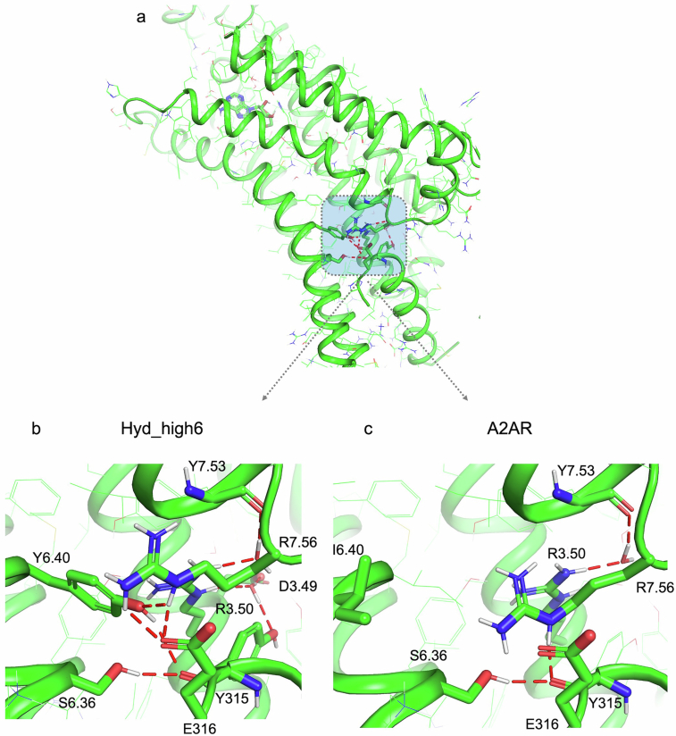
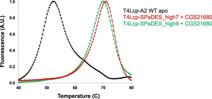
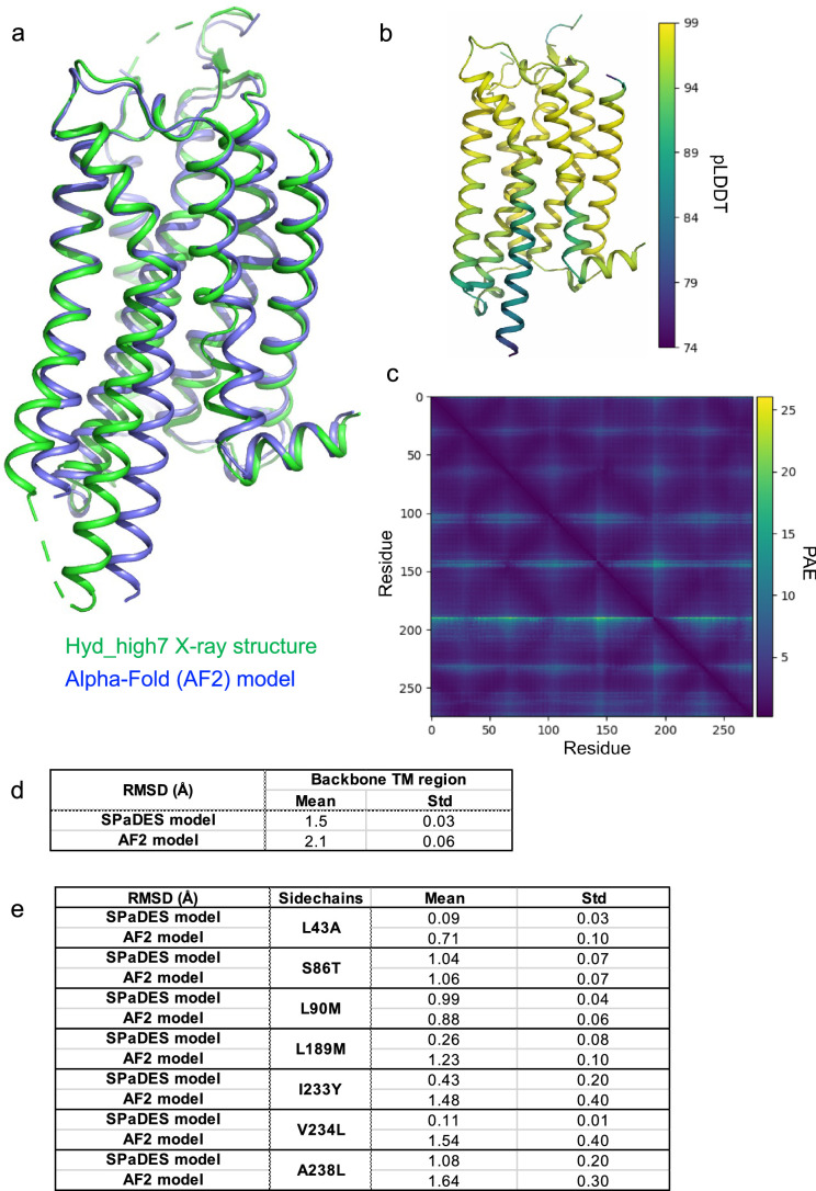
Protein catalysis and allostery require the atomic-level orchestration and motion of residues and ligand, solvent and protein effector molecules. However, the ability to design protein activity through precise protein–solvent cooperative interactions has not yet been demonstrated. Here we report the design of 14 membrane receptors that catalyse G protein nucleotide exchange through diverse engineered allosteric pathways mediated by cooperative networks of intraprotein, protein–ligand and –solvent molecule interactions. Consistent with predictions, the designed protein activities correlated well with the level of plasticity of the networks at flexible transmembrane helical interfaces. Several designs displayed considerably enhanced thermostability and activity compared with related natural receptors. The most stable and active variant crystallized in an unforeseen signalling-active conformation, in excellent agreement with the design models. The allosteric network topologies of the best designs bear limited similarity to those of natural receptors and reveal an allosteric interaction space larger than previously inferred from natural proteins. The approach should prove useful for engineering proteins with novel complex protein binding, catalytic and signalling activities.
Overview
The claims and statistics of this paper haven’t been checked yet.
Run the check — free, no account needed. It reads the open-access text and checks the sources it cites.
Integrity signals
Registry
Cites
Statistics
Figures
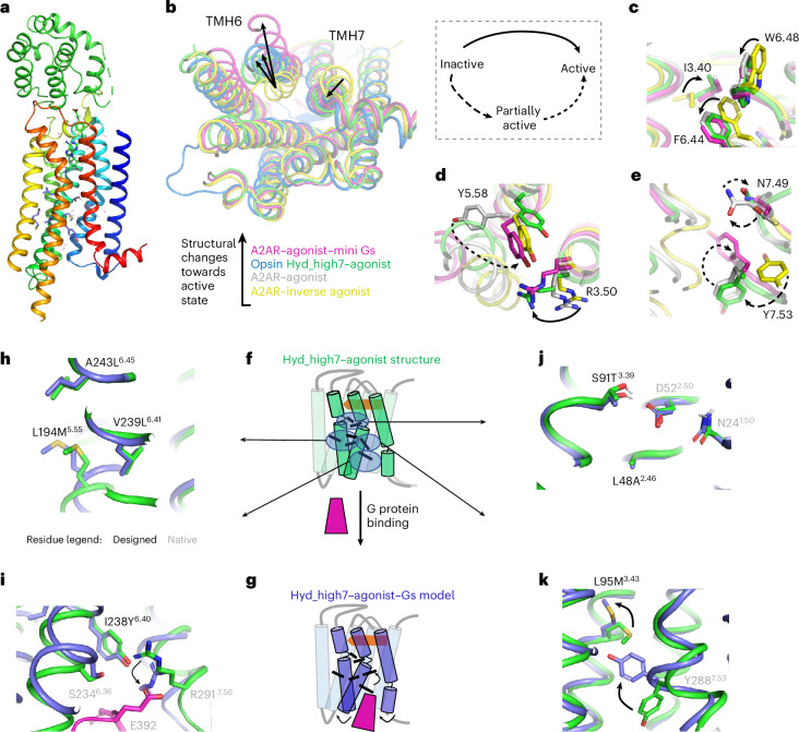
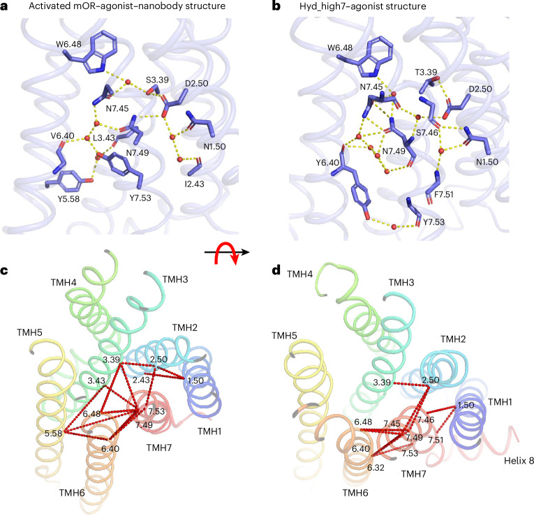
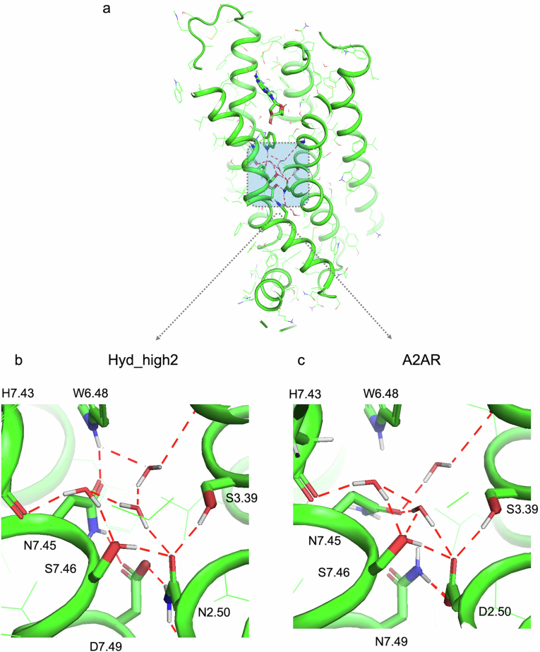
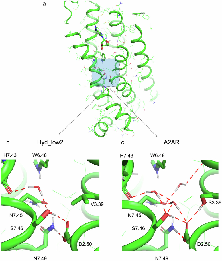
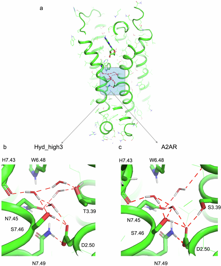
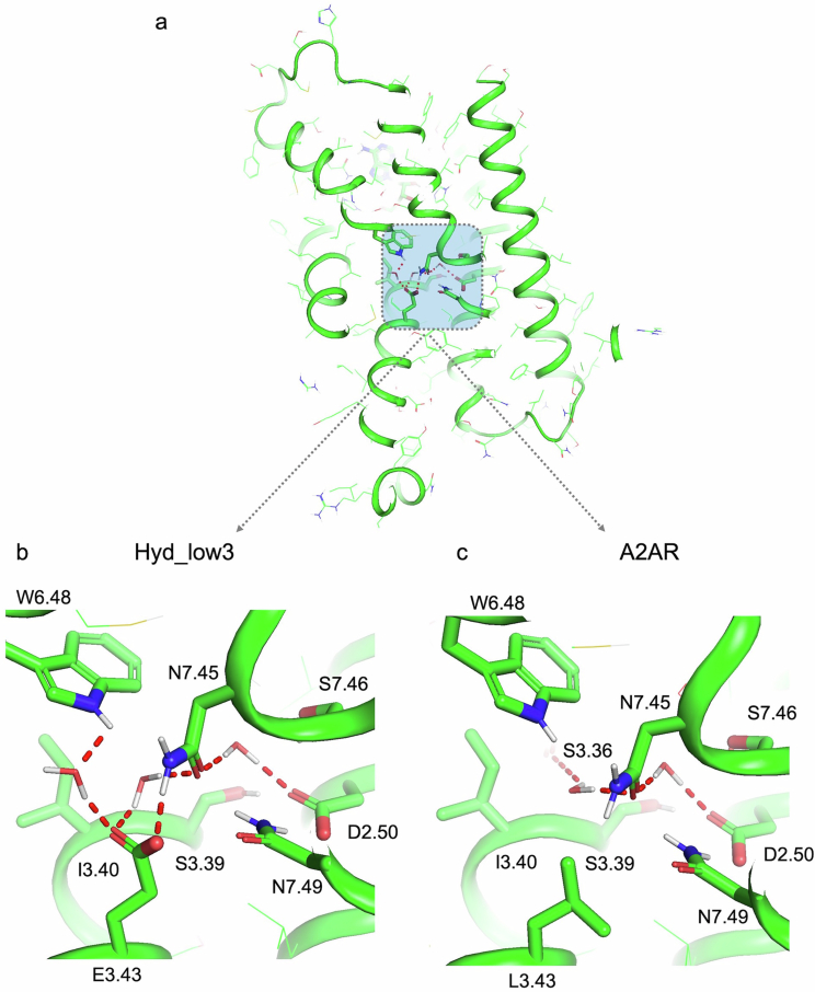
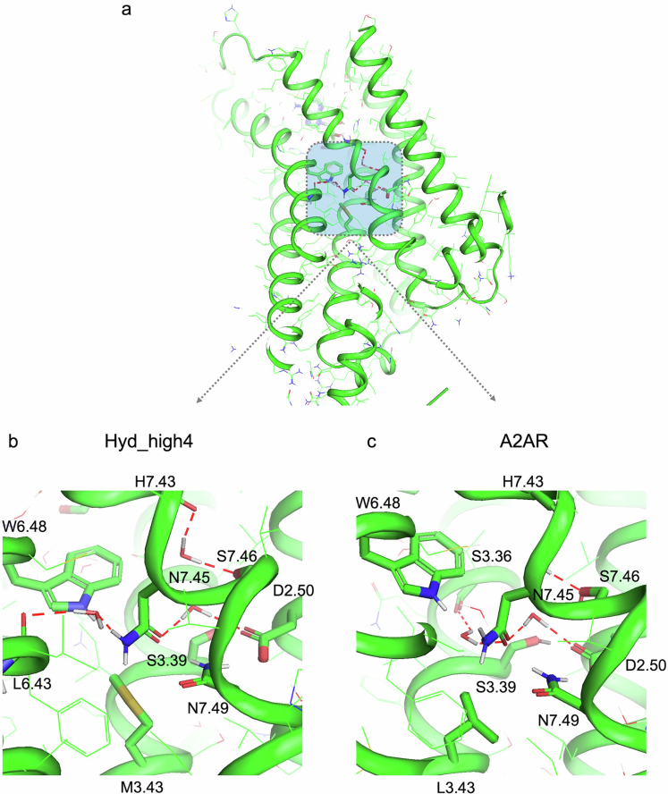
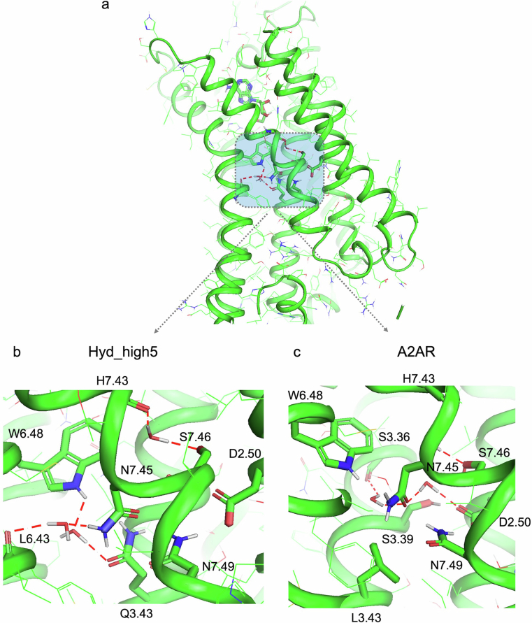
Verification
Claims the paper makes
The claims and statistics of this paper haven’t been checked yet.
Run the check — free, no account needed. It reads the open-access text and checks the sources it cites.
Self-check
Percentages, sums and reported differences re-computed from the paper’s own numbers.
The self-check needs the paper’s full text.
Citations
Cites
Highly accurate protein structure prediction with AlphaFold
Jumper, J., Evans, R., Pritzel, A., Green, T., Figurnov, M., Ronneberger, O., Tunyasuvunakool, K., Bates, R., Žídek, A., Potapenko, A., et al. (2021) · Nature · doi:10.1038/s41586-021-03819-2 · Cited by 43,764
Crystal structure of opsin in its G-protein-interacting conformation
Scheerer, P., Park, J. H., Hildebrand, P. W., Kim, Y. J., Krauß, N., Choe, H.-W., Hofmann, K. P., & Ernst, O. P. (2008) · Nature · doi:10.1038/nature07330 · Cited by 1,059
Structural waters define a functional channel mediating activation of the GPCR, rhodopsin
Angel, T. E., Gupta, S., Jastrzebska, B., Palczewski, K., & Chance, M. R. (2009) · Proceedings of the National Academy of Sciences · doi:10.1073/pnas.0901074106 · Cited by 203
Research works printed but not linked: 50
- Changeux, J. P. & Christopoulos, A. Allosteric modulation as a unifying mechanism for receptor function and regulation. Cell 166, 1084–1102 (2016). · DOI
- Yuan, S., Hu, Z., Filipek, S. & Vogel, H. W2466.48 opens a gate for a continuous intrinsic water pathway during activation of the adenosine A2A receptor. Angew. Chem. Int. Ed. 54, 556–559 (2015). · DOI
- Yuan, S., Filipek, S., Palczewski, K. & Vogel, H. Activation of G-protein-coupled receptors correlates with the formation of a continuous internal water pathway. Nat. Commun. 5, 4733 (2014). · DOI
- Setny, P. & Wiśniewska, M. D. Water-mediated conformational preselection mechanism in substrate binding cooperativity to protein kinase A. Proc. Natl Acad. Sci. USA 115, 3852–3857 (2018). · DOI
- Ye, L. et al. Mechanistic insights into allosteric regulation of the A2A adenosine G protein-coupled receptor by physiological cations. Nat. Commun. 9, 1372 (2018). · DOI
- Lu, P. et al. Accurate computational design of multipass transmembrane proteins. Science 359, 1042–1046 (2018). · DOI
- Boyken, S. E. et al. De novo design of protein homo-oligomers with modular hydrogen-bond network-mediated specificity. Science 352, 680–687 (2016). · DOI
- Jacobs, T. M. et al. Design of structurally distinct proteins using strategies inspired by evolution. Science 352, 687–690 (2016). · DOI
- Dou, J. et al. De novo design of a fluorescence-activating β-barrel. Nature 561, 485–491 (2018). · DOI · Paper page
- Angel, T. E., Chance, M. R. & Palczewski, K. Conserved waters mediate structural and functional activation of family A (rhodopsin-like) G protein-coupled receptors. Proc. Natl Acad. Sci. USA 106, 8555–8560 (2009). · DOI · Paper page
- Venkatakrishnan, A. J. et al. Diverse GPCRs exhibit conserved water networks for stabilization and activation. Proc. Natl Acad. Sci. USA 116, 3288–3293 (2019). · DOI
- Zarzycka, B., Zaidi, S. A., Roth, B. L. & Katritch, V. Harnessing ion-binding sites for GPCR pharmacology. Pharmacol. Rev. 71, 571–595 (2019). · DOI
- Feng, X., Ambia, J., Chen, K.-Y. M., Young, M. & Barth, P. Computational design of ligand-binding membrane receptors with high selectivity. Nat. Chem. Biol. 13, 715–723 (2017). · DOI
- Lai, J. K., Ambia, J., Wang, Y. & Barth, P. Enhancing structure prediction and design of soluble and membrane proteins with explicit solvent–protein interactions. Structure 25, 1758–1770.e8 (2017). · DOI
- Hauser, A. S. et al. GPCR activation mechanisms across classes and macro/microscales. Nat. Struct. Mol. Biol. 28, 879–888 (2021). · DOI
- Chen, K. M., Keri, D. & Barth, P. Computational design of G protein-coupled receptor allosteric signal transductions. Nat. Chem. Biol. 16, 77–86 (2020). · DOI
- Rasmussen, S. G. et al. Crystal structure of the β2 adrenergic receptor–Gs protein complex. Nature 477, 549–555 (2011). · DOI · Paper page
- Vickery, O. N. et al. Intracellular transfer of Na+ in an active-state G-protein-coupled receptor. Structure 26, 171–180.e2 (2018). · DOI
- Magnani, F., Shibata, Y., Serrano-Vega, M. J. & Tate, C. G. Co-evolving stability and conformational homogeneity of the human adenosine A2a receptor. Proc. Natl Acad. Sci. USA 105, 10744–10749 (2008). · DOI
- Carpenter, B., Nehme, R., Warne, T., Leslie, A. G. & Tate, C. G. Structure of the adenosine A2A receptor bound to an engineered G protein. Nature 536, 104–107 (2016). · DOI
- Ahuja, S. & Smith, S. O. Multiple switches in G protein-coupled receptor activation. Trends Pharmacol. Sci. 30, 494–502 (2009). · DOI
- Nygaard, R., Frimurer, T. M., Holst, B., Rosenkilde, M. M. & Schwartz, T. W. Ligand binding and micro-switches in 7TM receptor structures. Trends Pharmacol. Sci. 30, 249–259 (2009). · DOI
- Lebon, G. et al. Agonist-bound adenosine A2A receptor structures reveal common features of GPCR activation. Nature 474, 521–525 (2011). · DOI
- Huang, W. et al. Structural insights into micro-opioid receptor activation. Nature 524, 315–321 (2015). · DOI
- Venkatakrishnan, A. J. et al. Molecular signatures of G-protein-coupled receptors. Nature 494, 185–194 (2013). · DOI
- White, K. L. et al. Structural connection between activation microswitch and allosteric sodium site in GPCR signaling. Structure 26, 259–269.e5 (2018). · DOI
- Massink, A. et al. Sodium ion binding pocket mutations and adenosine A2A receptor function. Mol. Pharmacol. 87, 305–313 (2015). · DOI
- Nygaard, R., Valentin-Hansen, L., Mokrosinski, J., Frimurer, T. M. & Schwartz, T. W. Conserved water-mediated hydrogen bond network between TM-I, -II, -VI, and -VII in 7TM receptor activation. J. Biol. Chem. 285, 19625–19636 (2010). · DOI
- Mravic, M. et al. Packing of apolar side chains enables accurate design of highly stable membrane proteins. Science 363, 1418–1423 (2019). · DOI
- Joh, N. H. et al. De novo design of a transmembrane Zn2+-transporting four-helix bundle. Science 346, 1520–1524 (2014). · DOI
- Yin, J. et al. Structure of a D2 dopamine receptor–G-protein complex in a lipid membrane. Nature 584, 125–129 (2020). · DOI
- Ingwersen, J. et al. Dual roles of the adenosine A2a receptor in autoimmune neuroinflammation. J. Neuroinflammation 13, 48 (2016). · DOI
- Graber, S. G., Figler, R. A. & Garrison, J. C. Expression and purification of functional G protein alpha subunits using a baculovirus expression system. J. Biol. Chem. 267, 1271–1278 (1992). · DOI
- Xu, F. et al. Structure of an agonist-bound human A2A adenosine receptor. Science 332, 322–327 (2011). · DOI
- Chen, K. Y., Sun, J., Salvo, J. S., Baker, D. & Barth, P. High-resolution modeling of transmembrane helical protein structures from distant homologues. PLoS Comput. Biol. 10, e1003636 (2014). · DOI
- Jaakola, V. P. et al. The 2.6 angstrom crystal structure of a human A2A adenosine receptor bound to an antagonist. Science 322, 1211–1217 (2008). · DOI
- Shannon, P. et al. Cytoscape: a software environment for integrated models of biomolecular interaction networks. Genome Res. 13, 2498–2504 (2003). · DOI · Paper page
- Pándy-Szekeres, G. et al. GPCRdb in 2018: adding GPCR structure models and ligands. Nucleic Acids Res. 46, D440–D446. · DOI
- Evans, R. et al. Protein complex prediction with AlphaFold-Multimer. Preprint at bioRxiv https://doi.org/10.1101/2021.10.04.463034 (2022). · DOI · Paper page
- Paradis, J. S. et al. Computationally designed GPCR quaternary structures bias signaling pathway activation. Nat. Commun. 13, 6826 (2022). · DOI
- Matsumura, M. & Matthews, B. W. Control of enzyme activity by an engineered disulfide bond. Science 243, 792–794 (1989). · DOI
- Cellitti, J. et al. Exploring subdomain cooperativity in T4 lysozyme I: structural and energetic studies of a circular permutant and protein fragment. Protein Sci. 16, 842–851 (2007). · DOI
- Caffrey, M. & Cherezov, V. Crystallizing membrane proteins using lipidic mesophases. Nat. Protoc. 4, 706–731 (2009). · DOI
- Vonrhein, C. et al. Data processing and analysis with the autoPROC toolbox. Acta Crystallogr. D 67, 293–302 (2011). · DOI
- Kabsch, W. Integration, scaling, space-group assignment and post-refinement. Acta Crystallogr. D 66, 133–144 (2010). · DOI
- Evans, P. Scaling and assessment of data quality. Acta Crystallogr. D 62, 72–82 (2006). · DOI
- Winn, M. D. et al. Overview of the CCP4 suite and current developments. Acta Crystallogr. D 67, 235–242 (2011). · DOI · Paper page
- Segala, E. et al. Controlling the dissociation of ligands from the adenosine A2A receptor through modulation of salt bridge strength. J. Med. Chem. 59, 6470–6479 (2016). · DOI
- Murshudov, G. N. et al. REFMAC5 for the refinement of macromolecular crystal structures. Acta Crystallogr. D 67, 355–367 (2011). · DOI
- DiMaio, F. et al. Improved low-resolution crystallographic refinement with Phenix and Rosetta. Nat. Methods 10, 1102–1104 (2013). · DOI
Metadata
- Authors
- Chen, K.-Y. M., Lai, J. K., Rudden, L. S. P., Wang, J., Russell, A. M., Conners, K., Rutter, M. E., Condon, B., Tung, F., Kodandapani, L., Chau, B., Zhao, X., Benach, J., Baker, K., Hembre, E. J., & Barth, P.
- Affiliations
- École Polytechnique Fédérale de Lausanne (CH)
- Baylor College of Medicine (US)
- AbbVie (United States) (US)
- Eli Lilly (United States) (US)
- Corresponding author
- Barth, P.
- Venue
- Nature Chemistry
- Published
- 2025-01-23
- Volume
- 17
- Issue
- 3
- Pages
- 429-438
- ISSN
- 1755-4330 (ISSN-L)
- 1755-4349
- PMC
- PMC11882447
- OpenAlex
- W4406768741
- Cited by
- 14
- References
- 3
- Citations by year
- 2026: 9 · 2025: 5
- FWCI
- 4.21
- Type
- journal article
- Language
- en
- Open access
- hybrid
- doi.org
- Locations
- Nature Chemistry (publishedVersion, cc-by)
- Research Square (acceptedVersion, cc-by)
- PubMed Central (publishedVersion, cc-by)
- PubMed (publishedVersion)
- Infoscience (Ecole Polytechnique Fédérale de Lausanne) (submittedVersion)
- Funders
- U.S. Department of Health & Human Services | NIH | National Institute of General Medical Sciences
- Eli Lilly and Company
- Schweizerischer Nationalfonds zur Förderung der Wissenschaftlichen Forschung
- Topics
- Life Sciences › Biochemistry, Genetics and Molecular Biology › Molecular Biology › Receptor Mechanisms and Signaling
- Life Sciences › Biochemistry, Genetics and Molecular Biology › Molecular Biology › Protein Structure and Dynamics
- Physical Sciences › Chemistry › Organic Chemistry › Chemical Synthesis and Analysis
- Keywords
- cell surface receptors, G protein-coupled receptors, allosteric networks, protein-solvent interactions, protein design, thermal stability, protein-ligand interactions
- MeSH terms
- Allosteric Regulation, Humans, Ligands, Models, Molecular, Receptors, Cell Surface, Solvents, Protein Engineering, Signal Transduction
About these checks
Registry, retraction, Cites and statistics results are computed, not judged, with one exception: a printed reference that no index lists is read once by a model before it counts as a citation error. Claims are extracted by a model and kept only when their quote appears verbatim in the text; each cited source is then checked, with a model’s help, for whether it says what the claim relies on. Those verdicts can be wrong — read the claim’s quote and the cited source before relying on one.
Statistics and arithmetic are recomputed, not judged.
Confidence bands are shown only for fields calibrated on our annotated test set. None is calibrated yet, so no band is shown.
What counts as a claim ›How we measure confidence ›How we check this ›
Are you an author, or do you see a problem with this page? Request a review
