

The DOI matches its Crossref record.
Electrochemical Hydroxylation of C3–C12 n-Alkanes by Recombinant Alkane Hydroxylase (AlkB) and Rubredoxin-2 (AlkG) from Pseudomonas putida GPo1
Abstract
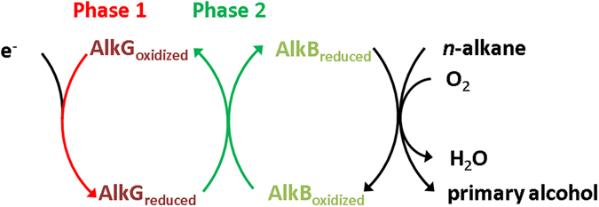
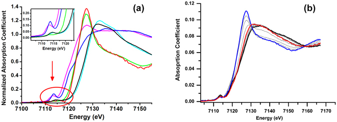
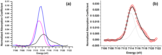
An unprecedented method for the efficient conversion of C 3–C 12 linear alkanes to their corresponding primary alcohols mediated by the membrane-bound alkane hydroxylase (AlkB) from Pseudomonas putida GPo1 is demonstrated. The X-ray absorption spectroscopy (XAS) studies support that electrons can be transferred from the reduced AlkG (rubredoxin-2, the redox partner of AlkB) to AlkB in a two-phase manner. Based on this observation, an approach for the electrocatalytic conversion from alkanes to alcohols mediated by AlkB using an AlkG immobilized screen-printed carbon electrode (SPCE) is developed. The framework distortion of AlkB–AlkG adduct on SPCE surface might create promiscuity toward gaseous substrates. Hence, small alkanes including propane and n-butane can be accommodated in the hydrophobic pocket of AlkB for C–H bond activation. The proof of concept herein advances the development of artificial C–H bond activation catalysts.
Overview
The claims and statistics of this paper haven’t been checked yet.
Run the check — free, no account needed. It reads the open-access text and checks the sources it cites.
Integrity signals
Registry
Cites
Statistics
Figures
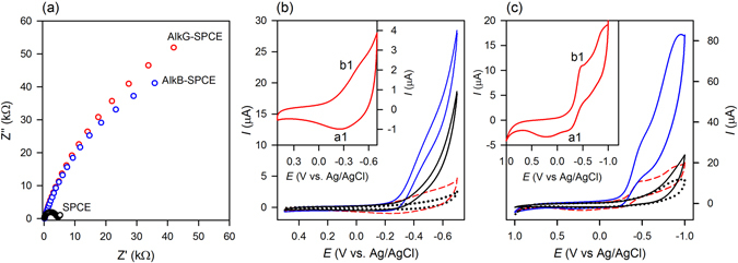
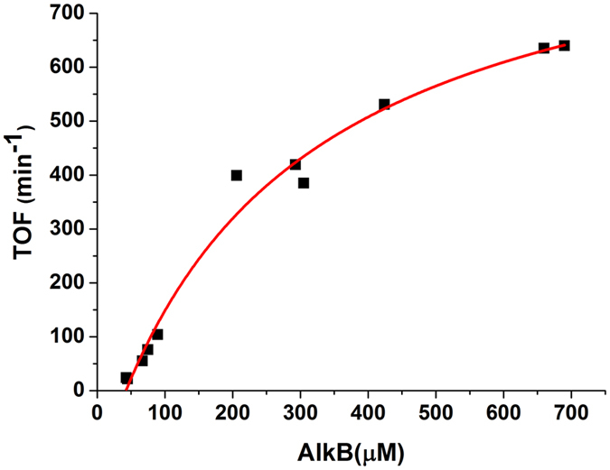
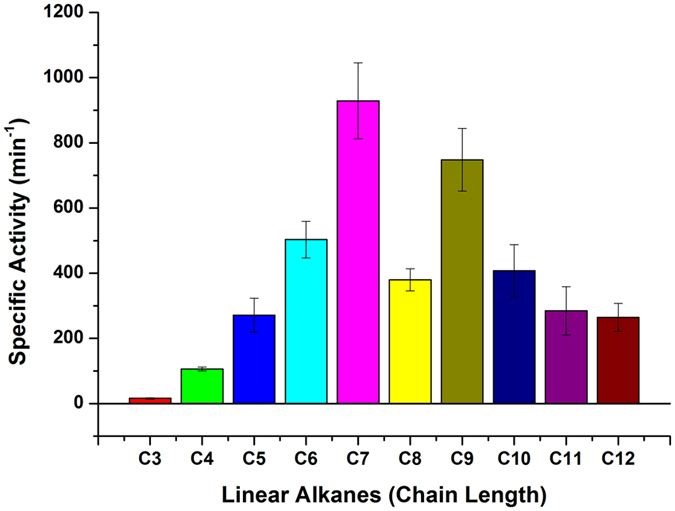
Verification
Claims the paper makes
The claims and statistics of this paper haven’t been checked yet.
Run the check — free, no account needed. It reads the open-access text and checks the sources it cites.
Claims other papers make about this one
Each is checked against this paper’s own text: does it say what the citing paper says it does?
In the citing paper
“As an integral membrane-bound diiron ω-hydroxylase, alkane monooxygenase from Pseudomonas putida Gpo1 (formerly known as Pseudomonas oleovorans) can selectively introduce O2 into the unreactive terminal methyl group of C5–C12 linear alkanes, generating primary alcohols (131, 132).”
From Toward the development of a molecular toolkit for the microbial remediation of per-and polyfluoroalkyl substances (2024) · cites this paper as [131]
Self-check
Percentages, sums and reported differences re-computed from the paper’s own numbers.
The self-check needs the paper’s full text.
Citations
Cites
No references could be resolved yet.
Research works printed but not linked: 60
- Thomas, J. M., Raja, R., Sankar, G. & Bell, R. G. Molecular-sieve catalysts for the selective oxidation of linear alkanes by molecular oxygen. Nature 398, 227–230 (1999). · DOI
- Munz, D. & Strassner, T. Alkane C-H functionalization and oxidation with molecular oxygen. Inorg. Chem. 54, 5043–5052 (2015). · DOI
- Shul’pin, G. B. New trends in oxidative functionalization of carbon-hydrogen bonds: a review. Catalysts 6 (2016). · DOI
- Van de Vyver, S. & Roman-Leshkov, Y. Emerging catalytic processes for the production of adipic acid. Catal. Sci. Technol. 3, 1465–1479 (2013). · DOI
- Sun, M. et al. Catalytic oxidation of light alkanes (C1–C4) by heteropoly compounds. Chem. Rev. 114, 981–1019 (2014). · DOI
- Austin, R. N. & Groves, J. T. Alkane-oxidizing metalloenzymes in the carbon cycle. Metallomics 3, 775–787 (2011). · DOI
- Rojo, F. Degradation of alkanes by bacteria. Environ. Microbiol. 11, 2477–2490 (2009). · DOI
- Ji, Y. R., Mao, G. N., Wang, Y. Y. & Bartlam, M. Structural insights into diversity and n-alkane biodegradation mechanisms of alkane hydroxylases. Front. Microbiol. 4, 1–13 (2013). · DOI
- van Beilen, J. B. & Funhoff, E. G. Alkane hydroxylases involved in microbial alkane degradation. Appl. Microbiol. Biotechnol. 74, 13–21 (2007). · DOI
- Shanklin, J. & Cahoon, E. B. Desaturation and related modifications of fatty acids. Annu. Rev. Plant Physiol. Plant Mol. Biol. 49, 611–641 (1998). · DOI
- Baptist, J. N., Gholson, R. K. & Coon, M. J. Hydrocarbon oxidation by a bacterial enzyme system. I. Products of octane oxidation. Biochim. Biophys. Acta 69, 40–47 (1963). · DOI
- Peterson, J. A., Basu, D. & Coon, M. J. Enzymatic omega-oxidation. I. Electron carriers in fatty acid and hydrocarbon hydroxylation. J. Biol. Chem. 241, 5162–5164 (1966). · DOI
- Peterson, J. A., Kusunose, M., Kusunose, E. & Coon, M. J. Enzymatic omega-oxidation. II. Function of rubredoxin as the electron carrier in omega-hydroxylation. J. Biol. Chem. 242, 4334–4340 (1967). · DOI
- Ruettinger, R. T., Olson, S. T., Boyer, R. F. & Coon, M. J. Identification of the omega-hydroxylase of Pseudomonas oleovorans as a nonheme iron protein requiring phospholipid for catalytic activity. Biochem. Biophys. Res. Commun 57, 1011–1017 (1974). · DOI
- Ueda, T., Lode, E. T. & Coon, M. J. Enzymatic omega-oxidation. 6. Isolation of homogeneous reduced diphosphopyridine nucleotide-rubredoxin reductase. J. Biol. Chem. 247, 2109–2116 (1972). · DOI
- Staijen, I. E., van Beilen, J. B. & Witholt, B. Expression, stability and performance of the three-component alkane mono-oxygenase of Pseudomonas oleovorans in Escherichia coli. Eur. J. Biochem. 267, 1957–1965 (2000). · DOI
- Lode, E. T. & Coon, M. J. Enzymatic omega-oxidation. V. Forms of Pseudomonas oleovorans rubredoxin containing one or two iron atoms: structure and function in omega-hydroxylation. J. Biol. Chem. 246, 791–802 (1971). · DOI
- Shanklin, J., Achim, C., Schmidt, H., Fox, B. G. & Munck, E. Mössbauer studies of alkane omega-hydroxylase: Evidence for a diiron cluster in an integral-membrane enzyme. Proc. Natl. Acad. Sci. USA 94, 2981–2986 (1997). · DOI · Paper page
- McKenna, E. J. & Coon, M. J. Enzymatic omega-oxidation. IV. Purification and properties of the omega-hydroxylase of Pseudomonas oleovorans. J. Biol. Chem. 245, 3882–3889 (1970). · DOI
- Austin, R. N., Born, D., Lawton, T. J. & Hamilton, G. E. In Hydrocarbon and Lipid Microbiology Protocols 1-15 (Humana Press, 2015).
- Alonso, H. & Roujeinikova, A. Characterization and two-dimensional crystallization of membrane component AlkB of the medium-chain alkane hydroxylase system from Pseudomonas putida GPo1. Appl. Environ. Microbiol. 78, 7946–7953 (2012). · DOI
- Alonso, H. et al. Structural and mechanistic insight into alkane hydroxylation by Pseudomonas putida AlkB. Biochem. J. 460, 283–293 (2014). · DOI
- Cooper, H. L. R. et al. Parallel and competitive pathways for substrate desaturation, hydroxylation, and radical rearrangement by the non-heme diiron hydroxylase AlkB. J. Am. Chem. Soc. 134, 20365–20375 (2012). · DOI
- Fu, H., Newcomb, M. & Wong, C. H. Pseudomonas olevorans monooxygenase catalyzed assymetric epoxidation of allyl alcohol derivatives and hydroxylation of a hypersensitive radical probe with the radical ring-opening rate exceeding the oxygen rebound rate. J. Am. Chem. Soc. 113, 5878–5880 (1991). · DOI
- Austin, R. N. et al. Cage escape competes with geminate recombination during alkane hydroxylation by the diiron oxygenase AlkB. Angew. Chem. Int. Ed. 47, 5232–5234 (2008). · DOI
- Austin, R. N., Chang, H. K., Zylstra, G. J. & Groves, J. T. The non-heme diiron alkane monooxygenase of Pseudomonas oleovorans (AlkB) hydroxylates via a substrate radical intermediate. J. Am. Chem. Soc. 122, 11747–11748 (2000). · DOI
- Ping, J. F., Wang, Y. X., Ying, Y. B. & Wu, J. Application of electrochemically reduced graphene oxide on screen-printed ion-selective electrode. Anal. Chem. 84, 3473–3479 (2012). · DOI
- Kong, F. Y. et al. A paper disk equipped with graphene/polyaniline/Au nanoparticles/glucose oxidase biocomposite modified screen-printed electrode: Toward whole blood glucose determination. Biosens. Bioelectronics 56, 77–82 (2014). · DOI
- Molinero-Abad, B., Alonso-Lomillo, M. A., Dominguez-Renedo, O. & Arcos-Martinez, M. J. Sulfite oxidase biosensors based on tetrathiafulvalene modified screen-printed carbon electrodes for sulfite determination in wine. Anal. Chim. Acta. 812, 41–44 (2014). · DOI
- Wang, J. Electrochemical glucose biosensors. Chem. Rev. 108, 814–825 (2008). · DOI · Paper page
- Willner, I. & Katz, E. Integration of layered redox proteins and conductive supports for bioelectronic applications. Angew. Chem. Int. Ed. 39, 1180–1218 (2000). · DOI
- Perry, A., Lian, L. Y. & Scrutton, N. S. Two-iron rubredoxin of Pseudomonas oleovorans: production, stability and characterization of the individual iron-binding domains by optical, CD and NMR spectroscopies. Biochem. J. 354, 89–98 (2001). · DOI
- Perry, A., Tambyrajah, W., Grossmann, J. G., Lian, L. Y. & Scrutton, N. S. Solution structure of the two-iron rubredoxin of Pseudomonas oleovorans determined by NMR spectroscopy and solution X-ray scattering and interactions with rubredoxin reductase. Biochemistry 43, 3167–3182 (2004). · DOI
- Li, Z. et al. X-ray absorption spectroscopic analysis of reductive [2Fe-2S] cluster degradation in hyperthermophilic archaeal succinate:caldariellaquinone oxidoreductase subunits. Biochemistry 42, 15003–15008 (2003). · DOI
- Tsang, H. T., Batie, C. J., Ballou, D. P. & Penner-Hahn, J. E. X-ray absorption spectroscopy of the [2-iron-2-sulfur] Rieske cluster in Pseudomonas cepacia phthalate dioxygenase. Determination of core dimensions and iron ligation. Biochemistry 28, 7233–7240 (1989). · DOI
- Lo, F. C. et al. The metal core structures in the recombinant Escherichia coli transcriptional factor SoxR. Chem.-Eur. J. 18, 2565–2577 (2012). · DOI
- Rudd, D. J. et al. Determination by X-ray absorption spectroscopy of the Fe-Fe separation in the oxidized form of the hydroxylase of methane monooxygenase alone and in the presence of MMOD. Inorg. Chem. 43, 4579–4589 (2004). · DOI
- Rudd, D. J., Sazinsky, M. H., Lippard, S. J., Hedman, B. & Hodgson, K. O. X-ray absorption spectroscopic study of the reduced hydroxylases of methane monooxygenase and toluene/o-xylene monooxygenase: differences in active site structure and effects of the coupling proteins MMOB and ToMOD. Inorg. Chem. 44, 4546–4554 (2005). · DOI
- Randall, C. R. et al. X-ray absorption pre-edge studies of high-spin iron(II) complexes. Inorg. Chem. 34, 1036–1039 (1995). · DOI
- Westre, T. E. et al. A multiplet analysis of Fe K-edge 1s → 3d pre-edge features of iron complexes. J. Am. Chem. Soc. 119, 6297–6314 (1997). · DOI
- Roe, A. L. et al. X-ray absorption spectroscopy of iron-tyrosinate proteins. J. Am. Chem. Soc. 106, 1676–1681 (1984). · DOI
- Shulman, R. G., Eisenberger, P., Blumberg, W. E. & Stombaugh, N. A. Determination of the iron-sulfur distances in rubredoxin by X-ray absorption spectroscopy. Proc. Natl. Acad. Sci. USA 72, 4003–4007 (1975). · DOI
- Shu, L. et al. EXAFS and Mössbauer characterization of the Diiron(III) site in stearoyl-acyl carrier protein Δ9– desaturase. J. Biol. Inorg. Chem. 3, 392–400 (1998). · DOI
- True, A. E., Scarrow, R. C., Randall, C. R., Holz, R. C. & Que, L. J. EXAFS studies of uteroferrin and its anion complexes. J. Am. Chem. Soc. 115, 4246–4255 (1993). · DOI
- Bai, Y. H. et al. X-ray structure of a mammalian stearoyl-CoA desaturase. Nature 524, 252–256 (2015). · DOI
- Scarrow, R. C. et al. EXAFS studies of binuclear iron proteins: hemerythrin and ribonucleotide reductase. J. Am. Chem. Soc. 109, 7857–7864 (1987). · DOI
- Shu, L., Liu, Y., Lipscomb, J. D. & Que, L. J. X-ray absorption spectroscopic studies of the methane monooxygenase hydroxylase component from Methylosinus trichosporium OB3b. J. Biol. Inorg. Chem. 1, 297–304 (1996). · DOI
- Prodromidis, M. I., Florou, A. B., Tzouwara-Karayanni, S. M. & Karayannis, M. I. The importance of surface coverage in the electrochemical study of chemically modified electrodes. Electroanalysis 12, 1498–1501 (2000). · DOI
- Wang, S. F. et al. Direct electrochemistry and electrocatalysis of heme proteins entrapped in agarose hydrogel films in room-temperature ionic liquids. Langmuir 21, 9260–9266 (2005). · DOI
- Bard, A. J. & Faulkner, L. R. Electrochemical Methods, Fundamentals, and Applications. 2nd ed., (Wiley, 2001).
- Johnson, E. L. & Hyman, M. R. Propane and n-butane oxidation by Pseudomonas putida GPo1. Appl. Environ. Microbiol. 72, 950–952 (2006). · DOI
- Koch, D. J., Chen, M. M., Van Beilen, J. B. & Arnold, F. H. In vivo evolution of butane oxidation by terminal alkane hydroxylases AlkB and CYP153A6. Appl. Environ. Microbiol. 75, 337–344 (2009). · DOI
- Peters, M. et al. Chemical technologies for exploiting and recycling carbon dioxide into the value chain. ChemSusChem 4, 1216–1240 (2011). · DOI
- Marimuthu, A., Zhang, J. & Linic, S. Tuning selectivity in propylene epoxidation by plasmon mediated photo-switching of Cu oxidation state. Science 339, 1590–1593 (2013). · DOI
- Ramu, R. et al. Regio-selective hydroxylation of gem-difluorinated octanes by alkane hydroxylase (AlkB). Tetrahedron Lett. 52, 2950–2953 (2011). · DOI
- Bernaudat, F. et al. Heterologous expression of membrane proteins: choosing the appropriate host. PLoS One 6, e29191, doi:10.1371/journal.pone.0029191 (2011). · DOI
- Walker, J. M. The Protein Protocols Handbook. 2nd ed., 421–428 (Humana Press, 2002). · DOI
- Ravel, B. & Newville, M. ATHENA, ARTEMIS, HEPHAESTUS: data analysis for X-ray absorption spectroscopy using IFEFFIT. J. Synchrotron Rad. 12, 537–541 (2005). · DOI
- Newville, M. IFEFFIT: interactive XAFS analysis and FEFF fitting. J. Synchrotron Rad. 8, 322–324 (2001). · DOI
- Ravel, B. ATOMS: crystallography for the X-ray absorption spectroscopist. J. Synchrotron Rad. 8, 314–316 (2001). · DOI
Cited by
Sorted by citations
Hu, M., & Scott, C. (2024) · Applied and Environmental Microbiology · doi:10.1128/aem.00157-24 · Cited by 63
Metadata
- Authors
- Tsai, Y.-F., Luo, W.-I., Chang, J.-L., Chang, C.-W., Chuang, H.-C., Ramu, R., Wei, G.-T., Zen, J.-M., & Yu, S. S.-F.
- ORCID
- Yu, Steve S.-F.
- Venue
- Scientific Reports
- Published
- 2017-08-21
- Volume
- 7
- Issue
- 1
- ISSN
- 2045-2322
- PMC
- PMC5566439
- Cited by
- 31
- References
- 0
- Language
- en
About these checks
Registry, retraction, Cites and statistics results are computed, not judged, with one exception: a printed reference that no index lists is read once by a model before it counts as a citation error. Claims are extracted by a model and kept only when their quote appears verbatim in the text; each cited source is then checked, with a model’s help, for whether it says what the claim relies on. Those verdicts can be wrong — read the claim’s quote and the cited source before relying on one.
Statistics and arithmetic are recomputed, not judged.
Confidence bands are shown only for fields calibrated on our annotated test set. None is calibrated yet, so no band is shown.
What counts as a claim ›How we measure confidence ›How we check this ›
Are you an author, or do you see a problem with this page? Request a review
How to cite
Cite CoChat for the checks on this page, and the paper for its own findings.
- This page
- CoChat. (2026). Citation check: Electrochemical Hydroxylation of C3–C12 n-Alkanes by Recombinant Alkane Hydroxylase (AlkB) and Rubredoxin-2 (AlkG) from Pseudomonas putida GPo1. https://app.cochat.ai/papers/10.1038/s41598-017-08610-w
- The paper
- Tsai, Y.-F., Luo, W.-I., Chang, J.-L., Chang, C.-W., Chuang, H.-C., Ramu, R., Wei, G.-T., Zen, J.-M., & Yu, S. S.-F. (2017). Electrochemical Hydroxylation of C3–C12 n-Alkanes by Recombinant Alkane Hydroxylase (AlkB) and Rubredoxin-2 (AlkG) from Pseudomonas putida GPo1. Scientific Reports. https://doi.org/10.1038/s41598-017-08610-w
