

Amygdala-centred functional connectivity affects daily cortisol concentrations: a putative link with anxiety
Open accessThe DOI matches its Crossref record; 58 of the 59 references it cites were found in a registry or an index.
Abstract
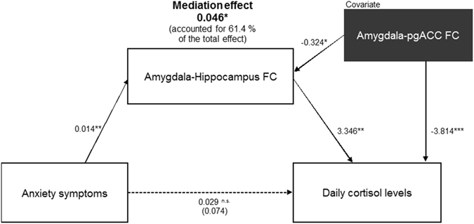
The amygdala plays a critical role in emotion. Its functional coupling with the hippocampus and ventromedial prefrontal cortex extending to a portion of the anterior cingulate cortex (ACC) is implicated in anxiogenesis and hypothalamic-pituitary-adrenal (HPA) system regulation. However, it remains unclear how amygdala-centred functional connectivity (FC) affects anxiety and cortisol concentrations in everyday life. Here, we investigate the relationship between daily cortisol concentrations (dCOR) and amygdala-centred FC during emotional processing in forty-one healthy humans. FC analyses revealed that higher dCOR predicted strengthened amygdala-centred FC with the hippocampus and cerebellum, but inhibited FC with the supramarginal gyrus and a perigenual part of the ACC (pgACC) when processing fearful faces (vs. neutral faces). Notably, the strength of amygdala-hippocampus FC mediated the positive relationship between cortisol and anxiety, specifically when the effect of amygdala-pgACC FC, a presumptive neural indicator of emotional control, was taken into account. Individuals with diminished connectivity between the amygdala and pgACC during fear-related processing might be more vulnerable to anxiogenesis as it pertains to greater circulating cortisol levels in everyday life. Individual functional patterns of amygdala-hippocampal-pgACC connectivity might provide a key to understand the complicate link between cortisol and anxiety-related behaviors.
Overview
The claims and statistics of this paper haven’t been checked yet.
Free: it reads the open-access text and checks the first 5 claims that cite a source we can check. Create a free account to check every claim.
Integrity signals
Registry
Cites
Statistics
Figures



Verification
Claims the paper makes
The claims and statistics of this paper haven’t been checked yet.
Free: it reads the open-access text and checks the first 5 claims that cite a source we can check. Create a free account to check every claim.
Claims other papers make about this one
Each is checked against this paper’s own text: does it say what the citing paper says it does?
The source backs part of the claim, not all of it — or backs it for a different population, condition or comparison.
In the citing paper
“However, under conditions of prolonged stress, persistent elevation of cortisol can disrupt this regulatory system, leading to hypersecretion, sensitized stress responses, and adrenal exhaustion [16].”
From Chronic Stress and Autoimmunity: The Role of HPA Axis and Cortisol Dysregulation (2025) · cites this paper as [16]
Experimental — accuracy not yet measuredThe source backs part of the claim, not all of it — or backs it for a different population, condition or comparison.
In the citing paper
“This system is regulated by a negative feedback mechanism, in which both ACTH and cortisol act as inhibitors of CRH and ACTH secretion, thereby maintaining homeostasis [16,17].”
From Chronic Stress and Autoimmunity: The Role of HPA Axis and Cortisol Dysregulation (2025) · cites this paper as [16]
Experimental — accuracy not yet measured
Self-check
Percentages, sums and reported differences re-computed from the paper’s own numbers.
The self-check needs the paper’s full text.
Citations
Cites
The assessment and analysis of handedness: The Edinburgh inventory
Oldfield, R. C. (1971) · Neuropsychologia · doi:10.1016/0028-3932(71)90067-4 · Cited by 35,900
(1998) · The Journal of Clinical Psychiatry · Cited by 19,139
(1998) · PubMed · Cited by 10,216
Maldjian, J. A., Laurienti, P. J., Kraft, R. A., & Burdette, J. H. (2003) · NeuroImage · doi:10.1016/s1053-8119(03)00169-1 · Cited by 5,085
Conn : A Functional Connectivity Toolbox for Correlated and Anticorrelated Brain Networks
Whitfield-Gabrieli, S., & Nieto-Castanon, A. (2012) · Brain Connectivity · doi:10.1089/brain.2012.0073 · Cited by 4,885
The Hopkins Symptom Checklist (HSCL): A self-report symptom inventory
Derogatis, L. R., Lipman, R. S., Rickels, K., Uhlenhuth, E. H., & Covi, L. (1974) · Behavioral Science · doi:10.1002/bs.3830190102 · Cited by 4,604
Functional neuroanatomy of emotion: A meta-analysis of emotion activation studies in PET and fMRI
Phan, K. L., Wager, T., Taylor, S. F., & Liberzon, I. (2002) · NeuroImage · doi:10.1006/nimg.2002.1087 · Cited by 3,502 · cited 2×
“The eligibility criteria were (1) right-handedness as defined by the Edinburgh Handedness Inventory47, (2) no Axis-I psychiatric disorders or substance-abuse history as determined using the Mini-International Neuropsychiatric Interview48, (3) no major medical illnesses, (4) no regular intake of psychotropics, steroids…”
Etkin, A., & Wager, T. D. (2007) · American Journal of Psychiatry · doi:10.1176/appi.ajp.2007.07030504 · Cited by 3,391
Two receptor systems for corticosterone in rat brain: Microdistribution and differential occupation
REUL, J. M. H. M., & KLOET, E. R. D. (1985) · Endocrinology · doi:10.1210/endo-117-6-2505 · Cited by 2,580
Miller, G. E., Chen, E., & Zhou, E. S. (2007) · Psychological Bulletin · doi:10.1037/0033-2909.133.1.25 · Cited by 2,467
Research works printed but not linked: 1
- Nakano, K., Stress management. 2nd ed.; Kongo Press: Tokyo, Japan. (2016).
Cited by
Stress, hypothalamic–pituitary–adrenal axis, hypothalamic–pituitary–gonadal axis, and aggression.
Mbiydzenyuy, N. E., & Qulu, L.-A. (2024) · Metabolic Brain Disease · doi:10.1007/s11011-024-01393-w · Cited by 255
“Increased cortisol levels can enhance the amygdala’s reactivity to threat cues, leading to heightened emotional responses and a propensity for aggressive behavior (Hakamata et al. 2017).”
BackgroundEarly Social Adversity, Altered Brain Functional Connectivity, and Mental Health
Holz, N. E., Berhe, O., Sacu, S., Schwarz, E., Tesarz, J., Heim, C. C., & Tost, H. (2022) · Biological Psychiatry · doi:10.1016/j.biopsych.2022.10.019 · Cited by 142
“4 to mPFC and the adjacent perigenual anterior cingulate cortex (pACC, Figure 2A) (20), which has been linked to alterations in HPA axis function (21-23), differences in emotion regulation (24), internalizing psychopathology (25), trauma-related proinflammatory states (26), and inflammation-associated mood deteriorati…”
BackgroundChronic Stress and Autoimmunity: The Role of HPA Axis and Cortisol Dysregulation
Nunez, S. G., Rabelo, S. P., Subotic, N., Caruso, J. W., & Knezevic, N. N. (2025) · International Journal of Molecular Sciences · doi:10.3390/ijms26209994 · Cited by 138
“However, under conditions of prolonged stress, persistent elevation of cortisol can disrupt this regulatory system, leading to hypersecretion, sensitized stress responses, and adrenal exhaustion [16].”
Amygdala substructure volumes in Major Depressive Disorder
Roddy, D., Kelly, J. R., Farrell, C., Doolin, K., Roman, E., Nasa, A., Frodl, T., Harkin, A., O'Mara, S., O'Hanlon, E., et al. (2021) · NeuroImage: Clinical · doi:10.1016/j.nicl.2021.102781 · Cited by 75
“Greater cortisol levels have been reported to be correlated with increased amygdala-centred FC in responses to fearful faces (Hakamata et al., 2017), and in young adults with a history of depression, cortisol levels correlated with increased amygdala connectivity to cognitive control network regions (Peters et al.”
BackgroundHormonal Cycles, Brain Network Connectivity, and Windows of Vulnerability to Affective Disorder
Andreano, J. M., Touroutoglou, A., Dickerson, B., & Barrett, L. F. (2018) · Trends in Neurosciences · doi:10.1016/j.tins.2018.08.007 · Cited by 75
“On the hormonal level, connectivity within the salience network [80], as well as between the amygdala and hippocampus, is associated with the magnitude of cortisol responses [78,81].”
Metadata
- Authors
- Hakamata, Y., Komi, S., Moriguchi, Y., Izawa, S., Motomura, Y., Sato, E., Mizukami, S., Kim, Y., Hanakawa, T., Inoue, Y., & Tagaya, H.
- Affiliations
- National Center of Neurology and Psychiatry (JP)
- Kitasato University Hospital (JP)
- National Institute of Occupational Safety and Health (JP)
- Kyorin University (JP)
- Kitasato University (JP)
- Corresponding author
- Hakamata, Yuko
- ORCID
- Venue
- Scientific Reports
- Published
- 2017-08-16
- Volume
- 7
- Issue
- 1
- Pages
- 8313-8313
- ISSN
- 2045-2322 (ISSN-L)
- PMC
- PMC5559590
- OpenAlex
- W2744964154
- Cited by
- 76
- References
- 59
- Citations by year
- 2026: 4 · 2025: 8 · 2024: 17 · 2023: 8 · 2022: 16 · 2021: 7 · 2020: 7 · 2019: 3 · 2018: 6
- FWCI
- 2.38
- Type
- journal article
- Language
- en
- Open access
- gold
- doi.org
- Licence
- cc-by
- creativecommons.org/licenses/by/4.0 (tdm)
- creativecommons.org/licenses/by/4.0 (vor)
- Locations
- Scientific Reports (publishedVersion, cc-by)
- PubMed Central (publishedVersion, cc-by)
- PubMed (publishedVersion)
- DOAJ (DOAJ: Directory of Open Access Journals) (submittedVersion, cc-by-sa)
- Funders
- Sanofi
- Astellas Pharma
- Japan Society for the Promotion of Science (17J40250, 26120008)
- Topics
- Life Sciences › Neuroscience › Behavioral Neuroscience › Stress Responses and Cortisol
- Life Sciences › Neuroscience › Cognitive Neuroscience › Functional Brain Connectivity Studies
- Life Sciences › Neuroscience › Cognitive Neuroscience › Memory and Neural Mechanisms
- Keywords
- amygdala, functional connectivity, anxiety, pregenual anterior cingulate cortex, fear processing, fearful faces, emotional control, hypothalamic-pituitary-adrenal axis, cerebellum, supramarginal gyrus
- MeSH terms
- Adult, Amygdala, Anxiety, Brain Mapping, Depression, Fear, Female, Humans, Hydrocortisone, Male, Neural Pathways, Young Adult, Connectome
About these checks
Registry, retraction, Cites and statistics results are computed, not judged, with one exception: a printed reference that no index lists is read once by a model before it counts as a citation error. Claims are extracted by a model and kept only when their quote appears verbatim in the text; each cited source is then checked, with a model’s help, for whether it says what the claim relies on. Those verdicts can be wrong — read the claim’s quote and the cited source before relying on one.
Statistics and arithmetic are recomputed, not judged.
Confidence bands are shown only for fields calibrated on our annotated test set. None is calibrated yet, so no band is shown.
What counts as a claim ›How we measure confidence ›How we check this ›
Are you an author, or do you see a problem with this page? Request a review
How to cite
Cite CoChat for the checks on this page, and the paper for its own findings.
- This page
- CoChat. (2026). Citation check: Amygdala-centred functional connectivity affects daily cortisol concentrations: a putative link with anxiety. https://app.cochat.ai/papers/10.1038/s41598-017-08918-7
- The paper
- Hakamata, Y., Komi, S., Moriguchi, Y., Izawa, S., Motomura, Y., Sato, E., Mizukami, S., Kim, Y., Hanakawa, T., Inoue, Y., & Tagaya, H. (2017). Amygdala-centred functional connectivity affects daily cortisol concentrations: a putative link with anxiety. Scientific Reports. https://doi.org/10.1038/s41598-017-08918-7
Data sources
Bibliographic data: Semantic Scholar, OpenAlex, Crossref.
Contains information from the Semantic Scholar Academic Graph Datasets, made available under the ODC Attribution License (ODC-BY 1.0).
Semantic Scholar’s data is described in: Kinney, R., et al. (2023). The Semantic Scholar Open Data Platform. arXiv:2301.10140.
