

The DOI matches its Crossref record; 1 of the 1 references it cites were found in a registry or an index.
Elastolytic activity of cysteine cathepsins K, S, and V promotes vascular calcification
Open accessAbstract
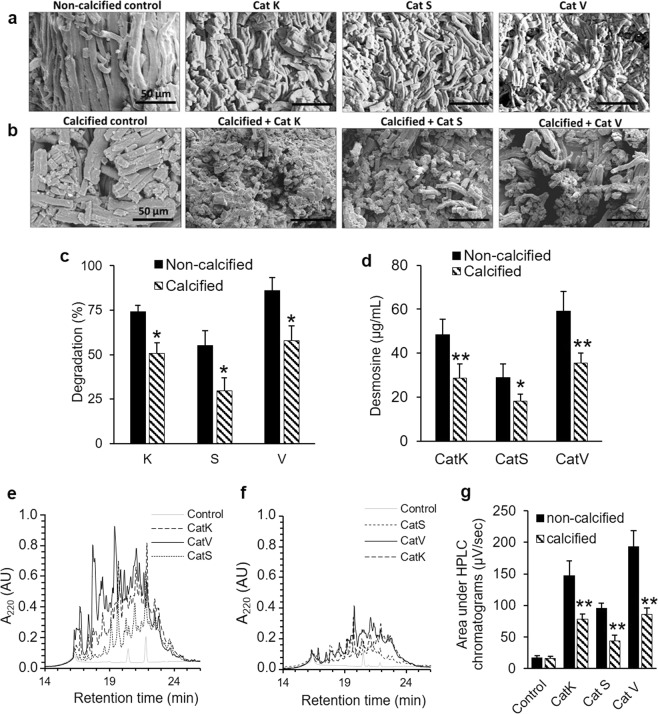
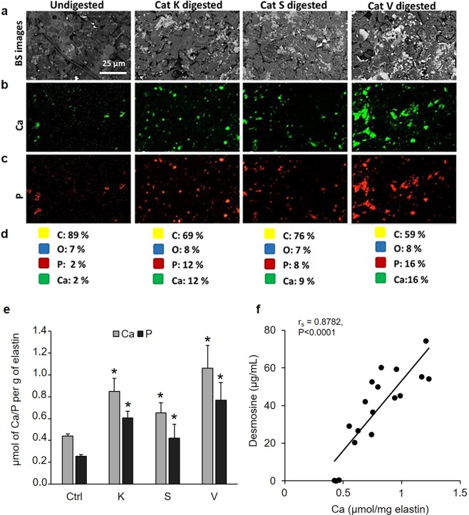
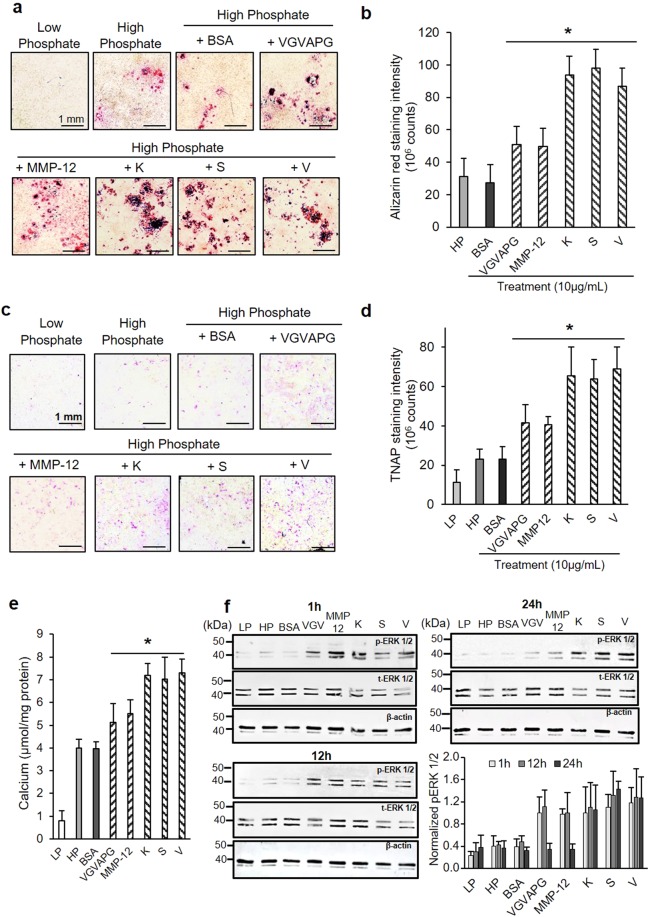
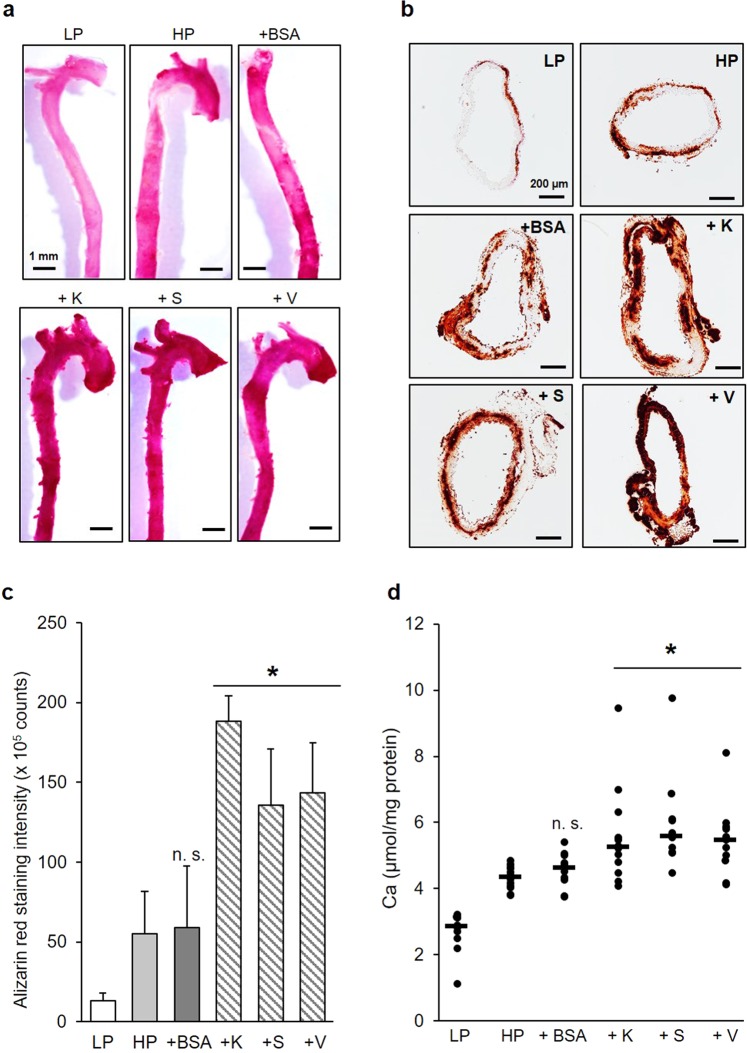
Elastin plays an important role in maintaining blood vessel integrity. Proteolytic degradation of elastin in the vascular system promotes the development of atherosclerosis, including blood vessel calcification. Cysteine cathepsins have been implicated in this process, however, their role in disease progression and associated complications remains unclear. Here, we showed that the degradation of vascular elastin by cathepsins (Cat) K, S, and V directly stimulates the mineralization of elastin and that mineralized insoluble elastin fibers were ~25–30% more resistant to CatK, S, and V degradation when compared to native elastin. Energy dispersive X-ray spectroscopy investigations showed that insoluble elastin predigested by CatK, S, or V displayed an elemental percentage in calcium and phosphate up to 8-fold higher when compared to non-digested elastin. Cathepsin-generated elastin peptides increased the calcification of MOVAS-1 cells acting through the ERK1/2 pathway by 34–36%. We made similar observations when cathepsin-generated elastin peptides were added to ex vivo mouse aorta rings. Altogether, our data suggest that CatK-, S-, and V-mediated elastolysis directly accelerates the mineralization of the vascular matrix by the generation of nucleation points in the elastin matrix and indirectly by elastin-derived peptides stimulating the calcification by vascular smooth muscle cells. Both processes inversely protect against further extracellular matrix degradation.
Overview
The claims and statistics of this paper haven’t been checked yet.
Run the check — free, no account needed. It reads the open-access text and checks the sources it cites.
Integrity signals
Registry
Cites
Statistics
Figures
Verification
Claims the paper makes
The claims and statistics of this paper haven’t been checked yet.
Run the check — free, no account needed. It reads the open-access text and checks the sources it cites.
Claims other papers make about this one
Each is checked against this paper’s own text: does it say what the citing paper says it does?
The source backs part of the claim, not all of it — or backs it for a different population, condition or comparison.
In the citing paper
“Moreover, glycine-rich sites on elastin particles are calcification nidi, making elastin-containing biomaterials susceptible to calcification upon implantation [110].”
From Immunogenicity of decellularized extracellular matrix scaffolds: a bottleneck in tissue engineering and regenerative medicine (2023) · cites this paper as [110]
Experimental — accuracy not yet measured
Self-check
Percentages, sums and reported differences re-computed from the paper’s own numbers.
The self-check needs the paper’s full text.
Citations
Cites
Dhingra, R. (2007) · Archives of Internal Medicine · doi:10.1001/archinte.167.9.879 · Cited by 861
Research works printed but not linked: 57
- Budoff, M. J. et al. Long-term prognosis associated with coronary calcification: observations from a registry of 25,253 patients. J. Am. Coll. Cardiol. 49, 1860–1870 (2007). · DOI
- Demer, L. L. & Tintut, Y. Vascular calcification: pathobiology of a multifaceted disease. Circulation 117, 2938–2948 (2008). · DOI
- Vliegenthart, R. et al. Coronary calcification improves cardiovascular risk prediction in the elderly. Circulation 112, 572–577 (2005). · DOI
- Zhu, D., Mackenzie, N. C. W., Farquharson, C. & Macrae, V. E. Mechanisms and clinical consequences of vascular calcification. Front. Endocrinol. 3, 95 (2012). · DOI
- Stary, H. C. Natural history of calcium deposits in atherosclerosis progression and regression. Z. Kardiol. 89(Suppl 2), 28–35 (2000). · DOI
- Wallin, R., Wajih, N., Greenwood, G. T. & Sane, D. C. Arterial calcification: a review of mechanisms, animal models, and the prospects for therapy. Med. Res. Rev. 21, 274–301 (2001). · DOI
- Simpson, C. L. et al. Toward cell therapy for vascular calcification: osteoclast-mediated demineralization of calcified elastin. Cardiovasc. Pathol. Off. J. Soc. Cardiovasc. Pathol. 16, 29–37 (2007). · DOI
- Lutgens, S. P. M., Cleutjens, K. B. J. M., Daemen, M. J. A. P. & Heeneman, S. Cathepsin cysteine proteases in cardiovascular disease. FASEB J. Off. Publ. Fed. Am. Soc. Exp. Biol. 21, 3029–3041 (2007).
- Newby, A. C. Proteinases and plaque rupture: unblocking the road to translation. Curr. Opin. Lipidol. 25, 358–366 (2014). · DOI
- Cheng, X. W., Huang, Z., Kuzuya, M., Okumura, K. & Murohara, T. Cysteine protease cathepsins in atherosclerosis-based vascular disease and its complications. Hypertens. Dallas Tex 1979 58, 978–986 (2011).
- Fonović, M. & Turk, B. Cysteine cathepsins and extracellular matrix degradation. Biochim. Biophys. Acta 1840, 2560–2570 (2014). · DOI
- Yasuda, Y. et al. Cathepsin V, a Novel and Potent Elastolytic Activity Expressed in Activated Macrophages. J. Biol. Chem. 279, 36761–36770 (2004). · DOI
- Samokhin, A. O., Wong, A., Saftig, P. & Brömme, D. Role of cathepsin K in structural changes in brachiocephalic artery during progression of atherosclerosis in apoE-deficient mice. Atherosclerosis 200, 58–68 (2008). · DOI
- Samokhin, A. O., Lythgo, P. A., Gauthier, J. Y., Percival, M. D. & Brömme, D. Pharmacological inhibition of cathepsin S decreases atherosclerotic lesions in Apoe −/− mice. J. Cardiovasc. Pharmacol. 56, 98–105 (2010). · DOI
- Lutgens, E. et al. Disruption of the cathepsin K gene reduces atherosclerosis progression and induces plaque fibrosis but accelerates macrophage foam cell formation. Circulation 113, 98–107 (2006). · DOI
- Izumi, Y. et al. Impact of circulating cathepsin K on the coronary calcification and the clinical outcome in chronic kidney disease patients. Heart Vessels 31, 6–14 (2016). · DOI
- Aikawa, E. et al. Arterial and aortic valve calcification abolished by elastolytic cathepsin S deficiency in chronic renal disease. Circulation 119, 1785–1794 (2009). · DOI
- Wopenka, B. & Pasteris, J. D. A mineralogical perspective on the apatite in bone. Mater. Sci. Eng. C 25, 131–143 (2005). · DOI
- Khan, A. F. et al. Raman Spectroscopy of Natural Bone and Synthetic Apatites. Appl. Spectrosc. Rev. 48, 329–355 (2013). · DOI
- Simionescu, A., Philips, K. & Vyavahare, N. Elastin-derived peptides and TGF-beta1 induce osteogenic responses in smooth muscle cells. Biochem. Biophys. Res. Commun. 334, 524–532 (2005). · DOI
- Simionescu, A., Simionescu, D. T. & Vyavahare, N. R. Osteogenic responses in fibroblasts activated by elastin degradation products and transforming growth factor-beta1: role of myofibroblasts in vascular calcification. Am. J. Pathol. 171, 116–123 (2007). · DOI
- Mackenzie, N. C. W. et al. MOVAS-1 cell line: a new in vitro model of vascular calcification. Int. J. Mol. Med. 27, 663–668 (2011).
- Ding, H.-T., Wang, C.-G., Zhang, T.-L. & Wang, K. Fibronectin enhances in vitro vascular calcification by promoting osteoblastic differentiation of vascular smooth muscle cells via ERK pathway. J. Cell. Biochem. 99, 1343–1352 (2006). · DOI
- Feng, W. et al. Apocynin attenuates angiotensin II-induced vascular smooth muscle cells osteogenic switching via suppressing extracellular signal-regulated kinase 1/2. Oncotarget 7, 83588–83600 (2016). · DOI
- Freise, C., Bobb, V. & Querfeld, U. Collagen XIV and a related recombinant fragment protect human vascular smooth muscle cells from calcium-/phosphate-induced osteochondrocytic transdifferentiation. Exp. Cell Res. 358, 242–252 (2017). · DOI
- Sukhova, G. K., Shi, G. P., Simon, D. I., Chapman, H. A. & Libby, P. Expression of the elastolytic cathepsins S and K in human atheroma and regulation of their production in smooth muscle cells. J. Clin. Invest. 102, 576–583 (1998). · DOI
- Platt, M. O. et al. Expression of cathepsin K is regulated by shear stress in cultured endothelial cells and is increased in endothelium in human atherosclerosis. Am. J. Physiol. Heart Circ. Physiol. 292, H1479–1486 (2007). · DOI
- Sukhova, G. K. et al. Deficiency of cathepsin S reduces atherosclerosis in LDL receptor-deficient mice. J. Clin. Invest. 111, 897–906 (2003). · DOI
- Kitamoto, S. et al. Cathepsin L deficiency reduces diet-induced atherosclerosis in low-density lipoprotein receptor-knockout mice. Circulation 115, 2065–2075 (2007). · DOI
- Wu, M., Rementer, C. & Giachelli, C. M. Vascular Calcification: An Update on Mechanisms and Challenges in Treatment. Calcif. Tissue Int. 93, 365–373 (2013). · DOI
- Wierzbicki, A. S., Viljoen, A. & Chambers, J. B. Aortic stenosis and lipids: does intervention work? Curr. Opin. Cardiol. 25, 379–384 (2010). · DOI
- Novaro, G. M. et al. Effect of hydroxymethylglutaryl coenzyme a reductase inhibitors on the progression of calcific aortic stenosis. Circulation 104, 2205–2209 (2001). · DOI
- Kendrick, J., Kestenbaum, B. & Chonchol, M. Phosphate and Cardiovascular Disease. Adv. Chronic Kidney Dis. 18, 113–119 (2011). · DOI
- Ix, J. H. et al. Serum Phosphorus Concentrations and Arterial Stiffness among Individuals with Normal Kidney Function to Moderate Kidney Disease in MESA. Clin. J. Am. Soc. Nephrol. 4, 609–615 (2009). · DOI
- Tonelli, M. et al. Relation between serum phosphate level and cardiovascular event rate in people with coronary disease. Circulation 112, 2627–2633 (2005). · DOI
- Montalcini, T., Gorgone, G. & Pujia, A. Serum calcium level is related to both intima-media thickness and carotid atherosclerosis: a neglect risk factor in obese/overweight subjects. J. Transl. Med. 10, 114 (2012). · DOI
- Vyavahare, N., Ogle, M., Schoen, F. J. & Levy, R. J. Elastin calcification and its prevention with aluminum chloride pretreatment. Am. J. Pathol. 155, 973–982 (1999). · DOI
- Urry, D. W., Krivacic, J. R. & Haider, J. Calcium ion effects a notable change in elastin conformation by interacting at neutral sites. Biochem. Biophys. Res. Commun. 43, 6–11 (1971). · DOI
- Bertazzo, S. et al. Nano-analytical electron microscopy reveals fundamental insights into human cardiovascular tissue calcification. Nat. Mater. 12, 576–583 (2013). · DOI
- Barroso, B., Abello, N. & Bischoff, R. Study of human lung elastin degradation by different elastases using high-performance liquid chromatography/mass spectrometry. Anal. Biochem. 358, 216–224 (2006). · DOI
- Maurice, P. et al. Elastin fragmentation and atherosclerosis progression: the elastokine concept. Trends Cardiovasc. Med. 23, 211–221 (2013). · DOI
- Heinz, A. et al. The action of neutrophil serine proteases on elastin and its precursor. Biochimie 94, 192–202 (2012). · DOI
- Heinz, A. et al. Degradation of tropoelastin by matrix metalloproteinases–cleavage site specificities and release of matrikines. FEBS J. 277, 1939–1956 (2010). · DOI
- Figueiredo, J.-L. et al. Selective Cathepsin S Inhibition Attenuates Atherosclerosis in Apolipoprotein E–Deficient Mice with Chronic Renal Disease. Am. J. Pathol. 185, 1156–1166 (2015). · DOI
- Mochizuki, S., Brassart, B. & Hinek, A. Signaling pathways transduced through the elastin receptor facilitate proliferation of arterial smooth muscle cells. J. Biol. Chem. 277, 44854–44863 (2002). · DOI
- Rusciani, A. et al. Elastin peptides signaling relies on neuraminidase-1-dependent lactosylceramide generation. PloS One 5, e14010 (2010). · DOI
- Blanchevoye, C. et al. Interaction between the elastin peptide VGVAPG and human elastin binding protein. J. Biol. Chem. 288, 1317–1328 (2013). · DOI
- Kirschke, H., Barrett, A. J. & Rawlings, N. D. Lysosomal cysteine proteases. (Oxford University Press, 1998). · DOI
- Reiser, J., Adair, B. & Reinheckel, T. Specialized roles for cysteine cathepsins in health and disease. J. Clin. Invest. 120, 3421–3431 (2010). · DOI
- Tsubokawa, T. et al. Cathepsin and calpain inhibitor E64d attenuates matrix metalloproteinase-9 activity after focal cerebral ischemia in rats. Stroke 37, 1888–1894 (2006). · DOI
- Christensen, J. & Shastri, V. P. Matrix-metalloproteinase-9 is cleaved and activated by cathepsin K. BMC Res. Notes 8, 322 (2015). · DOI
- Ikawati, M., Kawaichi, M. & Oka, C. Loss of HtrA1 serine protease induces synthetic modulation of aortic vascular smooth muscle cells. PloS One 13, e0196628 (2018). · DOI
- Kim, Y.-M. et al. Neutrophil Elastase Is Produced by Pulmonary Artery Smooth Muscle Cells and Is Linked to Neointimal Lesions. Am. J. Pathol. 179, 1560–1572 (2011). · DOI
- Yao, J. et al. Serine Protease Activation Essential for Endothelial-Mesenchymal Transition in Vascular Calcification. Circ. Res. 117, 758–769 (2015). · DOI
- Qin, X., Corriere, M. A., Matrisian, L. M. & Guzman, R. J. Matrix metalloproteinase inhibition attenuates aortic calcification. Arterioscler. Thromb. Vasc. Biol. 26, 1510–1516 (2006). · DOI
- Panwar, P. et al. Effects of Cysteine Proteases on the Structural and Mechanical Properties of Collagen Fibers. J. Biol. Chem. 288, 5940–5950 (2013). · DOI
- Hortells, L., Sosa, C., Millán, Á. & Sorribas, V. Critical Parameters of the In Vitro Method of Vascular Smooth Muscle Cell Calcification. PloS One 10, e0141751 (2015). · DOI
Cited by
Kasravi, M., Ahmadi, A., Babajani, A., Mazloomnejad, R., Hatamnejad, M. R., Shariatzadeh, S., Bahrami, S., & Niknejad, H. (2023) · Biomaterials Research · doi:10.1186/s40824-023-00348-z · Cited by 316
Metadata
- Authors
- Andrault, P.-M., Panwar, P., Mackenzie, N. C. W., & Brömme, D.
- Affiliations
- University of British Columbia (CA)
- Corresponding author
- Brömme, Dieter
- Venue
- Scientific Reports
- Published
- 2019-07-04
- Volume
- 9
- Issue
- 1
- Pages
- 9682-9682
- ISSN
- 2045-2322 (ISSN-L)
- PMC
- PMC6609650
- OpenAlex
- W2954568689
- Cited by
- 43
- References
- 1
- Citations by year
- 2026: 5 · 2025: 5 · 2024: 5 · 2023: 8 · 2022: 5 · 2021: 3 · 2020: 11 · 2019: 1
- FWCI
- 1.58
- Type
- journal article
- Language
- en
- Open access
- gold
- doi.org
- Locations
- Scientific Reports (publishedVersion, cc-by)
- Europe PMC (PubMed Central) (publishedVersion, cc-by)
- PubMed Central (publishedVersion, cc-by)
- PubMed (publishedVersion)
- DOAJ (DOAJ: Directory of Open Access Journals) (submittedVersion)
- Funders
- Gouvernement du Canada | Canadian Institutes of Health Research (MOP89974, MOP201209)
- Topics
- Life Sciences › Biochemistry, Genetics and Molecular Biology › Cancer Research › Protease and Inhibitor Mechanisms
- Life Sciences › Biochemistry, Genetics and Molecular Biology › Genetics › Dermatological and Skeletal Disorders
- Life Sciences › Biochemistry, Genetics and Molecular Biology › Cell Biology › Calpain Protease Function and Regulation
- Keywords
- elastolytic activity, cysteine cathepsins, cathepsin K, cathepsin S, cathepsin V, vascular calcification, elastin degradation, elastin peptides, ERK signaling, vascular smooth muscle cells, atherosclerosis, extracellular matrix degradation
- MeSH terms
- Animals, Aorta, Cathepsins, Cells, Cultured, Elastin, Mice, Inbred C57BL, Muscle, Smooth, Vascular, Myocytes, Smooth Muscle, Mice, Proteolysis, Vascular Calcification
About these checks
Registry, retraction, Cites and statistics results are computed, not judged, with one exception: a printed reference that no index lists is read once by a model before it counts as a citation error. Claims are extracted by a model and kept only when their quote appears verbatim in the text; each cited source is then checked, with a model’s help, for whether it says what the claim relies on. Those verdicts can be wrong — read the claim’s quote and the cited source before relying on one.
Statistics and arithmetic are recomputed, not judged.
Confidence bands are shown only for fields calibrated on our annotated test set. None is calibrated yet, so no band is shown.
What counts as a claim ›How we measure confidence ›How we check this ›
Are you an author, or do you see a problem with this page? Request a review
How to cite
Cite CoChat for the checks on this page, and the paper for its own findings.
- This page
- CoChat. (2026). Citation check: Elastolytic activity of cysteine cathepsins K, S, and V promotes vascular calcification. https://app.cochat.ai/papers/10.1038/s41598-019-45918-1
- The paper
- Andrault, P.-M., Panwar, P., Mackenzie, N. C. W., & Brömme, D. (2019). Elastolytic activity of cysteine cathepsins K, S, and V promotes vascular calcification. Scientific Reports. https://doi.org/10.1038/s41598-019-45918-1
