

The DOI matches its Crossref record.
Attenuation of oxidative stress and chromosomal aberrations in cultured macrophages and pulmonary cells following self-sustained high temperature synthesis of asbestos
Abstract
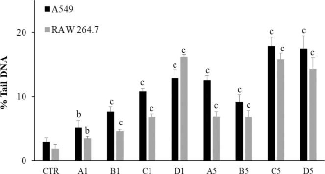
Inhalation of asbestos fibres can cause lung and pleural diseases in humans and constitutes a severe public health threat worldwide. The aim of the present study was to assess the biological effects induced in both pulmonary cells (A549) and monocyte/macrophage (RAW 264.7) cell lines by combustion slags obtained from asbestos through a self-sustained high-temperature synthesis (SHS) reaction. The SHS reaction involves rapid thermal treatment and displays great ability to neutralise asbestos. Cytotoxicity, redox status imbalance, lipid peroxide production, DNA strand breaks (comet assay) and chromosomal aberrations (cytokinesis block micronucleus test) were evaluated in cells exposed either to untreated asbestos fibres or to grinded SHS-generated slags of different granulometry, tested in cultured cells at varying doses and for varying exposure times. Our results show that asbestos fibres cause redox status imbalance, especially in monocyte/macrophage cell lines. Moreover, they promote lipid peroxidation and trigger genomic alterations. When the cells were exposed to slag powders, which are the products of SHS asbestos treatment, generation of lipid peroxides and induction of DNA strand breaks still persisted, due to the high content in iron and other metals detected in these samples. However, there was an attenuation of redox status imbalance and an absence of chromosomal aberrations, which probably reflects the loss of the asbestos fibrous structure following SHS reaction, as demonstrated by electron microscopy analyses. In conclusions, SHS-treated asbestos wastes can potentially have deleterious health effects due to the oxidative stress induced by inhaled powders but they loose the asbestos ability to induce chromosomal alterations.
Overview
The claims and statistics of this paper haven’t been checked yet.
Run the check — free, no account needed. It reads the open-access text and checks the sources it cites.
Integrity signals
Registry
Cites
Statistics
Figures
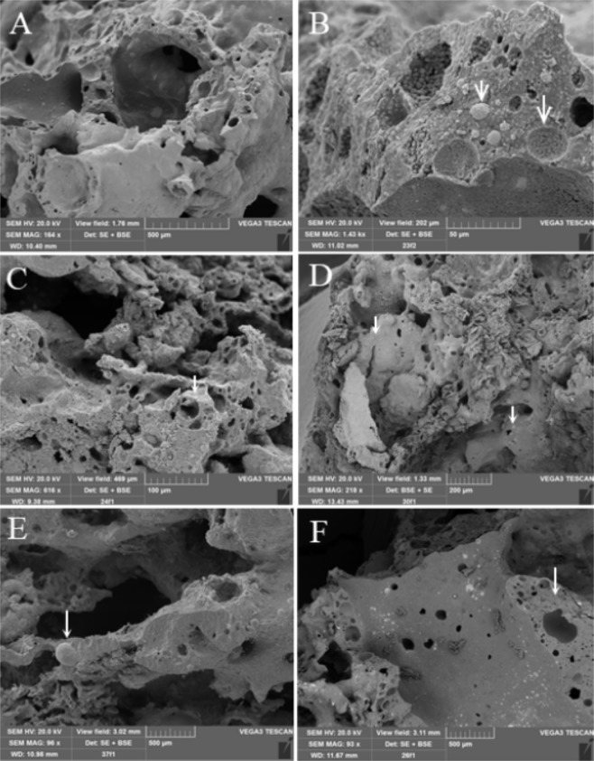
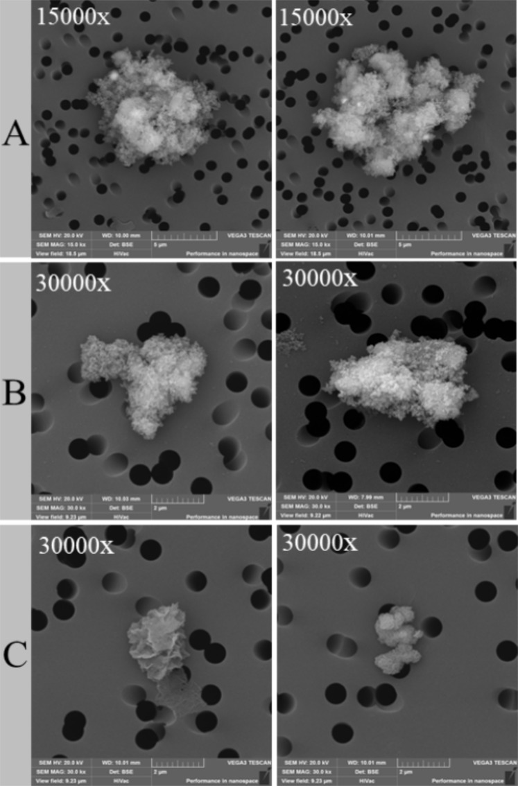
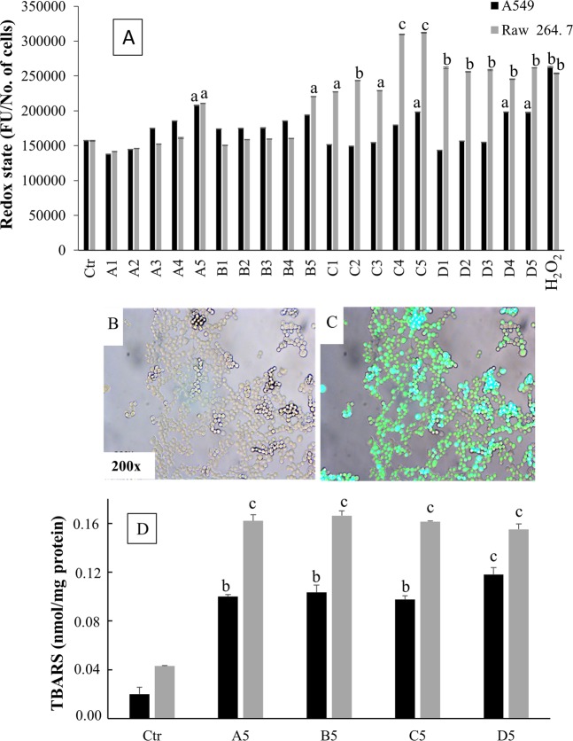
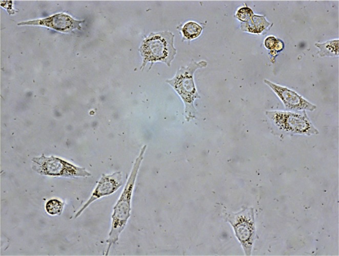
Verification
Claims the paper makes
The claims and statistics of this paper haven’t been checked yet.
Run the check — free, no account needed. It reads the open-access text and checks the sources it cites.
Claims other papers make about this one
Each is checked against this paper’s own text: does it say what the citing paper says it does?
In the citing paper
“Asbestos carcinogenesis depends on numerous factors, including inhaled dose, dimension, physical durability, surface features, crystallinity, and chemical composition.”
From Evaluation of the genotoxic and transformation potential induced by asbestos compared to cleavage fragments (2025) · cites this paper as [20]
In the citing paper
“All these factors have been linked to the formation of RNOs, the release of cytokines and growth factors promoting chronic inflammation, leukocyte invasion, and DNA damage20–22.”
From Evaluation of the genotoxic and transformation potential induced by asbestos compared to cleavage fragments (2025) · cites this paper as [20]
In the citing paper
“According to previous studies, DNA damage was evaluated by single-cell gel electrophoresis20.”
From Evaluation of the genotoxic and transformation potential induced by asbestos compared to cleavage fragments (2025) · cites this paper as [20]
Self-check
Percentages, sums and reported differences re-computed from the paper’s own numbers.
The self-check needs the paper’s full text.
Citations
Cites
No references could be resolved yet.
Research works printed but not linked: 37
- International Agency for Research on Cancer. Arsenic, Metals, Fibres and Dusts A Review of Human Carcinogens. Lyon, IARC Monographs on the Evaluation of Carcinogenic Risks to Humans vol. 100C, 219–309 (2012).
- Vainio, H. Epidemics of asbestos-related diseases something old, something new. Scand. J. Work Environ. Health 41, 1–4 (2015). · DOI
- Noonan, C. W. Environmental asbestos exposure and risk of mesothelioma. Ann. Transl. Med. 5, 234 (2017). · DOI
- Gaggero, L. et al. Airborne asbestos fibres monitoring in tunnel excavation. J. Environ. Manag. 196, 583–593 (2017).
- Sanchez, V. C., Pietruska, J. R., Miselis, N. R., Hurt, R. H. & Kane, A. B. Biopersistence and potential adverse health impacts of fibrous nanomaterials: what have we learned from asbestos? Wiley Interdiscip. Rev. Nanomed. Nanobiotechnol. 1, 511–529 (2009). · DOI · Paper page
- Furuya, S., Chimed-Ochir, O., Takahashi, K., David, A. & Takala, J. Global Asbestos Disaster. Int. J. Environ. Res. Public Health 16, 15 (2018). · DOI
- International Labour Organization (ILO) Resolution Concerning Asbestos. In Proceedings of the 95th Session of the International Labour Conference, Geneva, Switzerland, 31 May–16 June (2006).
- Takala, J. Eliminating occupational cancer. Ind. Health 53, 307–309 (2015). · DOI
- Marsili, D. et al. Prevention of Asbestos-Related Disease in Countries Currently Using Asbestos. Int. J. Environ. Res. Public Health 13, 494 (2016). · DOI
- Plescia, P. et al. Mechanochemical treatment to recycling asbestos containing waste. Waste Manag. 23, 209–218 (2003). · DOI
- Gaggero, L. & Ferretti, M. The Self-sustained High temperature Synthesis (SHS) technology as novel approach in the management of asbestos waste. J. Environ. Manag. 15, 246–256 (2018).
- Gaggero, L., Ferretti, M. & Caratto, V. Self-sustained combustion synthesis and asbestos-bearing waste: scaling up from laboratory towards pre-industrial size plant. Energy Procedia 97, 515–522 (2016). · DOI
- Kumar, R. K., O’Grady, R., Li, W. & Velan, G. M. Mitogenic activity for fibroblasts induced by silica and titanium dioxide particles in vitro and in vivo. Int. J. Exp. Pathol. 73, 573–583 (1992).
- Melloni, B. et al. Effect of exposure to silica on human alveolar macrophages in supporting growth activity in type II epithelial cells. Thorax. 51, 781–786 (1996). · DOI
- Huang, S. X., Jaurand, M. C., Kamp, D. W., Whysner, J. & Hei, T. K. Role of mutagenicity in asbestos fiber-induced carcinogenicity and other diseases. J. Toxicol. Environ. Health B Crit. Rev. 14, 179–245 (2011). · DOI · Paper page
- Stuart, B. O. Deposition and clearance of inhaled particles. Environ. Health Perspect. 55, 369–390 (1984). · DOI
- Terzano, C., Di Stefano, F., Conti, V., Graziani, E. & Petroianni, A. Air pollution ultrafine particles: toxicity beyond the lung. Eur. Rev. Med. Pharmacol. Sci. 14, 809–821 (2010).
- Miserocchi, G., Sancini, G., Mantegazza, F. & Chiappino, G. Translocation pathways for inhaled asbestos fibers. Environ. Health 24, 7–4 (2008).
- Ishida, T. et al. Live–cell imaging of macrophage phagocytosis of asbestos fibers under fluorescence microscopy. Genes Environ. 41, 14 (2019). · DOI
- Yoshida, R. et al. Urinary 8-oxo-7, 8-dihydro-2’-deoxyguanosine and biopyrrins levels among construction workers with asbestos exposure history. Ind. Health 39, 186–188 (2001). · DOI
- Santini, M. T. et al. Environmental fine particulate matter (PM 2.5) activates the RAW 264.7 macrophage cell line even at very low concentrations as revealed by 1H NMR. Chem. Res. Toxicol. 17, 63–74 (2004). · DOI
- Hooshmand, S. et al. Evidence for anti-inflammatory and antioxidative properties of dried plum polyphenols in macrophage RAW 264.7 cells. Food Funct. 6, 1719–1725 (2015). · DOI
- Luzhna, L., Kathiria, P. & Kovalchuk, O. Micronuclei in genotoxicity assessment: from genetics to epigenetics and beyond. Front. Genet. 11, 4–131 (2013).
- Schoonen, M. A. A. et al. Mineral-Induced Formation of Reactive Oxygen Species. Rev. Mineral. Geochem. 64, 179–221 (2006). · DOI
- Visalli, G., Baluce, B., Bertuccio, M., Picerno, I. & Di Pietro, A. Mitochondrial-mediated apoptosis pathway in alveolar epithelial cells exposed to the metals in combustion-generated particulate matter. J. Toxicol. Environ. Health A 78, 697–709 (2015).
- Visalli, G. et al. The role of the iron catalyst in the toxicity of multi-walled carbon nanotubes (MWCNTs). J. Trace Elem. Med. Biol. 43, 153–160 (2017). · DOI
- Lien, H. L., Yu, C. H., Kamali, S. & Sahu, R. S. Bimetallic Fe/Al system: an all-in-one solid-phase Fenton reagent for generation of hydroxyl radicals under oxic conditions. Sci. Total Environ. 673, 480–488 (2019). · DOI
- Munir, Z. A. Synthesis of high temperature materials by Self-Propagating Combustion methods. Amer. Ceram. Soc. Bull. 67, 342–349 (1988).
- Cardile, V. et al. Fluoro-edenite fibers induce expression of Hsp70 and inflammatory response. Int. J. Environ. Res. Public Health 4, 195–202 (2007). · DOI
- Musumeci, G., Cardile, V., Fenga, C., Caggia, S. & Loreto, C. Mineral fibre toxicity: expression of retinoblastoma (Rb) and phospho-retinoblastoma (pRb) protein in alveolar epithelial and mesothelial cell lines exposed to fluoro-edenite fibres. Cell. Biol. Toxicol. 27, 217–225 (2011). · DOI
- Kalyanaraman, B. et al. Measuring reactive oxygen and nitrogen species with fluorescent probes: challenges and limitations. Free Radic. Biol. Med. 52, 1–6 (2012). · DOI
- Ohkawa, H., Ohishi, N. & Yagi, K. Assay for lipid peroxides in animal tissues bythiobarbituric acid reaction. Anal. Biochem. 95, 351–358 (1979). · DOI
- Tice, R. R. et al. Single cell gel/comet assay: guidelines for in vitro and in vivo genetic toxicology testing. Environ. Mol. Mutagen. 35, 206–222 (2000). · DOI
- Ozcagli, E. et al. Effects of 3-monochloropropane-1,2-diol (3-MCPD) and its metabolites on DNA damageand repair under in vitro conditions. Food Chem. Toxicol. 89, 1–7 (2016). · DOI
- La Maestra, S. et al. DNA damage in exfoliated cells and histopathological alterations in the urinary tract of mice exposed to cigarette smoke and treated with chemopreventive agents. Carcinogenesis 34, 183–189 (2012). · DOI
- Fenech, M. Cytokinesis-block micronucleus cytome assay. Nat. Protoc. 2, 1084–1104 (2007). · DOI
- Kirsch-Volders. M. et al. Report from the in vitro micronucleus assay working group. Mutat. Res. 540, 153–163 (2003). Erratum in: Mutat. Res. 564, 97–100 (2004). · DOI
Cited by
Sorted by citations
La Maestra, S., Militello, G. M., Alberti, S., Benvenuti, M., & Gaggero, L. (2025) · Scientific Reports · doi:10.1038/s41598-025-86325-z · Cited by 10
Self-citation
Metadata
- Authors
- La Maestra, S., Micale, R. T., Ferretti, M., Izzotti, A., & Gaggero, L.
- Venue
- Scientific Reports
- Published
- 2020-05-22
- Volume
- 10
- Issue
- 1
- ISSN
- 2045-2322
- PMC
- PMC7244567
- Cited by
- 14
- References
- 0
- Language
- en
About these checks
Registry, retraction, Cites and statistics results are computed, not judged, with one exception: a printed reference that no index lists is read once by a model before it counts as a citation error. Claims are extracted by a model and kept only when their quote appears verbatim in the text; each cited source is then checked, with a model’s help, for whether it says what the claim relies on. Those verdicts can be wrong — read the claim’s quote and the cited source before relying on one.
Statistics and arithmetic are recomputed, not judged.
Confidence bands are shown only for fields calibrated on our annotated test set. None is calibrated yet, so no band is shown.
What counts as a claim ›How we measure confidence ›How we check this ›
Are you an author, or do you see a problem with this page? Request a review
