

The DOI matches its Crossref record; 1 of the 1 references it cites were found in a registry or an index.
Establishment of an experimental model for MHC homo-to-hetero transplantation
Open accessAbstract
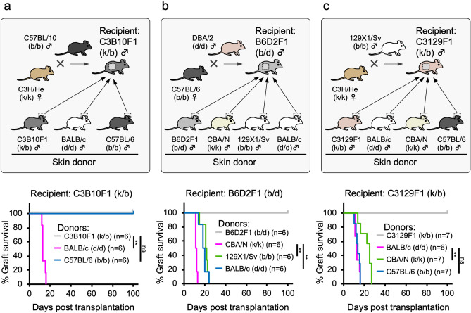
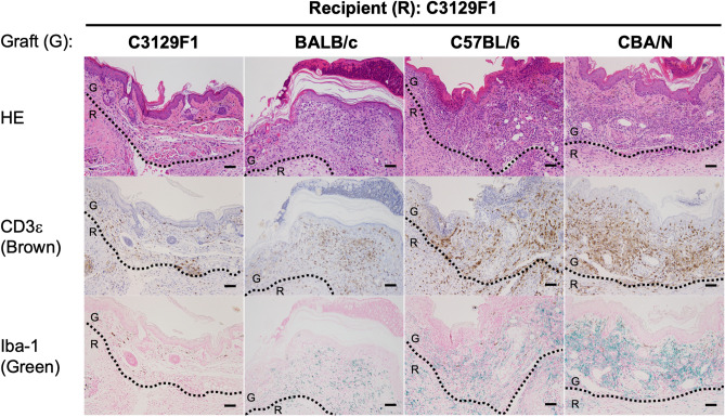
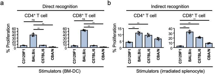
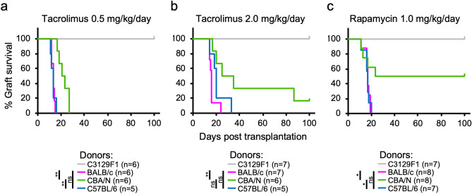
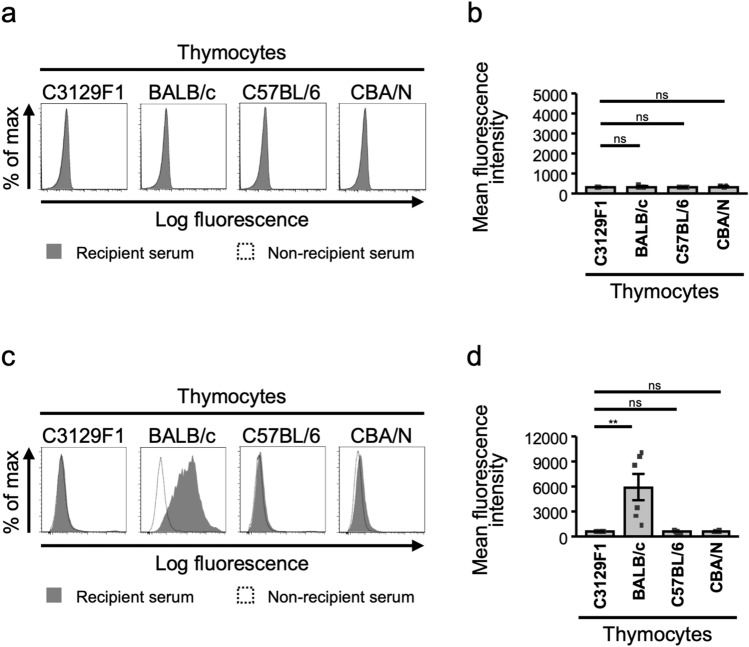
Preventing rejection is a major challenge in transplantation medicine, even when using pluripotent stem cell-derived grafts. In iPS cell (iPSC)-based transplantation, to reduce the risk of rejection, it is thought to be optimal that preparing the cells from donors whose human leukocyte antigen-haplotype are homozygous. Generally, this approach is referred to as major histocompatibility complex (MHC) homo-to-hetero transplantation, which is MHC-matched but minor antigen-mismatched. To investigate the immune response in the MHC homo-to-hetero transplantation, we established a murine experimental system in which MHC-matched but minor antigen-mismatched tissue (skin) grafts were transplanted into MHC-heterozygous recipients. Unexpectedly, only minor antigen-mismatched grafts were rejected at the same time points as rejection of fully allogeneic grafts. A vigorous anti-donor type T cell response was detected in vitro and conventional immunosuppressants targeting T cell activation had limited effects on controlling rejection. However, anti-donor antibodies were not detected only in the minor antigen-mismatched transplantation. This murine transplantation model can be used to further analyze immunological subjects for MHC homo-to-hetero iPSC-based transplantation.
Overview
The claims and statistics of this paper haven’t been checked yet.
Run the check — free, no account needed. It reads the open-access text and checks the sources it cites.
Integrity signals
Registry
Cites
Statistics
Figures
Verification
Claims the paper makes
The claims and statistics of this paper haven’t been checked yet.
Run the check — free, no account needed. It reads the open-access text and checks the sources it cites.
Claims other papers make about this one
Each is checked against this paper’s own text: does it say what the citing paper says it does?
The source backs part of the claim, not all of it — or backs it for a different population, condition or comparison.
In the citing paper
“There are various antigens other than MHC molecules, so-called minor histocompatibility antigens, which interact with MHC molecules to activate T cells.”
From Immunogenicity of decellularized extracellular matrix scaffolds: a bottleneck in tissue engineering and regenerative medicine (2023) · cites this paper as [5]
Experimental — accuracy not yet measuredThe cited source says what the claim attributes to it.
In the citing paper
“Minor antigen-mismatched grafts could be potent enough to cause rejection in MHC-matched grafts [5].”
From Immunogenicity of decellularized extracellular matrix scaffolds: a bottleneck in tissue engineering and regenerative medicine (2023) · cites this paper as [5]
Self-check
Percentages, sums and reported differences re-computed from the paper’s own numbers.
The self-check needs the paper’s full text.
Citations
Cites
Taylor, C. J., Peacock, S., Chaudhry, A. N., Bradley, J. A., & Bolton, E. M. (2012) · Cell stem cell · doi:10.1016/j.stem.2012.07.014 · Cited by 428
Research works printed but not linked: 17
- Ingulli, E. Mechanism of cellular rejection in transplantation. Pediatr. Nephrol.25, 61–74 (2010). · DOI · Paper page
- De Bueger, M. & Goulmy, E. Human minor histocompatibility antigens. Transpl. Immunol.1, 28–38 (1993). · DOI
- Nakatsuji, N., Nakajima, F. & Tokunaga, K. HLA-haplotype banking and iPS cells. Nat. Biotechnol.26, 739–740 (2008). · DOI
- Morizane, A. et al. MHC matching improves engraftment of iPSC-derived neurons in non-human primates. Nat. Commun.8, 1–12 (2017). · DOI · Paper page
- Kawamura, T. et al. Cardiomyocytes derived from MHC-homozygous induced pluripotent stem cells exhibit reduced allogeneic immunogenicity in MHC-matched non-human primates. Stem Cell Rep.6, 312–320 (2016). · DOI · Paper page
- Opelz, G. & Döhler, B. Effect of human leukocyte antigen compatibility on kidney graft survival: Comparative analysis of two decades. Transplantation84, 137–143 (2007). · DOI
- Jones, N. D. et al. Differential susceptibility of heart, skin, and islet allografts to T cell-mediated rejection. J. Immunol.166, 2824–2830 (2001). · DOI
- Neofytou, E., O’Brien, C. G., Couture, L. A. & Wu, J. C. Hurdles to clinical translation of human induced pluripotent stem cells. J. Clin. Invest.125, 2551–2557 (2015). · DOI
- Becker, L. E., Morath, C. & Suesal, C. Immune mechanisms of acute and chronic rejection. Clin. Biochem.49, 320–323 (2016). · DOI
- Vannaprasaht, S. et al. Personalized tacrolimus doses determined by CYP3A5 genotype for induction and maintenance phases of kidney transplantation. Clin. Ther.35, 1762–1769 (2013). · DOI
- Flechner, S. M. et al. Kidney transplantation without calcineurin inhibitor drugs: A prospective, randomized trial of sirolimus versus cyclosporine. Transplantation74, 1070–1076 (2002). · DOI
- Colvin, R. B. & Smith, R. N. Antibody-mediated organ-allograft rejection. Nat. Rev. Immunol.5, 807–817 (2005). · DOI
- Wiltshire, T. et al. Genome-wide single-nucleotide polymorphism analysis defines haplotype patterns in mouse. Proc. Natl. Acad. Sci. USA100, 3380–3385 (2003). · DOI
- Piazza, A. et al. Impact of donor-specific antibodies on chronic rejection occurrence and graft loss in renal transplantation: Posttransplant analysis using flow cytometric techniques1. Transplantation71, 1106–1112 (2001). · DOI
- Goldberg, E. H., Goble, R. & Jackman, S. H. Monoclonal antibodies defining the skin-selective alloantigens, Skn. Immunogenetics31, 393–395 (1990). · DOI
- Zhang, Q. & Reed, E. F. Non-MHC antigenic targets of the humoral immune response in transplantation. Curr. Opin. Immunol.22, 682–688 (2010). · DOI
- Sasaki, H. et al. New immunosuppressive cell therapy to prolong survival of induced pluripotent stem cell-derived allografts. Transplantation99, 2301–2310 (2015). · DOI
Cited by
Sorted by citations
Kasravi, M., Ahmadi, A., Babajani, A., Mazloomnejad, R., Hatamnejad, M. R., Shariatzadeh, S., Bahrami, S., & Niknejad, H. (2023) · Biomaterials Research · doi:10.1186/s40824-023-00348-z · Cited by 316
“Minor antigen-mismatched grafts could be potent enough to cause rejection in MHC-matched grafts [5].”
Background
Metadata
- Authors
- Murata, T., Wada, H., Otsuka, R., Sasaki, A., Tsuji, H., Itoh, M., Eguchi, N., Kawai, T., & Seino, K.-I.
- Affiliations
- Hokkaido University (JP)
- Massachusetts General Hospital (US)
- Corresponding author
- Seino, Ken-ichiro
- Venue
- Scientific Reports
- Published
- 2020-08-11
- Volume
- 10
- Issue
- 1
- Pages
- 13560-13560
- ISSN
- 2045-2322 (ISSN-L)
- PMC
- PMC7421494
- OpenAlex
- W3048669947
- Cited by
- 10
- References
- 1
- Citations by year
- 2025: 3 · 2024: 1 · 2023: 4 · 2022: 2
- FWCI
- 1.27
- Type
- journal article
- Language
- en
- Open access
- gold
- doi.org
- Locations
- Scientific Reports (publishedVersion, cc-by)
- PubMed Central (publishedVersion, cc-by)
- PubMed (publishedVersion)
- Hokkaido University Collection of Scholarly and Academic Papers (Hokkaido University) (submittedVersion, cc-by)
- Institutional Repositories DataBase (IRDB) (submittedVersion, cc-by)
- Funders
- Ministry of Education, Culture, Sports, Science and Technology
- Japan Agency for Medical Research and Development
- TERUMO LIFE SCIENCE FOUNDATION
- Topics
- Health Sciences › Medicine › Transplantation › Organ and Tissue Transplantation Research
- Health Sciences › Medicine › Surgery › Transplantation: Methods and Outcomes
- Life Sciences › Biochemistry, Genetics and Molecular Biology › Molecular Biology › Pluripotent Stem Cells Research
- Keywords
- graft rejection, skin grafts, immunosuppressants, anti-donor antibodies, murine transplantation model, HLA haplotype matching
- MeSH terms
- Animals, Female, Graft Rejection, Graft Survival, Histocompatibility Antigens, Isoantibodies, Male, Mice, Inbred BALB C, Mice, Inbred CBA, Mice, Inbred C57BL, Models, Theoretical, Tissue Donors, Transplantation, Heterologous, Transplantation, Homologous, Skin Transplantation, Mice
About these checks
Registry, retraction, Cites and statistics results are computed, not judged, with one exception: a printed reference that no index lists is read once by a model before it counts as a citation error. Claims are extracted by a model and kept only when their quote appears verbatim in the text; each cited source is then checked, with a model’s help, for whether it says what the claim relies on. Those verdicts can be wrong — read the claim’s quote and the cited source before relying on one.
Statistics and arithmetic are recomputed, not judged.
Confidence bands are shown only for fields calibrated on our annotated test set. None is calibrated yet, so no band is shown.
What counts as a claim ›How we measure confidence ›How we check this ›
Are you an author, or do you see a problem with this page? Request a review
