

The DOI matches its Crossref record; 1 of the 1 references it cites were found in a registry or an index.
UV-C mediated accumulation of pharmacologically significant phytochemicals under light regimes in in vitro culture of Fagonia indica (L.)
Abstract
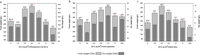
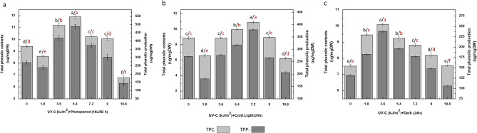
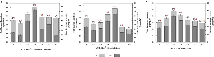
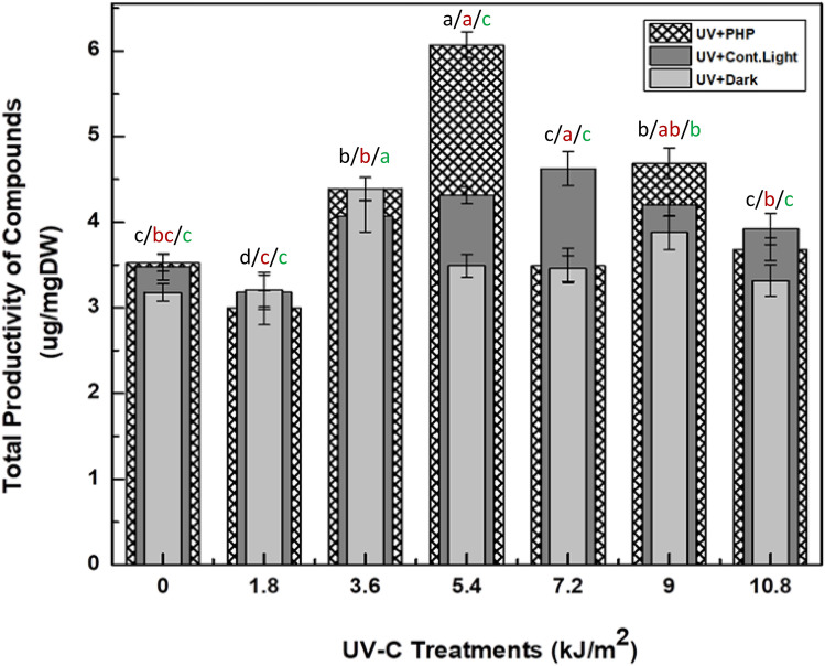
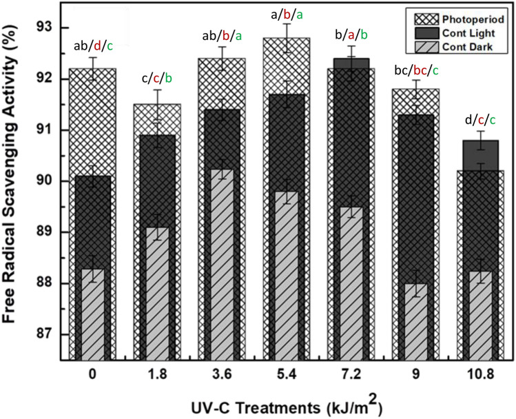
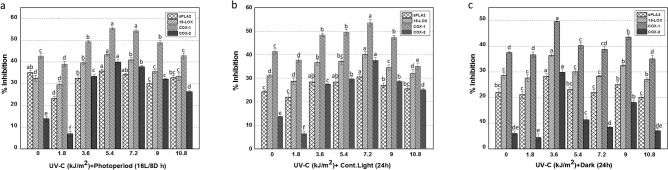
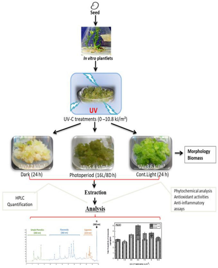
Fagonia indica(L.) is an important medicinal plant with multitude of therapeutic potentials. Such application has been attributed to the presence of various pharmacological important phytochemicals. However, the inadequate biosynthesis of such metabolites in intact plants has hampered scalable production. Thus, herein, we have established an in vitro based elicitation strategy to enhance such metabolites in callus culture of F. indica. Cultures were exposed to various doses of UV radiation (UV-C) and grown in different photoperiod regimes and their impact was evaluated on biomass accumulation, biosynthesis of phytochemicals along antioxidant expression. Cultures grown under photoperiod (16L/8D h) after exposure to UV-C (5.4 kJ/m 2) accumulated optimal biomass (438.3 g/L FW; 16.4 g/L DW), phenolics contents (TPC: 11.8 μgGAE/mg) and flavonoids contents (TFC: 4.05 μgQE/mg). Similarly, HPLC quantification revealed that total production (6.967 μg/mg DW) of phytochemicals wherein kaempferol (1.377 μg/mg DW), apigenin (1.057 μg/mg DW), myricetin (1.022 μg/mg DW) and isorhamnetin (1.022 μg/mg DW) were recorded highly accumulated compounds in cultures at UV-C (5.4 kJ/m 2) dose than other UV-C radiations and light regimes.. The antioxidants activities examined as DPPH (92.8%), FRAP (182.3 µM TEAC) and ABTS (489.1 µM TEAC) were also recorded highly expressed by cultures under photoperiod after treatment with UV-C dose 5.4 kJ/m 2. Moreover, same cultures also expressed maximum % inhibition towards phospholipase A2 (sPLA2: 35.8%), lipoxygenase (15-LOX: 43.3%) and cyclooxygenases (COX-1: 55.3% and COX-2: 39.9%) with 1.0-, 1.3-, 1.3- and 2.8-fold increased levels as compared with control, respectively. Hence, findings suggest that light and UV can synergistically improve the metabolism of F. indica and could be used to produce such valuable metabolites on commercial scale.
Overview
The claims and statistics of this paper haven’t been checked yet.
Run the check — free, no account needed. It reads the open-access text and checks the sources it cites.
Integrity signals
Registry
Cites
Statistics
Figures
Verification
Claims the paper makes
The claims and statistics of this paper haven’t been checked yet.
Run the check — free, no account needed. It reads the open-access text and checks the sources it cites.
Claims other papers make about this one
Each is checked against this paper’s own text: does it say what the citing paper says it does?
In the citing paper
“In recent times, available reports have suggested that CQDs exhibit anticancer potential in various types of cancer.22”
Self-check
Percentages, sums and reported differences re-computed from the paper’s own numbers.
The self-check needs the paper’s full text.
Citations
Cites
Antioxidant activity of some algerian medicinal plants extracts containing phenolic compounds
Djeridane, A., Yousfi, M., Nadjemi, B., Boutassouna, D., Stocker, P., & Vidal, N. (2006) · Food Chemistry · doi:10.1016/j.foodchem.2005.04.028 · Cited by 1,792
Research works printed but not linked: 91
- Khan, T., Ullah, M. A., Garros, L., Hano, C. & Abbasi, B. H. Synergistic effects of melatonin and distinct spectral lights for enhanced production of anti-cancerous compounds in callus cultures of Fagonia indica. J. Photochem. Photobiol. B 190, 163–171 (2019). · DOI
- Khan, T., Khan, T., Hano, C. & Abbasi, B. H. Effects of chitosan and salicylic acid on the production of pharmacologically attractive secondary metabolites in callus cultures of Fagonia indica. Ind. Crops Prod. 129, 525–535 (2019). · DOI
- Khan, T., Abbasi, B. H. & Khan, M. A. The interplay between light, plant growth regulators and elicitors on growth and secondary metabolism in cell cultures of Fagonia indica. J. Photochem. Photobiol. B 185, 153–160 (2018). · DOI
- Farheen, R., Siddiqui, B. S., Mahmood, I., Simjee, S. U. & Majeed, S. Triterpenoids and triterpenoid saponins from the aerial parts of Fagonia indica Burm. Phytochem. Lett. 13, 256–261 (2015). · DOI
- Zhang, W., Krohn, K., Draeger, S. & Schulz, B. Bioactive isocoumarins isolated from the endophytic fungus Microdochium bolleyi. J. Nat. Prod. 71, 1078–1081 (2008). · DOI
- Lam, M., Carmichael, A. R. & Griffiths, H. R. An aqueous extract of Fagonia cretica induces DNA damage, cell cycle arrest and apoptosis in breast cancer cells via FOXO3a and p53 expression. PLoS ONE 7, e40152 (2012). · DOI
- Khan, T., Abbasi, B. H., Khan, M. A. & Shinwari, Z. K. Differential effects of thidiazuron on production of anticancer phenolic compounds in callus cultures of Fagonia indica. Appl. Biochem. Biotechnol. 179, 46–58 (2016). · DOI
- Balasundram, N., Sundram, K. & Samman, S. Phenolic compounds in plants and agri-industrial by-products: antioxidant activity, occurrence, and potential uses. Food Chem. 99, 191–203 (2006). · DOI
- Cheynier, V. Phenolic compounds: from plants to foods. Phytochem. Rev. 11, 153–177 (2012). · DOI
- Freeman, B. C. & Beattie, G. A. An overview of plant defenses against pathogens and herbivores. Plant Health Instr. (2008). · DOI
- Dai, J. & Mumper, R. J. Plant phenolics: extraction, analysis and their antioxidant and anticancer properties. Molecules 15, 7313–7352 (2010). · DOI
- Zoratti, L., Karppinen, K., Luengo Escobar, A., Häggman, H. & Jaakola, L. Light-controlled flavonoid biosynthesis in fruits. Front. Plant Sci. 5, 534 (2014). · DOI
- Soto-Vaca, A., Gutierrez, A., Losso, J. N., Xu, Z. & Finley, J. W. Evolution of phenolic compounds from color and flavor problems to health benefits. J. Agric. Food Chem. 60, 6658–6677 (2012). · DOI
- Alfermann, A. & Petersen, M. Natural product formation by plant cell biotechnology. Plant Cell Tissue Organ Cult. 43, 199–205 (1995). · DOI
- Gaj, M. D. Factors influencing somatic embryogenesis induction and plant regeneration with particular reference to Arabidopsis thaliana (L.) Heynh. Plant Growth Regul. 43, 27–47 (2004). · DOI
- Zhong, J.-J. Biochemical engineering of the production of plant-specific secondary metabolites by cell suspension cultures. In Plant Cells 1–26 (Springer, Berlin, Heidelberg, 2001). · DOI
- Hussain, M. S. et al. Current approaches toward production of secondary plant metabolites. J. Pharm. Bioallied Sci. 4, 10 (2012). · DOI
- Matkowski, A. Plant in vitro culture for the production of antioxidants—a review. Biotechnol. Adv. 26, 548–560 (2008). · DOI
- Gorelick, J. & Bernstein, N. Elicitation: an underutilized tool in the development of medicinal plants as a source of therapeutic secondary metabolites. In Advances in Agronomy Vol. 124, 201–230 (Academic Press, 2014). · DOI
- Roberts, S. C. & Shuler, M. L. Large-scale plant cell culture. Curr. Opin. Biotechnol. 8, 154–159 (1997). · DOI
- Mulabagal, V. & Tsay, H.-S. Plant cell cultures-an alternative and efficient source for the production of biologically important secondary metabolites. Int. J. Appl. Sci. Eng. 2, 29–48 (2004).
- Vasconsuelo, A. & Boland, R. Molecular aspects of the early stages of elicitation of secondary metabolites in plants. Plant Sci. 172, 861–875 (2007). · DOI
- Gangopadhyay, M. et al. UVC-priming mediated modulation of forskolin biosynthesis key genes against Macrophomina root rot of Coleus forskohlii—a tissue culture based sustainable approach. Phytochem. Lett. 17, 36–44 (2016). · DOI
- Xuan, T. D., Khanh, T. D., Khang, D. T., Quan, N. T. & Elzaawely, A. A. Changes in chemical composition, total phenolics and antioxidant activity of Alpinia (Alpinia zerumbet) leaves exposed to UV. Int. Lett. Natl. Sci. 55, 25–34 (2016).
- Freitas, A. et al. Effect of UV-C radiation on bioactive compounds of pineapple (Ananas comosus L. Merr.) by-products. J. Sci. Food Agric. 95, 44–52 (2015). · DOI · Paper page
- Li, D. et al. ABA and UV-C effects on quality, antioxidant capacity and anthocyanin contents of strawberry fruit (Fragaria ananassa Duch.). Postharvest Biol. Technol. 90, 56–62 (2014). · DOI
- Yin, X. et al. Insights into the mechanisms underlying ultraviolet-C induced resveratrol metabolism in grapevine (V. amurensis Rupr.) cv.“Tonghua-3”. Front. Plant Sci. 7, 503 (2016). · DOI
- Anjum, S., Abbasi, B. H., Doussot, J., Favre-Réguillon, A. & Hano, C. Effects of photoperiod regimes and ultraviolet-C radiations on biosynthesis of industrially important lignans and neolignans in cell cultures of Linum usitatissimum L. (Flax). J. Photochem. Photobiol. B Biol. 167, 216–227 (2017). · DOI
- Shama, G. & Alderson, P. UV hormesis in fruits: a concept ripe for commercialisation. Trends Food Sci. Technol. 16, 128–136 (2005). · DOI
- Turtoi, M. Ultraviolet light treatment of fresh fruits and vegetables surface: a review. J. Agroaliment. Process. Technol. 19, 325–337 (2013).
- Nazir, M. et al. Interactive effect of melatonin and UV-C on phenylpropanoid metabolite production and antioxidant potential in callus cultures of purple basil (Ocimum basilicum L. var. s purpurascens). Molecules 25, 1072 (2020). · DOI
- Ullah, M. A. et al. Effect of ultraviolet-C radiation and melatonin stress on biosynthesis of antioxidant and antidiabetic metabolites produced in in vitro callus cultures of Lepidium sativum L. Int. J. Mol. Sci. 20, 1787 (2019). · DOI
- Versari, A., Parpinello, G. P., Tornielli, G. B., Ferrarini, R. & Giulivo, C. Stilbene compounds and stilbene synthase expression during ripening, wilting, and UV treatment in grape cv. Corvina. J. Agric. Food Chem. 49, 5531–5536 (2001). · DOI
- Yousefzadi, M. et al. The effect of light on gene expression and podophyllotoxin biosynthesis in Linum album cell culture. Plant Physiol. Biochem. 56, 41–46 (2012). · DOI
- Xu, A., Zhan, J.-C. & Huang, W.-D. Combined elicitation of chitosan and ultraviolet C enhanced stilbene production and expression of chitinase and β-1, 3-glucanase in Vitis vinifera cell suspension cultures. Plant Cell Tissue Organ Cult. (PCTOC) 124, 105–117 (2016). · DOI
- Jaakola, L. & Hohtola, A. Effect of latitude on flavonoid biosynthesis in plants. Plant Cell Environ. 33, 1239–1247 (2010). · DOI
- Yang, B., Zheng, J., Laaksonen, O., Tahvonen, R. & Kallio, H. Effects of latitude and weather conditions on phenolic compounds in currant (Ribes spp.) cultivars. J. Agric. Food Chem. 61, 3517–3532 (2013). · DOI
- Taylor, A. Some effects of photoperiod on the biosynthesis of phenylpropane derivatives in Xanthium. Plant Physiol. 40, 273 (1965). · DOI
- Carvalho, I. S., Cavaco, T., Carvalho, L. M. & Duque, P. Effect of photoperiod on flavonoid pathway activity in sweet potato (Ipomoea batatas (L.) Lam.) leaves. Food Chem. 118, 384–390 (2010). · DOI
- Nawkar, G. et al. UV-induced cell death in plants. Int. J. Mol. Sci. 14, 1608–1628 (2013). · DOI
- Katerova, Z., Todorova, D., Tasheva, K. & Sergiev, I. Influence of ultraviolet radiation on plant secondary metabolite production. Genet. Plant Physiol. 2, 113–144 (2012).
- Zacchini, M. & de Agazio, M. Spread of oxidative damage and antioxidative response through cell layers of tobacco callus after UV-C treatment. Plant Physiol. Biochem. 42, 445–450 (2004). · DOI
- Kok, B. On the inhibition of photosynthesis by intense light. Biochem. Biophys. Acta 21, 234–244 (1956). · DOI
- Lichtenthaler, H. K. et al. Effect of high light and high light stress on composition, function and structure of the photosynthetic apparatus. In Effects of Stress on Photosynthesis 353–370 (Springer, Dordrecht, 1983). · DOI
- Critchley, C. & Smillie, R. Leaf chlorophyll fluorescence as an indicator of high light stress (photoinhibition) in Cucumis sativus L. Funct. Plant Biol. 8, 133–141 (1981). · DOI
- Cleland, R. & Critchley, C. Studies on the mechanism of photoinhibition in higher plants. II. Inactivation by high light of photosystem II reaction center function in isolated spinach thylakoids and O2 evolving particles. Photobiochem. Photobiophys. 67, 1161–1165 (1985).
- Šiffel, P., Lebedev, N. & Šesták, Z. Changes in chloroplast absorption and fluorescence spectra during ontogeny of primary bean leaves. Photosynthetica 19, 127–137 (1985).
- Lichtenthaler, H. K. & Rinderle, U. The role of chlorophyll fluorescence in the detection of stress conditions in plants. CRC Crit. Rev. Anal. Chem. 19, S29–S85 (1988). · DOI
- Xu, A., Zhan, J.-C. & Huang, W.-D. Effects of ultraviolet C, methyl jasmonate and salicylic acid, alone or in combination, on stilbene biosynthesis in cell suspension cultures of Vitis vinifera L. cv. Cabernet Sauvignon. Plant Cell Tissue Organ Cult. (PCTOC) 122, 197–211 (2015). · DOI
- Lavola, A., Aphalo, P. J., Lahti, M. & Julkunen-Tiitto, R. Nutrient availability and the effect of increasing UV-B radiation on secondary plant compounds in Scots pine. Environ. Exp. Bot. 49, 49–60 (2003). · DOI
- Matsuura, H. N., de Costa, F., Yendo, A. C. A. & Fett-Neto, A. G. in Biotechnology for Medicinal Plants 171–190 (Springer, Berlin, 2013). · DOI
- Ali, M. & Abbasi, B. H. Light-induced fluctuations in biomass accumulation, secondary metabolites production and antioxidant activity in cell suspension cultures of Artemisia absinthium L. J. Photochem. Photobiol. B 140, 223–227 (2014). · DOI
- Yamamoto, S., Hayashi, S., Furusaki, S. & Shioya, S. 5-Aminolevulinic acid promotes callus growth and paclitaxel production in light-grown Taxus cuspidata suspension cultures. Eng. Life Sci. 15, 116–121 (2015). · DOI
- Fazal, H., Abbasi, B. H., Ahmad, N., Ali, M. & Ali, S. Sucrose induced osmotic stress and photoperiod regimes enhanced the biomass and production of antioxidant secondary metabolites in shake-flask suspension cultures of Prunella vulgaris L. Plant Cell Tissue Organ Cult. (PCTOC) 124, 573–581 (2016). · DOI
- Kovacs, E. & Keresztes, A. Effect of gamma and UV-B/C radiation on plant cells. Micron 33, 199–210 (2002). · DOI
- Jordan, B. R. Molecular response of plant cells to UV-B stress. Funct. Plant Biol. 29, 909–916 (2002). · DOI
- Jenkins, G. I. UV and blue light signal transduction in the regulation of flavonoid biosynthesis gene expression in Arabidopsis. In Cellular Integration of Signalling Pathways in Plant Development 71–82 (Springer, Berlin, Heidelberg, 1998). · DOI
- Nigro, F., Ippolito, A., Lattanzio, V., Di Venere, D. & Salerno, M. Effect of ultraviolet-C light on postharvest decay of strawberry. J. Plant Pathol. 82, 29–37 (2000).
- Tiecher, A., de Paula, L. A., Chaves, F. C. & Rombaldi, C. V. UV-C effect on ethylene, polyamines and the regulation of tomato fruit ripening. Postharvest Biol. Technol. 86, 230–239 (2013). · DOI
- Urban, L., Charles, F., de Miranda, M. R. A. & Aarrouf, J. Understanding the physiological effects of UV-C light and exploiting its agronomic potential before and after harvest. Plant Physiol. Biochem. 105, 1–11 (2016). · DOI
- Agati, G. et al. Functional roles of flavonoids in photoprotection: new evidence, lessons from the past. Plant Physiol. Biochem. 72, 35–45 (2013). · DOI
- Carbonell-Bejerano, P. et al. Solar ultraviolet radiation is necessary to enhance grapevine fruit ripening transcriptional and phenolic responses. BMC Plant Biol. 14, 183 (2014). · DOI
- Park, M.-H. & Kim, J.-G. Low-dose UV-C irradiation reduces the microbial population and preserves antioxidant levels in peeled garlic (Allium sativum L.) during storage. Postharvest Biol. Technol. 100, 109–112 (2015). · DOI
- Urban, L. et al. UV-C light and pulsed light as alternatives to chemical and biological elicitors for stimulating plant natural defenses against fungal diseases. Sci. Hortic. 235, 452–459 (2018). · DOI
- Waszczak, C., Carmody, M. & Kangasjärvi, J. Reactive oxygen species in plant signaling. Annu. Rev. Plant Biol. 69, 209–236 (2018). · DOI
- Mittler, R. ROS are good. Trends Plant Sci. 22, 11–19 (2017). · DOI · Paper page
- Sharma, K., Li, Y. & Schenk, P. M. UV-C-mediated lipid induction and settling, a step change towards economical microalgal biodiesel production. Green Chem. 16, 3539–3548 (2014). · DOI
- Wang, J. W. & Wu, J. Y. Effective elicitors and process strategies for enhancement of secondary metabolite production in hairy root cultures. In Biotechnology of Hairy Root Systems 55–89 (Springer, Berlin, Heidelberg, 2013). · DOI
- Moon, S. H. et al. A rich source of potential bioactive compounds with anticancer activities by Catharanthus roseus cambium meristematic stem cell cultures. J. Ethnopharmacol. 217, 107–117 (2018). · DOI
- Rivera-Pastrana, D. M., Gardea, A. A., Yahia, E. M., Martínez-Téllez, M. A. & González-Aguilar, G. A. Effect of UV-C irradiation and low temperature storage on bioactive compounds, antioxidant enzymes and radical scavenging activity of papaya fruit. J. Food Sci. Technol. 51, 3821–3829 (2014). · DOI
- Pinto, E. P. et al. The effect of postharvest application of UV-C radiation on the phenolic compounds of conventional and organic grapes (Vitis labrusca cv. ‘Concord’). Postharvest Biol. Technol. 120, 84–91 (2016). · DOI
- Windram, O. et al. Arabidopsis defense against Botrytis cinerea: chronology and regulation deciphered by high-resolution temporal transcriptomic analysis. Plant Cell 24, 3530–3557 (2012). · DOI
- Petit, A.-N. et al. Low responsiveness of grapevine flowers and berries at fruit set to UV-C irradiation. J. Exp. Bot. 60, 1155–1162 (2009). · DOI
- Ali, M. & Abbasi, B. H. Production of commercially important secondary metabolites and antioxidant activity in cell suspension cultures of Artemisia absinthium L. Ind. Crops Prod. 49, 400–406 (2013). · DOI
- Anjum, S. & Abbasi, B. H. Biomimetic synthesis of antimicrobial silver nanoparticles using in vitro-propagated plantlets of a medicinally important endangered species: Phlomis bracteosa. Int. J. Nanomed. 11, 1663 (2016).
- Erkan, M., Wang, S. Y. & Wang, C. Y. Effect of UV treatment on antioxidant capacity, antioxidant enzyme activity and decay in strawberry fruit. Postharvest Biol. Technol. 48, 163–171 (2008). · DOI
- Kim, D.-O., Chun, O. K., Kim, Y. J., Moon, H.-Y. & Lee, C. Y. Quantification of polyphenolics and their antioxidant capacity in fresh plums. J. Agric. Food Chem. 51, 6509–6515 (2003). · DOI
- Chen, H.-B. et al. Anti-inflammatory activity of coptisine free base in mice through inhibition of NF-κB and MAPK signaling pathways. Eur. J. Pharmacol. 811, 222–231 (2017). · DOI
- Elansary, H. O. et al. Bioactivities of traditional medicinal plants in Alexandria. Evid. Based Complement. Altern. Med. 2018, 1463579 (2018). · DOI
- Rathee, P. et al. Mechanism of action of flavonoids as anti-inflammatory agents: a review. Inflamm. Allergy Drug Targets (Former. Curr. Drug Targets Inflamm. Allergy) 8, 229–235 (2009).
- Bauer, R. & Tittel, G. Quality assessment of herbal preparations as a precondition of pharmacological and clinical studies. Phytomedicine 2, 193–198 (1996). · DOI
- Kumar, S. & Pandey, A. K. Chemistry and biological activities of flavonoids: an overview. Sci. World J. 2013, 162750 (2013). · DOI
- Ahmad, N. et al. Efficient regeneration and antioxidant potential in regenerated tissues of Piper nigrum L. Plant Cell Tissue Organ Cult. (PCTOC) 102, 129–134 (2010). · DOI
- Abbasi, B. H. et al. Shoot regeneration and free-radical scavenging activity in Silybum marianum L. Plant Cell Tissue Organ Cult. (PCTOC) 101, 371–376 (2010). · DOI
- Murashige, T. & Skoog, F. A revised medium for rapid growth and bio assays with tobacco tissue cultures. Physiol. Plant. 15, 473–497 (1962). · DOI
- Arias, J., Zapata, K., Rojano, B. & Arias, M. Effect of light wavelength on cell growth, content of phenolic compounds and antioxidant activity in cell suspension cultures of Thevetia peruviana. J. Photochem. Photobiol. B 163, 87–91 (2016). · DOI
- Benzie, I. F. & Strain, J. J. The ferric reducing ability of plasma (FRAP) as a measure of “antioxidant power”: the FRAP assay. Anal. Biochem. 239, 70–76 (1996). · DOI
- Tagliazucchi, D., Verzelloni, E., Bertolini, D. & Conte, A. In vitro bio-accessibility and antioxidant activity of grape polyphenols. Food Chem. 120, 599–606 (2010). · DOI
- Bourgeois, C. et al. Nettle (Urtica dioica L.) as a source of antioxidant and anti-aging phytochemicals for cosmetic applications. C. R. Chim. 19, 1090–1100 (2016). · DOI
- Wang, Y. et al. Simultaneous determination of rutin, luteolin, quercetin, and betulinic acid in the extract of Disporopsis pernyi (Hua) Diels by UPLC. J. Anal. Methods Chem. 2015, 130873 (2015). · DOI
- Usman, H. et al. Interactive Effects of wide-spectrum monochromatic lights on phytochemical production, antioxidant and biological activities of Solanum xanthocarpum callus cultures. Molecules 25, 2201 (2020). · DOI
Cited by
Sorted by citations
Mishra, S., das, K., Chatterjee, S., Sahoo, P., Kundu, S., Pal, M., Bhaumik, A., & Ghosh, C. K. (2023) · ACS Omega · doi:10.1021/acsomega.2c04964 · Cited by 65
Metadata
- Authors
- Abbasi, B. H., Khan, T., Khurshid, R., Nadeem, M., Drouet, S., & Hano, C.
- Venue
- Scientific Reports
- Published
- 2021-01-12
- Volume
- 11
- Issue
- 1
- ISSN
- 2045-2322
- PMC
- PMC7804141
- Cited by
- 32
- References
- 1
- Language
- en
About these checks
Registry, retraction, Cites and statistics results are computed, not judged, with one exception: a printed reference that no index lists is read once by a model before it counts as a citation error. Claims are extracted by a model and kept only when their quote appears verbatim in the text; each cited source is then checked, with a model’s help, for whether it says what the claim relies on. Those verdicts can be wrong — read the claim’s quote and the cited source before relying on one.
Statistics and arithmetic are recomputed, not judged.
Confidence bands are shown only for fields calibrated on our annotated test set. None is calibrated yet, so no band is shown.
What counts as a claim ›How we measure confidence ›How we check this ›
Are you an author, or do you see a problem with this page? Request a review
How to cite
Cite CoChat for the checks on this page, and the paper for its own findings.
- This page
- CoChat. (2026). Citation check: UV-C mediated accumulation of pharmacologically significant phytochemicals under light regimes in in vitro culture of Fagonia indica (L.). https://app.cochat.ai/papers/10.1038/s41598-020-79896-6
- The paper
- Abbasi, B. H., Khan, T., Khurshid, R., Nadeem, M., Drouet, S., & Hano, C. (2021). UV-C mediated accumulation of pharmacologically significant phytochemicals under light regimes in in vitro culture of Fagonia indica (L.). Scientific Reports. https://doi.org/10.1038/s41598-020-79896-6
