

The DOI matches its Crossref record; 34 of the 35 references it cites were found in a registry or an index.
Skin lesion classification of dermoscopic images using machine learning and convolutional neural network
Open accessAbstract
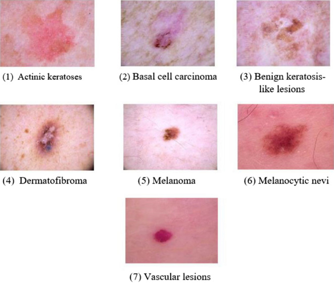
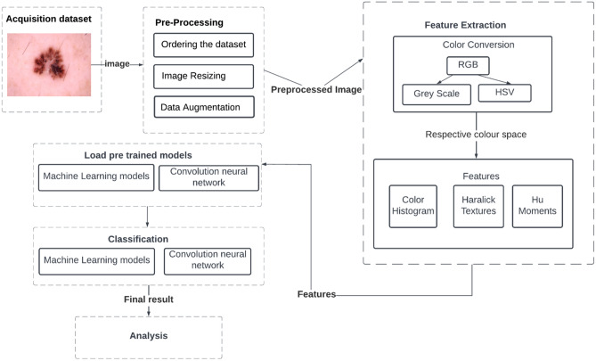
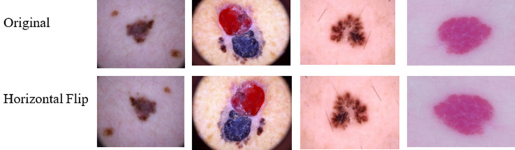
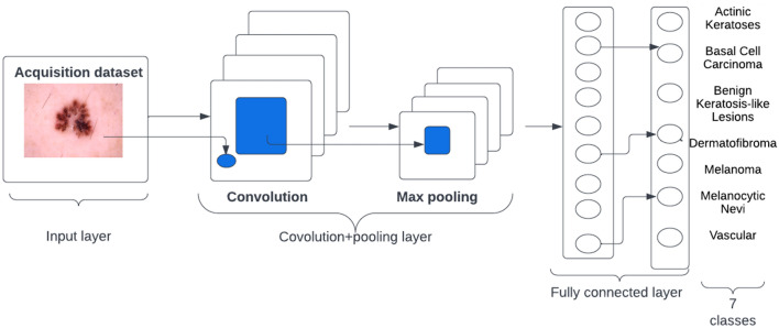
Detecting dangerous illnesses connected to the skin organ, particularly malignancy, requires the identification of pigmented skin lesions. Image detection techniques and computer classification capabilities can boost skin cancer detection accuracy. The dataset used for this research work is based on the HAM10000 dataset which consists of 10015 images. The proposed work has chosen a subset of the dataset and performed augmentation. A model with data augmentation tends to learn more distinguishing characteristics and features rather than a model without data augmentation. Involving data augmentation can improve the accuracy of the model. But that model cannot give significant results with the testing data until it is robust. The k-fold cross-validation technique makes the model robust which has been implemented in the proposed work. We have analyzed the classification accuracy of the Machine Learning algorithms and Convolutional Neural Network models. We have concluded that Convolutional Neural Network provides better accuracy compared to other machine learning algorithms implemented in the proposed work. In the proposed system, as the highest, we obtained an accuracy of 95.18% with the CNN model. The proposed work helps early identification of seven classes of skin disease and can be validated and treated appropriately by medical practitioners.
Overview
The claims and statistics of this paper haven’t been checked yet.
Run the check — free, no account needed. It reads the open-access text and checks the sources it cites.
Integrity signals
Registry
Cites
Statistics
Figures
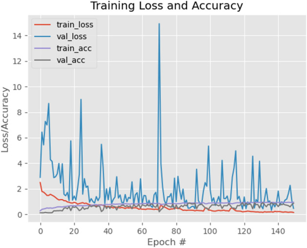
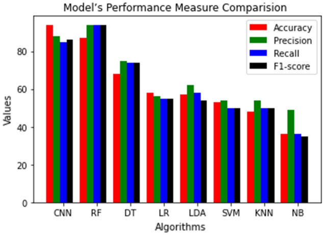
Verification
Claims the paper makes
The claims and statistics of this paper haven’t been checked yet.
Run the check — free, no account needed. It reads the open-access text and checks the sources it cites.
Claims other papers make about this one
Each is checked against this paper’s own text: does it say what the citing paper says it does?
The source backs part of the claim, not all of it — or backs it for a different population, condition or comparison.
In the citing paper
“However, most prior studies primarily focused on improving classification accuracy through increasingly complex architectures without sufficient attention to explaining model decisions (14–22).”
From Interpretable multimodal fusion for skin lesion classification using dermoscopic images and patient metadata (2026) · cites this paper as [14]
Experimental — accuracy not yet measuredThe source backs part of the claim, not all of it — or backs it for a different population, condition or comparison.
In the citing paper
“Later, CNN-based automatic feature extraction (14) and transfer learning with pretrained models (15, 18) improved representation learning.”
From Interpretable multimodal fusion for skin lesion classification using dermoscopic images and patient metadata (2026) · cites this paper as [14]
Experimental — accuracy not yet measuredThe source backs part of the claim, not all of it — or backs it for a different population, condition or comparison.
In the citing paper
“Most prior studies address this issue through conventional image-level augmentation (7, 14, 15, 17, 18, 20, 21, 23), which increases data volume but does not introduce genuine semantic diversity.”
From Interpretable multimodal fusion for skin lesion classification using dermoscopic images and patient metadata (2026) · cites this paper as [14]
Experimental — accuracy not yet measuredThe cited source says what the claim attributes to it.
In the citing paper
“Class imbalance remains a central challenge in seven-class skin lesion classification using HAM10000.”
From Interpretable multimodal fusion for skin lesion classification using dermoscopic images and patient metadata (2026) · cites this paper as [14]
The check wasn’t confident either way, so it gives no verdict — never disputed.
In the citing paper
“In the classification stage, most prior studies relied primarily on the fully connected layer for final classification (7, 14, 16–18, 20).”
From Interpretable multimodal fusion for skin lesion classification using dermoscopic images and patient metadata (2026) · cites this paper as [14]
Experimental — accuracy not yet measured
Self-check
Percentages, sums and reported differences re-computed from the paper’s own numbers.
The self-check needs the paper’s full text.
Citations
Cites
2 retracted
RETRACTED ARTICLE: Hybrid genetic algorithm and a fuzzy logic classifier for heart disease diagnosis
Gadekallu, T. R., Maddikunta, P. K. R., Lakshmanna, K., Rajput, D. S., Kaluri, R., & Srivastava, G. (2019) · Evolutionary Intelligence · doi:10.1007/s12065-019-00327-1 · Cited by 407
Self-citationRetractedRETRACTED ARTICLE: Skin lesion classification using decision trees and random forest algorithms
Dhivyaa, C. R., Sangeetha, K., -, M. B., Amaran, S., Vetriselvi, T., & Johnpaul, P. (2020) · Journal of Ambient Intelligence and Humanized Computing · doi:10.1007/s12652-020-02675-8 · Cited by 90
RetractedTschandl, P., Rosendahl, C., & Kittler, H. (2018) · Scientific Data · doi:10.1038/sdata.2018.161 · Cited by 3,628
Classification of Skin Disease Using Deep Learning Neural Networks with MobileNet V2 and LSTM
Srinivasu, P. N., SivaSai, J. G., Ijaz, M. F., Bhoi, A. K., Kim, W., & Kang, J. J. (2021) · Sensors · doi:10.3390/s21082852 · Cited by 763
Transfer learning using a multi-scale and multi-network ensemble for skin lesion classification
Mahbod, A., Schaefer, G., Wang, C., Dorffner, G., Ecker, R., & Ellinger, I. (2020) · Computer Methods and Programs in Biomedicine · doi:10.1016/j.cmpb.2020.105475 · Cited by 326
Skin cancer diagnosis based on optimized convolutional neural network
Zhang, N., Cai, Y.-X., Wang, Y.-Y., Tian, Y., Wang, X., & Badami, B. (2019) · Artificial Intelligence in Medicine · doi:10.1016/j.artmed.2019.101756 · Cited by 325
A multi-class skin Cancer classification using deep convolutional neural networks
Chaturvedi, S. S., Tembhurne, J. V., & Diwan, T. (2020) · Multimedia Tools and Applications · doi:10.1007/s11042-020-09388-2 · Cited by 297
Melanoma Recognition in Dermoscopy Images via Aggregated Deep Convolutional Features
Yu, Z., Jiang, X., Zhou, F., Qin, J., Ni, D., Chen, S., Lei, B., & Wang, T. (2018) · IEEE Transactions on Biomedical Engineering · doi:10.1109/tbme.2018.2866166 · Cited by 253
Khan, M. A., Akram, T., Zhang, Y., & Sharif, M. I. (2021) · Pattern Recognition Letters · doi:10.1016/j.patrec.2020.12.015 · Cited by 247
Convolutional Neural Networks Using MobileNet for Skin Lesion Classification
Sae-Lim, W., Wettayaprasit, W., & Aiyarak, P. (2019) · International Joint Conference on Computer Science and Software Engineering · doi:10.1109/jcsse.2019.8864155 · Cited by 233
Hameed, N., Shabut, A. M., Ghosh, M. K., & Hossain, M. A. (2019) · Expert Systems with Applications · doi:10.1016/j.eswa.2019.112961 · Cited by 217
Adegun, A. A., & Viriri, S. (2020) · IEEE Access · doi:10.1109/access.2020.3016651 · Cited by 212
DSNet: Automatic dermoscopic skin lesion segmentation
Hasan, M. K., Dahal, L., Samarakoon, P. N., Tushar, F. I., & Martí, R. (2020) · Computers in Biology and Medicine · doi:10.1016/j.compbiomed.2020.103738 · Cited by 210
Classification of Skin Lesions into Seven Classes Using Transfer Learning with AlexNet
Hosny, K. M., Kassem, M. A. A., & Fouad, M. M. (2020) · Journal of Imaging Informatics in Medicine · doi:10.1007/s10278-020-00371-9 · Cited by 181
Khan, M. A., Zhang, Y., Sharif, M., & Akram, T. (2021) · Computers & Electrical Engineering · doi:10.1016/j.compeleceng.2020.106956 · Cited by 173
Khan, M. A., Javed, M. Y., Sharif, M., Saba, T., & Rehman, A. U. (2019) · doi:10.1109/iccisci.2019.8716400 · Cited by 166
A DE-ANN Inspired Skin Cancer Detection Approach Using Fuzzy C-Means Clustering
Kumar, M., Alshehri, M., AlGhamdi, R. A., Sharma, P., & Deep, V. (2020) · Mobile Networks and Applications · doi:10.1007/s11036-020-01550-2 · Cited by 152
Hoang, L., Lee, S., Lee, E.-J., & Kwon, K. (2022) · Applied Sciences · doi:10.3390/app12052677 · Cited by 148
Abbas, S., Jalil, Z., Javed, A. R., Batool, I., Khan, M. Z., Noorwali, A., Gadekallu, T. R., & Akbar, A. (2021) · PeerJ Computer Science · doi:10.7717/peerj-cs.390 · Cited by 125
Polat, K., & Koç, K. O. (2020) · Journal of Artificial Intelligence and Systems · doi:10.33969/ais.2020.21006 · Cited by 124
Gundluru, N., Rajput, D. S., Lakshmanna, K., Kaluri, R., Shorfuzzaman, M., Uddin, M., & Khan, M. A. R. (2022) · Computational Intelligence and Neuroscience · doi:10.1155/2022/8512469 · Cited by 117
Self-citationChatterjee, S., Dey, D., & Munshi, S. (2019) · Computer Methods and Programs in Biomedicine · doi:10.1016/j.cmpb.2019.06.018 · Cited by 115
Risk-Aware Machine Learning Classifier for Skin Lesion Diagnosis
Mobiny, A., Singh, A., & Nguyen, H. V. (2019) · Journal of Clinical Medicine · doi:10.3390/jcm8081241 · Cited by 112
Skin Lesion Classification by Ensembles of Deep Convolutional Networks and Regularly Spaced Shifting
Thurnhofer‐Hemsi, K., López‐Rubio, E., Domínguez, E., & Elizondo, D. A. (2021) · IEEE Access · doi:10.1109/access.2021.3103410 · Cited by 104
Garcia-Arroyo, J. L., & García-Zapirain, B. (2018) · Computer Methods and Programs in Biomedicine · doi:10.1016/j.cmpb.2018.11.001 · Cited by 94
Sáez, A., Sánchez‐Monedero, J., Gutiérrez, P. A., & Hervás‐Martínez, C. (2015) · IEEE Transactions on Medical Imaging · doi:10.1109/tmi.2015.2506270 · Cited by 87
Nugroho, A. A., Slamet, İ., & Sugiyanto, S. (2019) · AIP conference proceedings · doi:10.1063/1.5141652 · Cited by 84
Diagnosis of Brain Tumor Using Light Weight Deep Learning Model with Fine-Tuning Approach
Shelatkar, T., Urvashi, D., Shorfuzzaman, M., Alsufyani, A., & Lakshmanna, K. (2022) · Computational and Mathematical Methods in Medicine · doi:10.1155/2022/2858845 · Cited by 83
Self-citationRajput, D. S., Basha, S. M., Xin, Q., Gadekallu, T. R., Kaluri, R., Lakshmanna, K., & Maddikunta, P. K. R. (2021) · Journal of Ambient Intelligence and Humanized Computing · doi:10.1007/s12652-021-03154-4 · Cited by 75
Self-citationHash-Based Deep Learning Approach for Remote Sensing Satellite Imagery Detection
Gadamsetty, S., Rupa, C., Ch, A., Iwendi, C., & Gadekallu, T. R. (2022) · Water · doi:10.3390/w14050707 · Cited by 68
Skin lesion classification enhancement using border-line features – The melanoma vs nevus problem
Pereira, P. M. M., Fonseca-Pinto, R., Paiva, R. P., Assunção, P., Tavora, L. M. N., Thomaz, L. A., & de Faria, S. M. M. (2019) · Biomedical Signal Processing and Control · doi:10.1016/j.bspc.2019.101765 · Cited by 54
Kernel sparse representation based model for skin lesions segmentation and classification
Moradi, N., & Mahdavi‐Amiri, N. (2019) · Computer Methods and Programs in Biomedicine · doi:10.1016/j.cmpb.2019.105038 · Cited by 50
Melanoma recognition in dermoscopy images using lesion's peripheral region information
Tajeddin, N. Z., & Asl, B. M. (2018) · Computer Methods and Programs in Biomedicine · doi:10.1016/j.cmpb.2018.05.005 · Cited by 39
Research works printed but not linked: 1
- Zaqout, I. Diagnosis of skin lesions based on dermoscopic images using image processing techniques. J. Pattern Recogn. Sel. Methods Appl. 2019, 189–204 (2019).
Cited by
Sorted by citations
Islam, T., Hafiz, M. S., Jim, J. R., Kabir, M. M., & Mridha, M. F. (2024) · Healthcare Analytics · doi:10.1016/j.health.2024.100340 · Cited by 234
A survey, review, and future trends of skin lesion segmentation and classification
Hasan, M. K., Ahamad, M. A., Yap, C. H., & Yang, G. (2023) · Computers in Biology and Medicine · doi:10.1016/j.compbiomed.2023.106624 · Cited by 161
Skin Lesion Classification and Detection Using Machine Learning Techniques: A Systematic Review
Debelee, T. G. (2023) · Diagnostics · doi:10.3390/diagnostics13193147 · Cited by 140
Musthafa, M. M., Mahesh, T. R., Kumar, V. V., & Guluwadi, S. (2024) · BMC Medical Imaging · doi:10.1186/s12880-024-01356-8 · Cited by 140
Alsahafi, Y. S., Kassem, M. A. A., & Hosny, K. M. (2023) · Journal Of Big Data · doi:10.1186/s40537-023-00769-6 · Cited by 127
Metadata
- Authors
- Shetty, B., Fernandes, R., Rodrigues, A. P., Chengoden, R., Bhattacharya, S., & Lakshmanna, K.
- Affiliations
- Nitte University (IN)
- Vellore Institute of Technology University (IN)
- Corresponding author
- Lakshmanna, Kuruva
- Venue
- Scientific Reports
- Published
- 2022-10-28
- Volume
- 12
- Issue
- 1
- Pages
- 18134-18134
- ISSN
- 2045-2322 (ISSN-L)
- PMC
- PMC9616944
- OpenAlex
- W4307511304
- Cited by
- 290
- References
- 35
- Citations by year
- 2026: 61 · 2025: 101 · 2024: 78 · 2023: 46 · 2022: 4
- FWCI
- 23.96
- Type
- journal article
- Language
- en
- Update notices
- correction (2022-12-19)
- Open access
- gold
- doi.org
- Locations
- Scientific Reports (publishedVersion, cc-by)
- Research Square (acceptedVersion, cc-by)
- PubMed Central (publishedVersion, cc-by)
- PubMed (publishedVersion)
- DOAJ (DOAJ: Directory of Open Access Journals) (submittedVersion, cc-by-sa)
- Funders
- VIT University (632014)
- Topics
- Health Sciences › Medicine › Oncology › Cutaneous Melanoma Detection and Management
- Physical Sciences › Computer Science › Artificial Intelligence › AI in cancer detection
- Physical Sciences › Computer Science › Computer Vision and Pattern Recognition › Advanced Neural Network Applications
- Keywords
- skin lesion classification, dermoscopic images, pigmented skin lesions, skin cancer detection, HAM10000 dataset, data augmentation, k-fold cross-validation, convolutional neural networks, machine learning classification
- MeSH terms
- Machine Learning, Algorithms, Humans, Skin Diseases, Skin Neoplasms, Neural Networks, Computer
About these checks
Registry, retraction, Cites and statistics results are computed, not judged, with one exception: a printed reference that no index lists is read once by a model before it counts as a citation error. Claims are extracted by a model and kept only when their quote appears verbatim in the text; each cited source is then checked, with a model’s help, for whether it says what the claim relies on. Those verdicts can be wrong — read the claim’s quote and the cited source before relying on one.
Statistics and arithmetic are recomputed, not judged.
Confidence bands are shown only for fields calibrated on our annotated test set. None is calibrated yet, so no band is shown.
What counts as a claim ›How we measure confidence ›How we check this ›
Are you an author, or do you see a problem with this page? Request a review
