

The DOI matches its Crossref record; 2 of the 2 references it cites were found in a registry or an index.
Detecting red-lesions from retinal fundus images using unique morphological features
Open accessAbstract
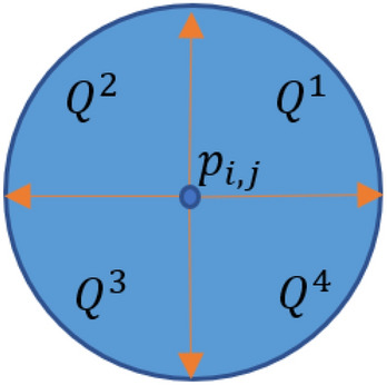
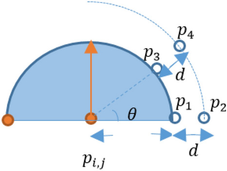
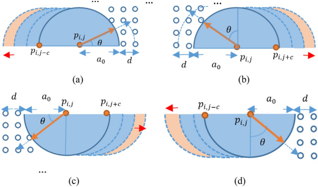
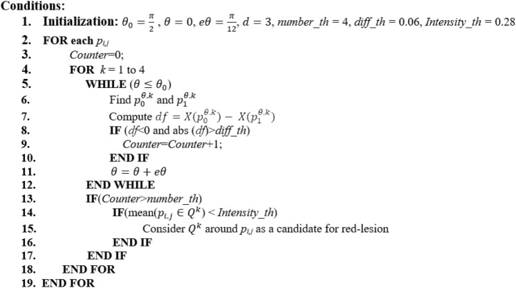
One of the most important retinal diseases is Diabetic Retinopathy (DR) which can lead to serious damage to vision if remains untreated. Red-lesions are from important demonstrations of DR helping its identification in early stages. The detection and verification of them is helpful in the evaluation of disease severity and progression. In this paper, a novel image processing method is proposed for extracting red-lesions from fundus images. The method works based on finding and extracting the unique morphological features of red-lesions. After quality improvement of images, a pixel-based verification is performed in the proposed method to find the ones which provide a significant intensity change in a curve-like neighborhood. In order to do so, a curve is considered around each pixel and the intensity changes around the curve boundary are considered. The pixels for which it is possible to find such curves in at least two directions are considered as parts of red-lesions. The simplicity of computations, the high accuracy of results, and no need to post-processing operations are the important characteristics of the proposed method endorsing its good performance.
Overview
The claims and statistics of this paper haven’t been checked yet.
Run the check — free, no account needed. It reads the open-access text and checks the sources it cites.
Integrity signals
Registry
Cites
Statistics
Figures
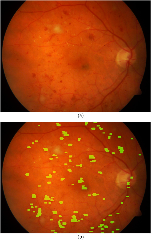
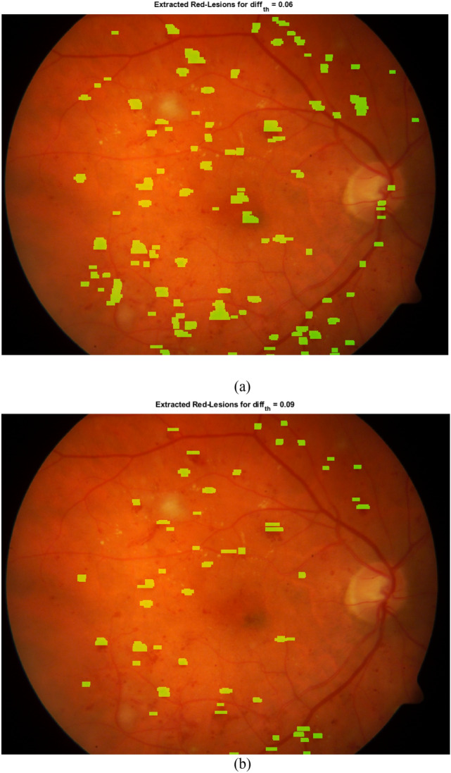
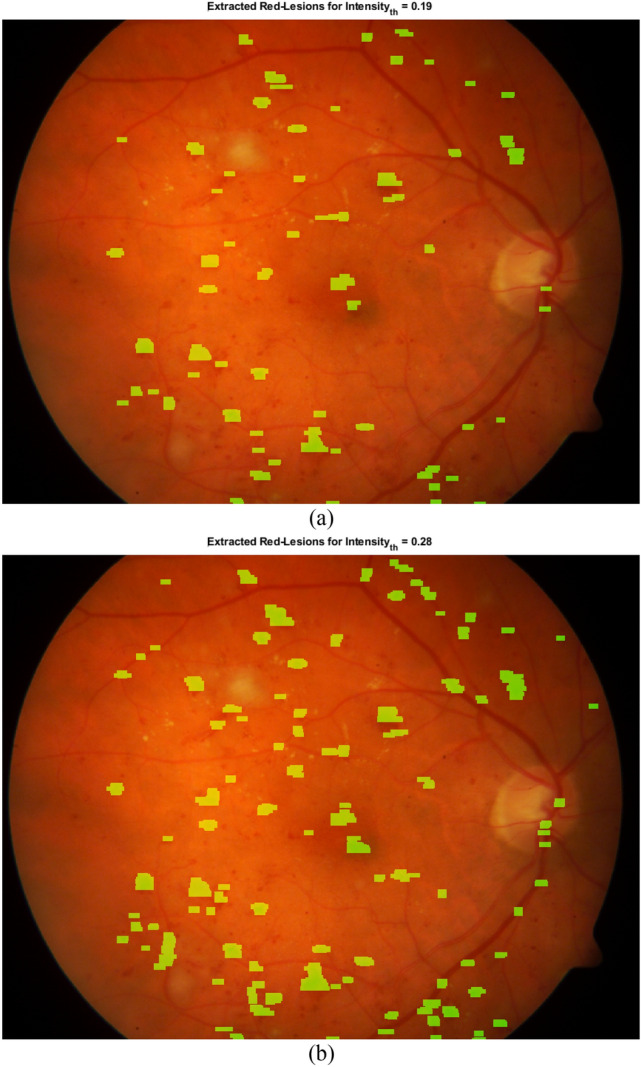
Verification
Claims the paper makes
The claims and statistics of this paper haven’t been checked yet.
Run the check — free, no account needed. It reads the open-access text and checks the sources it cites.
Claims other papers make about this one
Each is checked against this paper’s own text: does it say what the citing paper says it does?
The source backs part of the claim, not all of it — or backs it for a different population, condition or comparison.
In the citing paper
“YOLOv3 has been used to detect red lesions in RGB fundus images, achieving an average precision of 83.3% [55].”
From The YOLO Framework: A Comprehensive Review of Evolution, Applications, and Benchmarks in Object Detection (2024) · cites this paper as [55]
Experimental — accuracy not yet measured
Self-check
Percentages, sums and reported differences re-computed from the paper’s own numbers.
The self-check needs the paper’s full text.
Citations
Cites
van Grinsven, M. J. J. P., van Ginneken, B., Hoyng, C. B., Theelen, T., & Sanchez, C. I. (2016) · IEEE Transactions on Medical Imaging · doi:10.1109/tmi.2016.2526689 · Cited by 426
Li, J. Q., Welchowski, T., Schmid, M., Letow, J., Wolpers, C., Pascual-Camps, I., Holz, F. G., & Finger, R. P. (2020) · European Journal of Epidemiology · doi:10.1007/s10654-019-00560-z · Cited by 179
Research works printed but not linked: 37
- Shukla, U. V. & Tripathy, K. Diabetic Retinopathy (NIH Bookshelf, StatPearls, 2022).
- Monemian, M. & Rabbani, H. Red-lesion extraction in retinal fundus images by directional intensity changes’ analysis. Sci. Rep. 11, 18223 (2021). · DOI
- Oraa, R. R. et al. Entropy rate super-pixel classification for automatic red lesion detection in fundus images. Entropy 21(4), 417 (2019). · DOI
- Wu, B., Zhu, W., Shi, F., Zhu, S. & Chen, X. Automatic detection of micro-aneurysms in retinal fundus images. Comput. Med. Imaging Graph. 55, 106–112 (2017). · DOI
- Oraa, R. R., Garcia, M., Perez, J. O., Lopez-Galvez, M. I. & Hornero, R. Effective fundus image decomposition for the detection of red lesions and hard exudates to aid in the diagnosis of diabetic retinopathy. Sensors 20(22), 1–17 (2020). · DOI
- Wang, J. et al. Automated diabetic retinopathy grading and lesion detection based on the modified R-FCN object-detection algorithm. IET Comput. Vis. 14(1), 1–8 (2020). · DOI
- Long, S. et al. Micro-aneurysm detection in color fundus images using machine learning based on directional local contrast. Biomed. Eng. Online 19(1), 21 (2020). · DOI
- Orlando, J. I., Prokofyeva, E., de Fresno, M. & Blaschko, M. B. An ensemble deep learning based approach for red lesion detection in fundus images. Comput. Methods Prog. Biomed. 153, 115–127 (2018). · DOI
- Kar, S. S. & Maity, S. P. Automatic detection of retinal lesions for screening of diabetic retinopathy. IEEE Trans. Biomed. Eng. 65(3), 608–618 (2018). · DOI
- Wu, J. et al. Hemorrhage detection in fundus image based on 2D Gaussian fitting and human visual characteristics. Opt. Laser Technol. 21, 69–77 (2019). · DOI
- Aziz, T., Ilesanmi, A. E. & Charoenlarpnopparut, C. Efficient and accurate hemorrhages detection in retinal fundus images using smart window features. Appl. Sci. 13, 1 (2021).
- Qiao, L., Zhu, Y. & Zhou, H. Diabetic retinopathy detection using prognosis of microaneurysm and early diagnosis system for non-proliferative diabetic retinopathy based on deep learning algorithms. IEEE Access 8, 104292–104302 (2020). · DOI
- Ram, K., Joshi, G. D. & Sivaswamy, J. A Successive clutter-rejection-based approach for early detection of diabetic retinopathy. IEEE Trans. Biomed. Eng. 58(3), 664–673 (2011). · DOI
- Narasimha-Iyer, H. et al. Robust detection and classification of longitudinal changes in color retinal fundus images for monitoring diabetic retinopathy. IEEE Trans. Biomed. Eng. 53(6), 1084–1098 (2006). · DOI
- Zhang, B., Vijaya Kumar, B. V. K. & Zhang, D. Detecting diabetes mellitus and nonproliferative diabetic retinopathy using tongue color, texture, and geometry features. IEEE Trans. Biomed. Eng. 61(2), 491–501 (2014). · DOI
- Pires, R. Beyond lesion-based diabetic retinopathy: A direct approach for referral. IEEE J. Biomed. Health Inform. 21(1), 193–200 (2017). · DOI
- Zhou, Y., Wang, B., Huang, L., Cui, S. & Shao, L. A benchmark for studying diabetic retinopathy: Segmentation, grading, and transferability. IEEE Trans. Med. Imag. 40(3), 818–828 (2021). · DOI
- Wang, J., Bai, Y. & Xia, B. Simultaneous diagnosis of severity and features of diabetic retinopathy in fundus photography using deep learning. IEEE J. Biomed. Health Inform. 24(12), 3397–3407 (2020). · DOI
- Aghamohamadian-Sharbaf, M., Pourreza, H. R. & Banaee, T. A novel curvature-based algorithm for automatic grading of retinal blood vessel tortuosity. IEEE J. Biomed. Health Inform. 20(2), 586–595 (2016). · DOI
- Antal, B. & Hajdu, A. An ensemble-based system for microaneurysm detection and diabetic retinopathy grading. IEEE Trans. Biomed. Eng. 59(6), 1720–1726 (2012). · DOI
- Quellec, G. et al. Optimal wavelet transform for the detection of microaneurysms in retina photographs. IEEE Trans. Med. Imag. 27(9), 1230–1241 (2008). · DOI
- Momeni Pour, A., Seyedarabi, H., Abbasi Jahromi, S. H. & Javadzadeh, A. Automatic detection and monitoring of diabetic retinopathy using efficient convolutional neural networks and contrast limited adaptive histogram equalization. IEEE Access. 8, 136668–136673 (2020). · DOI
- Chen, W., Yang, B., Li, J. & Wang, J. An approach to detecting diabetic retinopathy based on integrated shallow convolutional neural networks. IEEE Access. 8, 178552–178562 (2020). · DOI
- Wang, J., Bai, Y. & Xia, B. Feasibility of diagnosing both severity and features of diabetic retinopathy in fundus photography. IEEE Access 7, 102589–102597 (2019). · DOI
- Bilal, A., Sun, G., Li, Y., Mazhar, S. & Khan, A. Q. Diabetic retinopathy detection and classification using mixed models for a disease grading database. IEEE Access. 9, 23544–23553 (2021). · DOI
- Gao, Z. et al. Diagnosis of diabetic retinopathy using deep neural networks. IEEE Access. 7, 3360–3370 (2019). · DOI
- Shankar, K., Zhang, Y., Liu, Y., Wu, L. & Chen, C. H. Hyperparameter tuning deep learning for diabetic retinopathy fundus image classification. IEEE Access. 8, 118164–118173 (2020). · DOI
- Costa, P., Galdran, A., Smailagic, A. & Campilho, A. A weakly-supervised framework for interpretable diabetic retinopathy detection on retinal images. IEEE Access. 6, 18747–18758 (2018). · DOI
- Tan, C. S. H., Chew, M. C. Y., Lim, L. W. Y. & Sadda, S. R. Advances in retinal imaging for diabetic retinopathy and diabetic macular edema. Indian J. Ophthalmol. 64(1), 76–83 (2016). · DOI
- Lam, C., Yu, C., Huang, L. & Rubin, D. Retinal lesion detection with deep learning using image patches. Invest. Ophthalmol. Vis. Sci. 59(1), 590–596 (2018). · DOI
- Alyoubi, W. L., Abulkhair, M. F. & Shalash, W. M. Diabetic retinopathy fundus image classification and lesions localization system using deep learning. Sensors. 21, 3704 (2021). · DOI · Paper page
- Das, S., Kharbanda, K., Suchetha, M., Raman, R. & Dhas, E. Deep learning architecture based on segmented fundus image features for classification of diabetic retinopathy. Biomed. Signal Process. Control. 68, 102600 (2021). · DOI
- Zago, G. T., Andreão, R. V., Dorizzi, B. & Salles, E. O. T. Diabetic retinopathy detection using red lesion localization and convolutional neural networks. Comput. Biol. Med. 116, 103537 (2020). · DOI
- Kauppi, T. et al. DIARETDB1 diabetic retinopathy database and evaluation protocol. in Proceedings of the 11th Conference on Medical Image Understanding and Analysis, Aberystwyth (2007). · DOI
- Pal, P., Kundu, S. & Dhara, A. K. Detection of red-lesions in retinal fundus images using YOLO V3. Curr. Eye. Res. 7(2), 49 (2020).
- Zhou, W. et al. Automated detection of red lesions using superpixel multichannel multifeature. Comput. Math. Methods Med. 2017, 9854825 (2017). · DOI
- Colomer, A., Igual, J. & Naranjo, V. Detection of early signs of diabetic retinopathy based on textural and morphological information in fundus images. Sensors 20, 1005 (2020). · DOI
Cited by
Ali, M. L., & Zhang, Z. (2024) · Computers · doi:10.3390/computers13120336 · Cited by 383
Metadata
- Authors
- Monemian, M., & Rabbani, H.
- Affiliations
- Isfahan University of Medical Sciences (IR)
- Corresponding author
- Rabbani, Hossein
- Venue
- Scientific Reports
- Published
- 2023-03-01
- Volume
- 13
- Issue
- 1
- Pages
- 3487-3487
- ISSN
- 2045-2322 (ISSN-L)
- PMC
- PMC9977778
- OpenAlex
- W4322733455
- Cited by
- 10
- References
- 2
- Citations by year
- 2026: 4 · 2025: 1 · 2024: 2 · 2023: 3
- FWCI
- 1.66
- Type
- journal article
- Language
- en
- Open access
- gold
- doi.org
- Locations
- Scientific Reports (publishedVersion, cc-by)
- PubMed Central (publishedVersion, cc-by)
- PubMed (publishedVersion)
- DOAJ (DOAJ: Directory of Open Access Journals) (submittedVersion, cc-by-sa)
- Topics
- Health Sciences › Medicine › Radiology, Nuclear Medicine and Imaging › Retinal Imaging and Analysis
- Physical Sciences › Computer Science › Computer Vision and Pattern Recognition › Medical Image Segmentation Techniques
- Health Sciences › Medicine › Ophthalmology › Retinal Diseases and Treatments
- Keywords
- diabetic retinopathy, fundus images, morphological features, lesion segmentation, early disease detection
- MeSH terms
- Abomasum, Animals, Diabetic Retinopathy, Fundus Oculi, Image Processing, Computer-Assisted, Patient Acuity
About these checks
Registry, retraction, Cites and statistics results are computed, not judged, with one exception: a printed reference that no index lists is read once by a model before it counts as a citation error. Claims are extracted by a model and kept only when their quote appears verbatim in the text; each cited source is then checked, with a model’s help, for whether it says what the claim relies on. Those verdicts can be wrong — read the claim’s quote and the cited source before relying on one.
Statistics and arithmetic are recomputed, not judged.
Confidence bands are shown only for fields calibrated on our annotated test set. None is calibrated yet, so no band is shown.
What counts as a claim ›How we measure confidence ›How we check this ›
Are you an author, or do you see a problem with this page? Request a review
