

The DOI matches its Crossref record; 92 of the 92 references it cites were found in a registry or an index.
Inhibition of SARS-CoV-2 3CL protease by the anti-viral chimeric protein RetroMAD1
Open accessPublished version of a preprint
Abstract
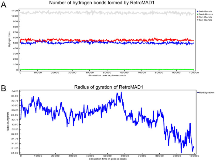
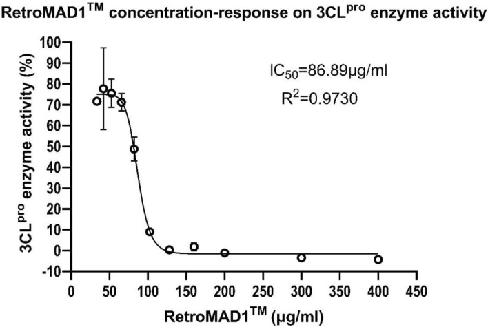
COVID-19 results from SARS-CoV-2, which mutates frequently, challenging current treatments. Therefore, it is critical to develop new therapeutic drugs against this disease. This study explores the interaction between SARS-CoV-2 3CL pro and RetroMAD1, a well-characterized coronavirus protein and potential drug target, using in-silico methods. The analysis through the HDOCK server showed stable complex formation with a binding energy of -12.3, the lowest among reference drugs. The RetroMAD1-3CL pro complex underwent a 100 ns molecular dynamics simulation (MDS) in an explicit solvation system, generating various trajectories, including RMSD, RMSF, hydrogen bonding, radius of gyration, and ligand binding energy. MDS results confirmed intact interactions within the RetroMAD1-3CL pro complex during simulations. In vitro experiments validated RetroMAD1's ability to inhibit 3CL pro enzyme activity and prevent SARS-CoV-2 infection in human bronchial cells. RetroMAD1 exhibited antiviral efficacy comparable to Remdesivir without cytotoxicity at effective concentrations. These results suggest RetroMAD1 as a potential drug candidate against SARS-CoV-2, warranting further in vivo and clinical studies to assess its efficiency.
Overview
The claims and statistics of this paper haven’t been checked yet.
Run the check — free, no account needed. It reads the open-access text and checks the sources it cites.
Integrity signals
Registry
Cites
Statistics
Figures
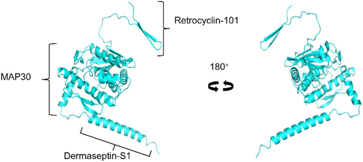
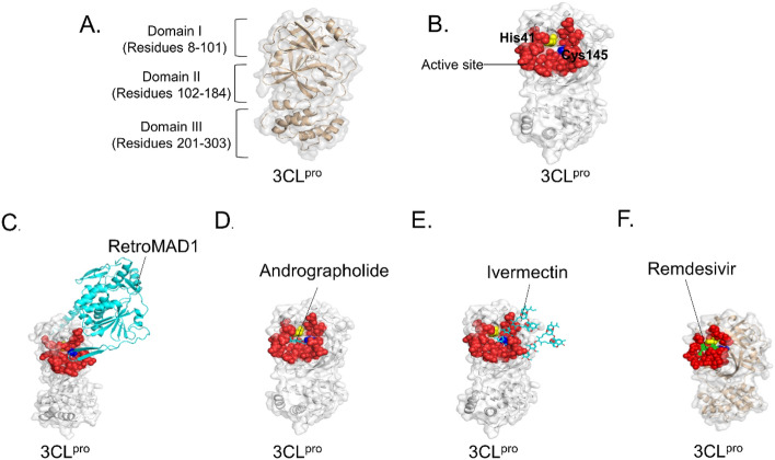
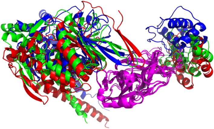
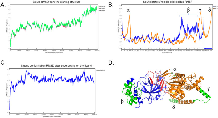
Verification
Claims the paper makes
The claims and statistics of this paper haven’t been checked yet.
Run the check — free, no account needed. It reads the open-access text and checks the sources it cites.
Self-check
Percentages, sums and reported differences re-computed from the paper’s own numbers.
The self-check needs the paper’s full text.
Citations
Cites
Highly accurate protein structure prediction with AlphaFold
Jumper, J., Evans, R., Pritzel, A., Green, T., Figurnov, M., Ronneberger, O., Tunyasuvunakool, K., Bates, R., Žídek, A., Potapenko, A., et al. (2021) · Nature · doi:10.1038/s41586-021-03819-2 · Cited by 43,764
A SIMPLE METHOD OF ESTIMATING FIFTY PER CENT ENDPOINTS12
REED, L. J., & MUENCH, H. (1938) · American Journal of Epidemiology · doi:10.1093/oxfordjournals.aje.a118408 · Cited by 19,926
SWISS-MODEL: homology modelling of protein structures and complexes
Waterhouse, A., Bertoni, M., Bienert, S., Studer, G., Tauriello, G., Gumienny, R., Heer, F. T., de Beer, T. A. P., Rempfer, C., Bordoli, L., et al. (2018) · Nucleic Acids Research · doi:10.1093/nar/gky427 · Cited by 13,067
ColabFold - Making protein folding accessible to all
Mirdita, M., Schütze, K., Moriwaki, Y., Heo, L., Ovchinnikov, S., & Steinegger, M. (2021) · doi:10.1101/2021.08.15.456425 · Cited by 9,391
Electrostatics of nanosystems: Application to microtubules and the ribosome
Baker, N., Sept, D., Joseph, S., Holst, M., & McCammon, J. A. (2001) · Proceedings of the National Academy of Sciences · doi:10.1073/pnas.181342398 · Cited by 7,125
Characteristics of SARS-CoV-2 and COVID-19
Hu, B., Guo, H., Zhou, P., & Shi, Z.-L. (2021) · Nature Reviews Microbiology · doi:10.1038/s41579-020-00459-7 · Cited by 5,291
MMseqs2 enables sensitive protein sequence searching for the analysis of massive data sets
Steinegger, M., & Söding, J. (2017) · Nature Biotechnology · doi:10.1038/nbt.3988 · Cited by 4,840
Kim, S., Chen, J., Cheng, T., Gindulyte, A., He, J., He, S., Li, Q., Shoemaker, B. A., Thiessen, P. A., Yu, B., et al. (2023) · Nucleic Acids Research · doi:10.1093/nar/gkac956 · Cited by 3,443
Repurposed Antiviral Drugs for Covid-19 — Interim WHO Solidarity Trial Results
WHO Solidarity Trial Consortium (2021) · New England Journal of Medicine · doi:10.1056/nejmoa2023184 · Cited by 2,745
Improvements to the APBS biomolecular solvation software suite
Jurrus, E. R., Engel, D. W., Star, K. T., Monson, K., Brandi, J., Felberg, L. E., Brookes, D. H., Wilson, L., Chen, J., Liles, K., et al. (2017) · Protein Science · doi:10.1002/pro.3280 · Cited by 2,519
Research works printed but not linked: 1
- Satpathy, R. In silico prediction of anti–SARS-CoV-2 effect of dermaseptin peptides from amphibian origin. Trends Peptide Protein Sci. 5, 1–9 (2020).
Metadata
- Authors
- Chan, L.-C., Mat Yassim, A. S., Ahmad Fuaad, A. A. H., Leow, T. C., Sabri, S., Radin Yahaya, R. S., & Abu Bakar, A. M. S.
- Affiliations
- Universiti Putra Malaysia (MY)
- Venue
- Scientific Reports
- Published
- 2023-11-17
- Volume
- 13
- Issue
- 1
- Pages
- 20178-20178
- ISSN
- 2045-2322 (ISSN-L)
- PMC
- PMC10656507
- OpenAlex
- W4388751591
- Cited by
- 5
- References
- 92
- Citations by year
- 2026: 1 · 2025: 1 · 2024: 3
- FWCI
- 1.03
- Type
- journal article
- Language
- en
- Open access
- gold
- doi.org
- Licence
- cc-by
- creativecommons.org/licenses/by/4.0 (tdm)
- creativecommons.org/licenses/by/4.0 (vor)
- Locations
- Scientific Reports (publishedVersion, cc-by)
- Research Square (acceptedVersion, cc-by)
- PubMed Central (publishedVersion, cc-by)
- PubMed (publishedVersion)
- DOAJ (DOAJ: Directory of Open Access Journals) (submittedVersion, cc-by-sa)
- Universiti Putra Malaysia Institutional Repository (Universiti Putra Malaysia) (submittedVersion, cc-by)
- Greater South Information System
- Funders
- Utah State University
- Topics
- Health Sciences › Medicine › Infectious Diseases › SARS-CoV-2 and COVID-19 Research
- Life Sciences › Pharmacology, Toxicology and Pharmaceutics › Drug Discovery › Computational Drug Discovery Methods
- Life Sciences › Biochemistry, Genetics and Molecular Biology › Molecular Biology › vaccines and immunoinformatics approaches
- Keywords
- SARS-CoV-2, COVID-19 pandemic, main protease, main protease inhibition, fusion protein, antiviral drug candidates, protein-protein complex stability, molecular dynamics simulation, human bronchial epithelial cells, cytotoxicity
- MeSH terms
- COVID-19, SARS-CoV-2, Antiviral Agents, Cysteine Endopeptidases, Humans, Peptide Hydrolases, Protease Inhibitors, Recombinant Fusion Proteins, Viral Nonstructural Proteins
About these checks
Registry, retraction, Cites and statistics results are computed, not judged, with one exception: a printed reference that no index lists is read once by a model before it counts as a citation error. Claims are extracted by a model and kept only when their quote appears verbatim in the text; each cited source is then checked, with a model’s help, for whether it says what the claim relies on. Those verdicts can be wrong — read the claim’s quote and the cited source before relying on one.
Statistics and arithmetic are recomputed, not judged.
Confidence bands are shown only for fields calibrated on our annotated test set. None is calibrated yet, so no band is shown.
What counts as a claim ›How we measure confidence ›How we check this ›
Are you an author, or do you see a problem with this page? Request a review
How to cite
Cite CoChat for the checks on this page, and the paper for its own findings.
- This page
- CoChat. (2026). Citation check: Inhibition of SARS-CoV-2 3CL protease by the anti-viral chimeric protein RetroMAD1. https://app.cochat.ai/papers/10.1038/s41598-023-47511-z
- The paper
- Chan, L.-C., Mat Yassim, A. S., Ahmad Fuaad, A. A. H., Leow, T. C., Sabri, S., Radin Yahaya, R. S., & Abu Bakar, A. M. S. (2023). Inhibition of SARS-CoV-2 3CL protease by the anti-viral chimeric protein RetroMAD1. Scientific Reports. https://doi.org/10.1038/s41598-023-47511-z
Data sources
Bibliographic data: Semantic Scholar, OpenAlex, Crossref.
Contains information from the Semantic Scholar Academic Graph Datasets, made available under the ODC Attribution License (ODC-BY 1.0).
Semantic Scholar’s data is described in: Kinney, R., et al. (2023). The Semantic Scholar Open Data Platform. arXiv:2301.10140.
