

The DOI matches its Crossref record.
Investigating the cytotoxicity of Aluminum-Doped zinc oxide nanoparticles in normal versus cancerous breast cells
Overview
The claims and statistics of this paper haven’t been checked yet.
Run the check — free, no account needed. It reads the open-access text and checks the sources it cites.
Integrity signals
Registry
Cites
Statistics
Figures
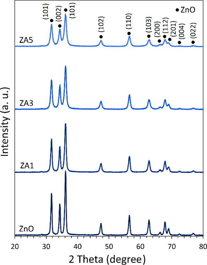
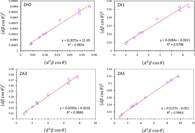
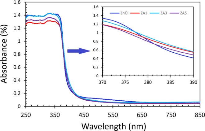
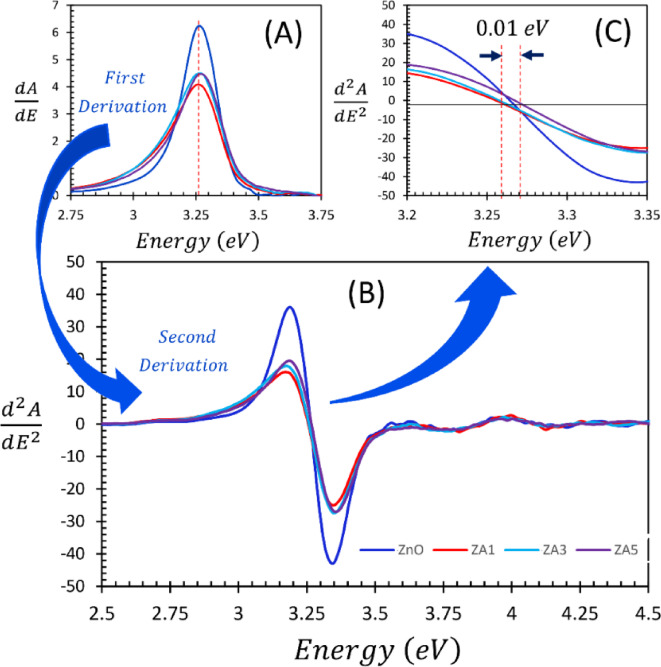
Verification
Claims the paper makes
The claims and statistics of this paper haven’t been checked yet.
Run the check — free, no account needed. It reads the open-access text and checks the sources it cites.
Claims other papers make about this one
Each is checked against this paper’s own text: does it say what the citing paper says it does?
In the citing paper
“From a safety perspective, CPP-containing and other strongly cationic nanoparticles increase the risk of dose-dependent cytotoxicity, hemolysis, complement activation and non-specific adsorption to plasma proteins and vascular endothelium, which can trigger undesired uptake in the blood circulation, rapid RES sequestration and increased exposure of healthy tissues [76].”
From Overcoming tumor microenvironment barriers: transformable and bioinspired nanomedicine strategies for deep tumor penetration (2026) · cites this paper as [76]
Self-check
Percentages, sums and reported differences re-computed from the paper’s own numbers.
The self-check needs the paper’s full text.
Citations
Cites
No references could be resolved yet.
Research works printed but not linked: 51
- Haleem, A., Javaid, M., Singh, R. P., Rab, S. & Suman, R. Applications of nanotechnology in medical field: a brief review. Global Health J. 7, 70–77 (2023). · DOI
- Kemp, J. A. & Kwon, Y. J. Cancer nanotechnology: current status and perspectives. Nano Convergence. 8, 34 (2021). · DOI
- Khorsand Zak, A., Khorsand Zak, H. & Esmaeilzadeh, J. Cytotoxicity studies of ultra-fine NiO nanoparticles on HeLa and GC-1spg cell lines: A promising materials for cancer treatment application. Ceram. Int. https://doi.org/10.1016/j.ceramint.2025.01.502 (2025). · DOI
- Al-darwesh, M. Y., Babakr, K. A. & Qader, I. N. Characterization and anticancer evaluation of zirconia nanoparticles synthesized via green route using sophora flavescens roots extract. Nano-Structures Nano-Objects. 39, 101245. https://doi.org/10.1016/j.nanoso.2024.101245 (2024). · DOI
- Al-darwesh, M. Y., Hamid, L. L., Ibrahim, S. S. & Mohammed, M. A. Construction and characterization of 5,6-O-Isopropylidene-L-Ascorbic acid-loaded silver and manganese dioxide nanoparticles: cytotoxicity study, antibacterial, and antioxidant activities. Mater. Chem. Phys. 335, 130517. https://doi.org/10.1016/j.matchemphys.2025.130517 (2025). · DOI
- Ibrahim, S. S., Al-darwesh, M. Y., Arkawazi, S. W., Babakr, K. A. & Qader, I. N. Synthesis, characterization, antioxidant, antibacterial activity, and molecular Docking studies of Zn (II) and Cu (II) complexes nanoparticles. J. Mol. Struct. 1326, 141120. https://doi.org/10.1016/j.molstruc.2024.141120 (2025). · DOI
- Al-darwesh, M. Y., El-Subeyhi, M., Hamid, L. L. & Mutter, T. Y. Nanocapsule formation of Ciprofloxacin-Loaded zirconia nanoparticles coated with Chitosan for antibacterial, antibiofilm, Anti-Inflammatory, and catalytic applications. J. Cluster Sci. 36, 121. https://doi.org/10.1007/s10876-025-02834-x (2025). · DOI
- Al-darwesh, M. Y., Babakr, K. A., Qader, I. N. & Mohammed, M. A. Antimicrobial, anti-inflammatory, and anticancer potential of green synthesis TiO2 nanoparticles using Sophora flavescens root extract. Chem. Pap. 79, 1207–1221. https://doi.org/10.1007/s11696-024-03853-0 (2025). · DOI
- Hannachi, E. et al. In vitro antimicrobial and anticancer peculiarities of ytterbium and cerium Co-doped zinc oxide nanoparticles. Biology 11, 1836 (2022). · DOI
- Rissi, N. C., Comparetti, E. J., Estevão, B. M., Mastelaro, V. R. & Zucolotto, V. Doped plasmonic zinc oxide nanoparticles with near-infrared absorption for antitumor activity. ACS Appl. Nano Mater. 4, 9779–9789 (2021). · DOI
- Mahmoudi Khatir, N. & Khorsand Zak, A. Antibacterial activity and structural properties of gelatin-based sol-gel synthesized Cu-doped ZnO nanoparticles; promising material for biomedical applications. Heliyon 10, https://doi.org/10.1016/j.heliyon.2024.e37022 (2024). · DOI
- Al-darwesh, M. Y., Ibrahim, S. S. & Mohammed, M. A. A review on plant extract mediated green synthesis of zinc oxide nanoparticles and their biomedical applications. Results Chem. 7, 101368. https://doi.org/10.1016/j.rechem.2024.101368 (2024). · DOI
- Racca, L. et al. Effective combination of biocompatible zinc oxide nanocrystals and high-energy shock waves for the treatment of colorectal cancer. Cancer Nanotechnol. 14, 37 (2023). · DOI
- Al-darwesh, M. Y., Ibrahim, S. S. & Hamid, L. L. Ficus carica latex mediated biosynthesis of zinc oxide nanoparticles and assessment of their antibacterial activity and biological safety. Nano-Structures Nano-Objects. 38, 101163. https://doi.org/10.1016/j.nanoso.2024.101163 (2024). · DOI
- Hamidian, K., Sarani, M., Barani, M. & Khakbaz, F. Cytotoxic performance of green synthesized ag and Mg dual doped ZnO NPs using Salvadora persica extract against MDA-MB-231 and MCF-10 cells. Arab. J. Chem. 15, 103792 (2022). · DOI
- Varlamova, E. G. & Turovsky, E. A. The main cytotoxic effects of Methylseleninic acid on various cancer cells. Int. J. Mol. Sci. 22, 6614 (2021). · DOI
- Rahimi, S., Chen, Y., Zareian, M., Pandit, S. & Mijakovic, I. Cellular and subcellular interactions of graphene-based materials with cancerous and non-cancerous cells. Adv. Drug Deliv. Rev. 189, 114467 (2022). · DOI
- Ahamed, M., Akhtar, M. J., Khan, M. M. & Alhadlaq, H. A. SnO2-doped zno/reduced graphene oxide nanocomposites: synthesis, characterization, and improved anticancer activity via oxidative stress pathway. Int. J. Nanomed. 16, 89–104 (2021). · DOI
- Biswas, A., Kar, U. & Jana, N. R. Cytotoxicity of ZnO nanoparticles under dark conditions via oxygen vacancy dependent reactive oxygen species generation. Phys. Chem. Chem. Phys. 24, 13965–13975 (2022). · DOI
- Aljabali, A. A. et al. Synthesis, characterization, and assessment of anti-cancer potential of ZnO nanoparticles in an in vitro model of breast cancer. Molecules 27, 1827 (2022). · DOI
- Khorsand Zak, A. et al. Sonochemical synthesis of hierarchical ZnO nanostructures. Ultrason. Sonochem. 20, 395–400. https://doi.org/10.1016/j.ultsonch.2012.07.001 (2013). · DOI
- Razali, R., Zak, A. K., Majid, W. H. A. & Darroudi, M. Solvothermal synthesis of microsphere ZnO nanostructures in DEA media. Ceram. Int. 37, 3657–3663. https://doi.org/10.1016/j.ceramint.2011.06.026 (2011). · DOI
- Yousefi, R., Jamali-Sheini, F. & Zak, A. K. A comparative study of the properties of ZnO nano/microstructures grown using two types of thermal evaporation Set-Up conditions. Chem. Vap. Deposition. 18, 215–220. https://doi.org/10.1002/cvde.201206979 (2012). · DOI
- Mahmoudian, M. R., Basirun, W. J., Alias, Y. & Khorsand Zak, A. Electrochemical characteristics of coated steel with poly(N-methyl pyrrole) synthesized in presence of ZnO nanoparticles. Thin Solid Films. 520, 258–265. https://doi.org/10.1016/j.tsf.2011.07.067 (2011). · DOI
- Khorsand Zak, A., Esmaeilzadeh, J. & Hashim, A. M. Exploring the gelatin-based sol-gel approach: A convenient route for fabricating high-quality pure and doped ZnO nanostructures. Ceram. Int. 50, 12649–12663. https://doi.org/10.1016/j.ceramint.2024.01.254 (2024). · DOI
- Zamiri, R. et al. Laser assisted fabrication of zno/ag and zno/au core/shell nanocomposites. Appl. Phys. A. 111, 487–493. https://doi.org/10.1007/s00339-012-7517-y (2013). · DOI
- Kumar Janakiraman, A. et al. Fabrication, Characterization and In Vitro Cell Cytotoxicity of ZnO Nanoparticles on MDA-MB 231 Breast Cancer Cell Line. Chemistry Select 8, (2023). · DOI
- Al-Enazi, N. M., Alsamhary, K., Kha, M. & Ameen, F. In vitro anticancer and antibacterial performance of biosynthesized ag and Ce co-doped ZnO NPs. Bioprocess Biosyst. Eng. 46, 89–103 (2023). · DOI
- Hannachi, E. et al. Fabrication, characterization, anticancer and antibacterial activities of ZnO nanoparticles doped with Y and Ce elements. J. Cluster Sci. 34, 1777–1788 (2023). · DOI
- Vidhya, M. S. et al. Anti-cancer applications of zr, co, Ni-doped ZnO thin nanoplates. Mater. Lett. 283, 128760 (2021). · DOI
- Rezaei, F. Y., Pircheraghi, G. & Nikbin, V. S. Antibacterial activity, cell wall damage, and cytotoxicity of zinc oxide nanospheres, nanorods, and nanoflowers. ACS Appl. Nano Mater. 7, 15242–15254 (2024). · DOI
- Kafi, Z., Kompany, A., Arabi, H. & Khorsand Zak, A. The effect of cobalt-doping on microstructure and dielectric properties of CaCu3Ti4O12 ceramics. J. Alloys Compd. 727, 168–176. https://doi.org/10.1016/j.jallcom.2017.07.236 (2017). · DOI
- Khorrami, G. H. & Kompany, A. Khorsand zak, A. A facile sol–gel approach to synthesize KNN nanoparticles at low temperature. Mater. Lett. 110, 172–175. https://doi.org/10.1016/j.matlet.2013.07.115 (2013). · DOI
- Kordi, F., Khorsand Zak, A., Darroudi, M. & Hazrati Saedabadi, M. Synthesis and characterizations of Ag-decorated graphene oxide nanosheets and their cytotoxicity studies. Chem. Pap. 73, 1945–1952. https://doi.org/10.1007/s11696-019-00747-4 (2019). · DOI
- Khorsand Zak, A. & Hashim, A. M. Synthesis, characterization and cytotoxicity studies of Au-decorated graphene oxide nanosheets. Ceram. Int. 49, 18577–18583. https://doi.org/10.1016/j.ceramint.2023.02.232 (2023). · DOI
- Khorsand Zak, A., Esmaeilzadeh, J. & Hashim, A. M. X-ray peak broadening and strain-driven preferred orientations of pure and Al-doped ZnO nanoparticles prepared by a green gelatin-based sol-gel method. J. Mol. Struct. 1303, 137537. https://doi.org/10.1016/j.molstruc.2024.137537 (2024). · DOI
- Khorsand Zak, A., Hashim, A. M. & Esmaeilzadeh, J. Role of ceo₂ and calcination temperature on the structural and optical properties of (ZnO) 1-x/ (CeO₂)ₓ nanocomposites in the UV–visible region. Ceram. Int. 50, 4167–4177. https://doi.org/10.1016/j.ceramint.2023.11.204 (2024). · DOI
- Khorsand Zak, A., Hashim, A. M. & Esmaeilzadeh, J. XPS studies and Kramers-Kronig analysis of the optical properties of ZnO/SnO2 nanocomposites synthesized by gelatin-based sol-gel method. Opt. Mater. 142, 114111. https://doi.org/10.1016/j.optmat.2023.114111 (2023). · DOI
- Mayekar, J. A review on the synthesis of doped and undoped ZnO thin films and Understanding effect of doping on the band gap of doped zinc oxide thin films on the basis of Burstein-moss effect. Int. J. Res. Analyt Rev. 9, 971–974 (2022).
- Ma, Y. et al. Size-dependent cytotoxicity and reactive oxygen species of cerium oxide nanoparticles in human retinal pigment epithelia cells. Int. J. Nanomed. 16, 5333–5341 (2021). · DOI
- Shubha, P., Ganesh, S. & Shyamsundar, S. Anti-proliferative activity of biosynthesized zinc oxide nanoparticles against breast cancer MCF-7 cells. Mater. Chem. Phys. 315, 128900. https://doi.org/10.1016/j.matchemphys.2024.128900 (2024). · DOI
- Ghaznavi, H. et al. Synthesis, characterization, and evaluation of copper-doped zinc oxide nanoparticles anticancer effects: in vitro and in vivo experiments. BMC Cancer. 25, 37. https://doi.org/10.1186/s12885-024-13398-w (2025). · DOI
- Akhtar, M. J., Alhadlaq, H. A., Alshamsan, A., Majeed Khan, M. A. & Ahamed, M. Aluminum doping tunes band gap energy level as well as oxidative stress-mediated cytotoxicity of ZnO nanoparticles in MCF-7 cells. Sci. Rep. 5, 13876. https://doi.org/10.1038/srep13876 (2015). · DOI
- Xie, S. et al. Low concentrations of zinc oxide nanoparticles cause severe cytotoxicity through increased intracellular reactive oxygen species. J. Biomed. Nanotechnol. 17, 2420–2432 (2021). · DOI
- Nawaz, A. et al. Zinc Oxide Nanoparticles: Pathways to Micropollutant Adsorption, Dye Removal, and Antibacterial Actions-A Study of Mechanisms, Challenges, and Future Prospects. J. Mol. Structure, 1312, 138545 (2024). · DOI
- Sun, A., Tang, B. Z., Tsim, K. W. K. & Wang, W. X. Unique interplay between Zn 2 + and nZnO determined the dynamic cellular stress in zebrafish cells. Environ. Science: Nano. 8, 2324–2335 (2021).
- Ali, S. et al. Progress in Nanomaterial-Driven Redox Reactions for Water Purification: A Critical Review. Desalination Water Treatment, 320, 100616 (2024). · DOI
- Zyoud, S. H. et al. Enhanced photocatalytic and antimicrobial properties of undoped and aluminum-doped zinc oxide nanosheets synthesized via novel laser-assisted chemical bath technique. Appl. Phys. A. 129, 750 (2023). · DOI
- Khayatian, S. A., Kompany, A., Shahtahmassebi, N. & Khorsand Zak, A. Enhanced photocatalytic performance of Al-Doped ZnO NPs-Reduced graphene oxide nanocomposite for removing of Methyl orange dye from water under Visible-Light irradiation. J. Inorg. Organomet. Polym Mater. 28, 2677–2688. https://doi.org/10.1007/s10904-018-0940-6 (2018). · DOI
- Chen, G. H. et al. Mitochondria-dependent oxidative stress mediates ZnO nanoparticle (ZnO NP)-induced mitophagy and lipotoxicity in freshwater teleost fish. Environ. Sci. Technol. 56, 2407–2420 (2022). · DOI
- Yang, X. et al. Redox-regulating nanotherapeutics potentiating ferroptosis-driven cancer treatment. Chemical Eng. J., 484, 149518 (2024). · DOI
Cited by
Sorted by citations
Sheng, J., Yuan, W., Zhang, M., Chen, Z., Qi, Y., Zhao, Y., & Zhang, S. (2026) · Journal of Nanobiotechnology · doi:10.1186/s12951-026-04028-7 · Cited by 35
Metadata
- Authors
- Khorsand Zak, A., Hashim, A. M., & Khorsand Zak, H.
- Venue
- Scientific Reports
- Published
- 2025-07-12
- Volume
- 15
- Issue
- 1
- ISSN
- 2045-2322
- PMC
- PMC12255732
- Cited by
- 14
- References
- 0
- Language
- en
About these checks
Registry, retraction, Cites and statistics results are computed, not judged, with one exception: a printed reference that no index lists is read once by a model before it counts as a citation error. Claims are extracted by a model and kept only when their quote appears verbatim in the text; each cited source is then checked, with a model’s help, for whether it says what the claim relies on. Those verdicts can be wrong — read the claim’s quote and the cited source before relying on one.
Statistics and arithmetic are recomputed, not judged.
Confidence bands are shown only for fields calibrated on our annotated test set. None is calibrated yet, so no band is shown.
What counts as a claim ›How we measure confidence ›How we check this ›
Are you an author, or do you see a problem with this page? Request a review
