

The DOI matches its Crossref record; 44 of the 44 references it cites were found in a registry or an index.
Whole-exome sequencing screening for candidate genes and potential pathogenic variants associated with early-onset high myopia in 47 Chinese families
Open accessPublished version of a preprint
Abstract
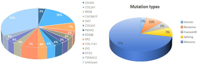
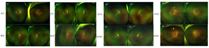
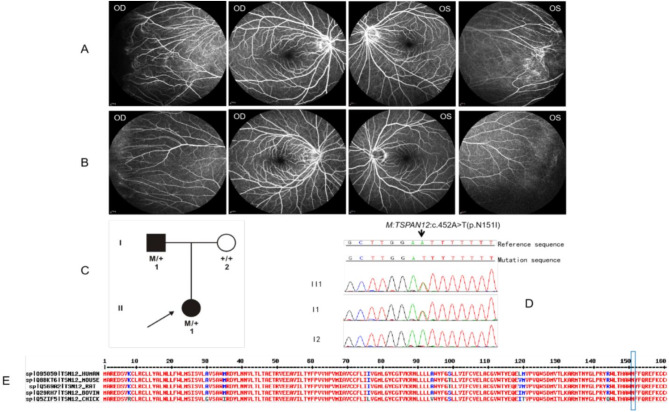
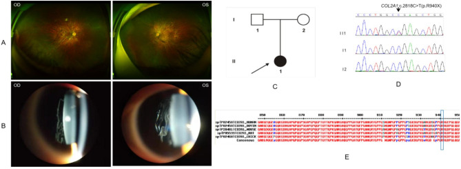
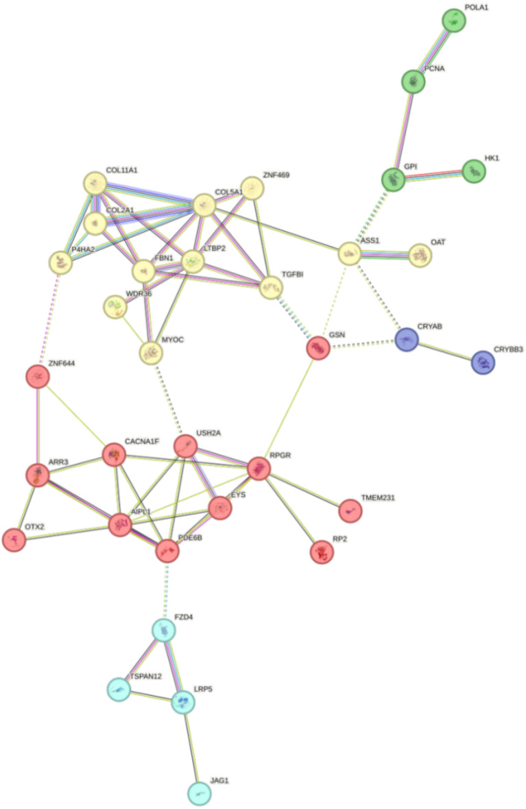
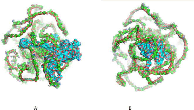
Early-onset high myopia (eoHM) occurs before school age and is an ideal model for monogenic studies of high myopia due to minimal environmental influence. This study screened for genes and variants associated with eoHM in 47 unrelated Chinese patients with eoHM. Protein-protein interaction (PPI) network analysis was conducted to detect interactions among candidate genes, and protein-protein docking was performed. In 28 patients (28/47, 59.6%), 32 potential pathogenic variants in 22 candidate genes were identified, including 24 novel variants. Among these 28 patients, 64.3% (18/28) carried pathogenic variants in RetNet genes. Of these, 12 patients (42.9%, 12/28) had pathogenic variants in six known genes (TSPAN12, CACNA1F, USH2A, RPGR, COL2A1, and COL11A1), which are responsible for retinal dystrophy and Stickler syndrome associated with eoHM. Additionally, 7 patients (25.0%, 7/28) carried pathogenic variants in seven candidate genes for ocular disease (POLA1, TMEM231, HK1, GSN, COL5A1, CRYBB3, and WDR), which were identified as potentially pathogenic in Chinese eoHM patients for the first time. Phenotype analysis showed that 11 patients presented with only high myopia, 10 patients had inherited retinal diseases (IRDs) with eoHM, and 7 patients had ocular-only Stickler syndrome (Ocular-STL) with eoHM. The initial clinical records of these 17 patients did not show recognizable signs of other primary diseases except for high myopia, and further specific clinical examinations confirmed the diagnosis of IRDs or Stickler syndrome through eoHM. The PPI network analysis identified 87 candidate genes associated with early-onset high myopia (eoHM), grouped into four functional clusters. Thirteen hub genes, including RPGR, COL5A1, CRYAB, and FBN1, were crucial for the pathogenesis of myopia. The network showed strong biological relevance with highly significant enrichment (p-value < 1.0e-16). Our study expands the list of candidate genes associated with eoHM and suggests that eoHM may be the first reason for children to visit an ophthalmology clinic and an important clue for clinicians to detect underlying ocular diseases. These findings highlight the complex interplay of these genes, providing valuable insights into the molecular mechanisms of myopia and potential targets for future therapeutic interventions.
Overview
The claims and statistics of this paper haven’t been checked yet.
Run the check — free, no account needed. It reads the open-access text and checks the sources it cites.
Integrity signals
Registry
Cites
Statistics
Figures
Verification
Claims the paper makes
The claims and statistics of this paper haven’t been checked yet.
Run the check — free, no account needed. It reads the open-access text and checks the sources it cites.
Self-check
Percentages, sums and reported differences re-computed from the paper’s own numbers.
The self-check needs the paper’s full text.
Citations
Cites
Highly accurate protein structure prediction with AlphaFold
Jumper, J., Evans, R., Pritzel, A., Green, T., Figurnov, M., Ronneberger, O., Tunyasuvunakool, K., Bates, R., Žídek, A., Potapenko, A., et al. (2021) · Nature · doi:10.1038/s41586-021-03819-2 · Cited by 43,764
Richards, S., Aziz, N., Bale, S., Bick, D., Das, S., Gastier-Foster, J., Grody, W. W., Hegde, M., Lyon, E., Spector, E., et al. (2015) · Genetics in Medicine · doi:10.1038/gim.2015.30 · Cited by 32,594
Ligand docking and binding site analysis with PyMOL and Autodock/Vina
Seeliger, D., & de Groot, B. L. (2010) · Journal of Computer-Aided Molecular Design · doi:10.1007/s10822-010-9352-6 · Cited by 2,623
Performance and Its Limits in Rigid Body Protein-Protein Docking
Desta, I. T., Porter, K. A., Xia, B., Kozakov, D., & Vajda, S. (2020) · Structure · doi:10.1016/j.str.2020.06.006 · Cited by 703
Morgan, I. G., & Rose, K. A. (2004) · Progress in Retinal and Eye Research · doi:10.1016/j.preteyeres.2004.06.004 · Cited by 685
Clinical features of hereditary progressive arthro-ophthalmopathy (Stickler syndrome): A survey
Stickler, G. B., Hughes, W., & Houchin, P. (2001) · Genetics in Medicine · doi:10.1097/00125817-200105000-00008 · Cited by 223
Exome Sequencing Identifies ZNF644 Mutations in High Myopia
Shi, Y., Li, Y., Zhang, D., Zhang, H., Li, Y., Lü, F., Liu, X., He, F., Gong, B., Li, C., et al. (2011) · PLoS Genetics · doi:10.1371/journal.pgen.1002084 · Cited by 198
Stickler syndrome, ocular-only variants and a key diagnostic role for the ophthalmologist
Snead, M. P., McNinch, A. M., Poulson, A. V., Bearcroft, P. W. P., Silverman, B. L., Gomersall, P. A., Parfect, V. H., & Richards, A. J. (2011) · Eye · doi:10.1038/eye.2011.201 · Cited by 188
Risk factors for high myopia: a 22‐year follow‐up study from childhood to adulthood
Pärssinen, O., & Kauppinen, M. (2018) · Acta Ophthalmologica · doi:10.1111/aos.13964 · Cited by 148
Mutations in SCO2 Are Associated with Autosomal-Dominant High-Grade Myopia
Tran-Viet, K.-N., Powell, C., Barathi, V. A., Klemm, T., Maurer‐Stroh, S., Limviphuvadh, V., Soler, V. J., Ho, C. E. H., Yanovitch, T. L., Schneider, G., et al. (2013) · The American Journal of Human Genetics · doi:10.1016/j.ajhg.2013.04.005 · Cited by 115
Research works printed but not linked: 1
- Rui, X. et al. Genotypes and phentypes of hereditary eye diseases associated with early-onset high myopia. Chin J Exp Ohthalmol 41(7), 662–674. https://doi.org/10.3760/cma.j.cn115989-20211216-00695 (2023). · DOI
Metadata
- Authors
- Rui, X., Li, H., Ma, R., Yang, S., lian, Y., Cheng, W., Ma, M., Rong, W., & Sheng, X.
- Affiliations
- Aier Eye Hospital Group (China) (CN)
- Ningxia Hui Autonomous Region Peoples Hospital (CN)
- Corresponding author
- Rong, Weining
- Sheng, Xunlun
- Venue
- Scientific Reports
- Published
- 2025-04-02
- Volume
- 15
- Issue
- 1
- Pages
- 11252-11252
- ISSN
- 2045-2322 (ISSN-L)
- PMC
- PMC11965419
- OpenAlex
- W4409148677
- Cited by
- 4
- References
- 44
- Citations by year
- 2026: 3 · 2025: 1
- FWCI
- 1.20
- Type
- journal article
- Language
- en
- Open access
- gold
- doi.org
- Licence
- cc-by-nc-nd
- creativecommons.org/licenses/by-nc-nd/4.0 (tdm)
- creativecommons.org/licenses/by-nc-nd/4.0 (vor)
- Locations
- Scientific Reports (publishedVersion, cc-by-nc-nd)
- Research Square (acceptedVersion, cc-by)
- PubMed Central (publishedVersion, cc-by-nc-nd)
- PubMed (publishedVersion)
- DOAJ (DOAJ: Directory of Open Access Journals) (submittedVersion, cc-by-sa)
- Funders
- National Natural Science Foundation of China (82260206)
- the key research and development project of Ningxia Hui Autonomous Region (2024BEG02017)
- the training project of the scientific innovation commanding talented person in Ningxia Hui Autonomous Region (2020GKLRLX13)
- Major achievement transformation project of Ningxia Hui Autonomous Region (2022CJE09011)
- Topics
- Health Sciences › Medicine › Ophthalmology › Retinal Development and Disorders
- Health Sciences › Medicine › Ophthalmology › Ophthalmology and Visual Impairment Studies
- Life Sciences › Biochemistry, Genetics and Molecular Biology › Genetics › Genomics and Rare Diseases
- Keywords
- candidate genes, pathogenic variants, inherited retinal disease, Stickler syndrome, protein-protein interaction network, hub genes, COL5A1, COL2A1, COL11A1, novel variants, Chinese family, genetic screening, protein-protein docking, alphaB-crystallin
- MeSH terms
- Exome Sequencing, East Asian People, Adolescent, Child, Child, Preschool, China, Female, Humans, Male, Myopia, Pedigree, Age of Onset, Genetic Predisposition to Disease, Asian People, Protein Interaction Maps
About these checks
Registry, retraction, Cites and statistics results are computed, not judged, with one exception: a printed reference that no index lists is read once by a model before it counts as a citation error. Claims are extracted by a model and kept only when their quote appears verbatim in the text; each cited source is then checked, with a model’s help, for whether it says what the claim relies on. Those verdicts can be wrong — read the claim’s quote and the cited source before relying on one.
Statistics and arithmetic are recomputed, not judged.
Confidence bands are shown only for fields calibrated on our annotated test set. None is calibrated yet, so no band is shown.
What counts as a claim ›How we measure confidence ›How we check this ›
Are you an author, or do you see a problem with this page? Request a review
How to cite
Cite CoChat for the checks on this page, and the paper for its own findings.
- This page
- CoChat. (2026). Citation check: Whole-exome sequencing screening for candidate genes and potential pathogenic variants associated with early-onset high myopia in 47 Chinese families. https://app.cochat.ai/papers/10.1038/s41598-025-95574-x
- The paper
- Rui, X., Li, H., Ma, R., Yang, S., lian, Y., Cheng, W., Ma, M., Rong, W., & Sheng, X. (2025). Whole-exome sequencing screening for candidate genes and potential pathogenic variants associated with early-onset high myopia in 47 Chinese families. Scientific Reports. https://doi.org/10.1038/s41598-025-95574-x
Data sources
Bibliographic data: Semantic Scholar, OpenAlex, Crossref.
Contains information from the Semantic Scholar Academic Graph Datasets, made available under the ODC Attribution License (ODC-BY 1.0).
Semantic Scholar’s data is described in: Kinney, R., et al. (2023). The Semantic Scholar Open Data Platform. arXiv:2301.10140.
