

The DOI matches its Crossref record; 3 of the 3 references it cites were found in a registry or an index.
Dynamics and activation of membrane-bound B cell receptor assembly
Open accessPublished version of a preprint
Abstract
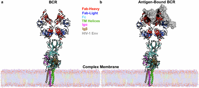
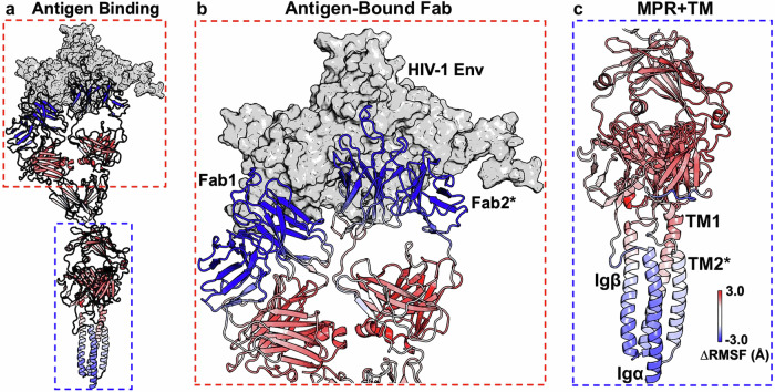
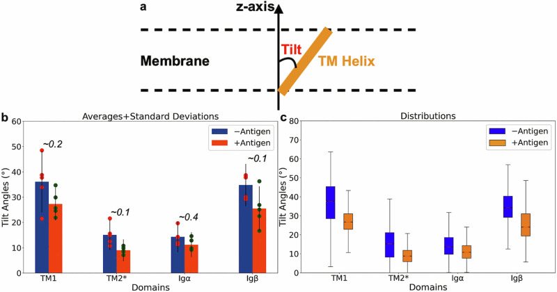
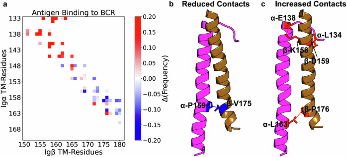
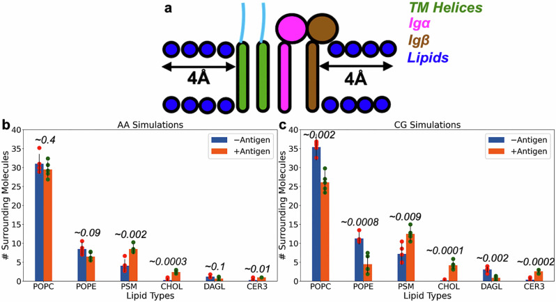
B-cell receptor (BCR) complexes are expressed on the surface of a B-cell and are critical in antigen recognition and modulating the adaptive immune response. Even though the relevance of antibodies has been known for almost a hundred years, the antigen-dependent activation mechanism of B-cells has remained elusive. Several models have been proposed for BCR activation, including cross-linking, conformation-induced oligomerization, and dissociation activation models. Recently, the first cryo-EM structures of the human B-cell antigen receptor of the IgM and IgG isotypes have been published that validates the asymmetric organization of the BCR complex. Here, we carry out extensive molecular dynamics simulations to probe the conformational changes upon antigen binding and the influence of the membrane lipids. We identify two critical dynamical events that could be associated with antigen-dependent activation of BCR. First, antigen binding causes increased flexibility in regions distal to the antigen binding site. Second, antigen binding alters the rearrangement of IgM transmembrane helices, including the relative interaction of Igα/Igβ that mediates intracellular signaling. Furthermore, these transmembrane rearrangements lead to changes in localized lipid composition. Our work indirectly supports the conformational-change induced models of BCR activation and contributes to the understanding of the antigen-dependent activation mechanism of BCRs.
Overview
The claims and statistics of this paper haven’t been checked yet.
Run the check — free, no account needed. It reads the open-access text and checks the sources it cites.
Integrity signals
Registry
Cites
Statistics
Figures
Verification
Claims the paper makes
The claims and statistics of this paper haven’t been checked yet.
Run the check — free, no account needed. It reads the open-access text and checks the sources it cites.
Self-check
Percentages, sums and reported differences re-computed from the paper’s own numbers.
The self-check needs the paper’s full text.
Citations
Cites
Highly accurate protein structure prediction with AlphaFold
Jumper, J., Evans, R., Pritzel, A., Green, T., Figurnov, M., Ronneberger, O., Tunyasuvunakool, K., Bates, R., Žídek, A., Potapenko, A., et al. (2021) · Nature · doi:10.1038/s41586-021-03819-2 · Cited by 43,764
Accurate structure prediction of biomolecular interactions with AlphaFold 3
Abramson, J., Adler, J., Dunger, J., Evans, R., Green, T., Pritzel, A., Ronneberger, O., Willmore, L., Ballard, A. J., Bambrick, J., et al. (2024) · Nature · doi:10.1038/s41586-024-07487-w · Cited by 15,861
Martini 3: A general purpose force field for coarse-grained molecular dynamics
Souza, P. C. T., Alessandri, R., Barnoud, J., Thallmair, S., Faustino, I., Grünewald, F., Patmanidis, I., Abdizadeh, H., Bruininks, B. M. H., Wassenaar, T. A., et al. (2021) · Nature Methods · doi:10.1038/s41592-021-01098-3 · Cited by 1,551
Research works printed but not linked: 59
- Robinson, R. Distinct B Cell Receptor Functions Are Determined by Phosphorylation. PLOS Biol. 4, e231 (2006). · DOI
- Harwood, N. E. & Batista, F. D. Early events in B cell activation. Annu. Rev. Immunol. 28, 185–210 (2010). · DOI
- Su, Q. et al. Cryo-EM structure of the human IgM B cell receptor. Science 377, 875–880 (2022). · DOI
- Treanor, B. B-cell receptor: from resting state to activate. Immunology 136, 21–27 (2012). · DOI
- Shelby, S. A., Castello-Serrano, I., Wisser, K. C., Levental, I. & Veatch, S. L. Membrane phase separation drives responsive assembly of receptor signaling domains. Nat. Chem. Biol. 19, 750–758 (2023). · DOI
- Harwood, N. E. & Batista, F. D. New insights into the early molecular events underlying B cell activation. Immunity 28, 609–619 (2008). · DOI
- Tolar, P. & Pierce, S. K. Unveiling the B cell receptor structure. Science 377, 819–820 (2022). · DOI
- Thyagarajan, R., Arunkumar, N. & Song, W. Polyvalent Antigens Stabilize B Cell Antigen Receptor Surface Signaling Microdomains. J. Immunol. 170, 6099–6106 (2003). · DOI
- Minguet, S., Dopfer, E.-P. & Schamel, W. W. A. Low-valency, but not monovalent, antigens trigger the B-cell antigen receptor (BCR). Int. Immunol. 22, 205–212 (2010). · DOI
- Kim, Y.-M. et al. Monovalent ligation of the B cell receptor induces receptor activation but fails to promote antigen presentation. PNAS 103, 3327–3332 (2006). · DOI
- Reth, M., Wienands, J. & Schamel, W. W. An unsolved problem of the clonal selection theory and the model of an oligomeric B-cell antigen receptor. Immunological Rev. 176, 10–18 (2000). · DOI
- Tolar, P. & Pierce, S. K. A conformation-induced oligomerization model for B cell receptor microclustering and signaling. Curr. Top. Microbiol. Immunol. 340, 155–169 (2010).
- Yang, J. & Reth, M. Oligomeric organization of the B-cell antigen receptor on resting cells. Nature 467, 465–469 (2010). · DOI
- Yang, J. & Reth, M. The dissociation activation model of B cell antigen receptor triggering. FEBS Lett. 584, 4872–4877 (2010). · DOI
- Yang, J. & Reth, M. Receptor Dissociation and B-Cell Activation. Curr. Top. Microbiol. Immunol. 393, 27–43 (2016).
- Huse, K. et al. Mechanism of CD79A and CD79B Support for IgM+ B Cell Fitness through B Cell Receptor Surface Expression. J. Immunol. 209, 2042–2053 (2022). · DOI
- Schamel, W. W. & Reth, M. Monomeric and oligomeric complexes of the B cell antigen receptor. Immunity 13, 5–14 (2000). · DOI
- Tolar, P., Sohn, H. W. & Pierce, S. K. The initiation of antigen-induced B cell antigen receptor signaling viewed in living cells by fluorescence resonance energy transfer. Nat. Immunol. 6, 1168–1176 (2005). · DOI
- Dong, Y. et al. Structural principles of B cell antigen receptor assembly. Nature 612, 156–161 (2022). · DOI · Paper page
- Bonsignori, M. et al. Two Distinct Broadly Neutralizing Antibody Specificities of Different Clonal Lineages in a Single HIV-1-Infected Donor: Implications for Vaccine Design. J. Virol. 86, 4688–4692 (2012). · DOI
- Bonsignori, M. et al. Inference of the HIV-1 VRC01 Antibody Lineage Unmutated Common Ancestor Reveals Alternative Pathways to Overcome a Key Glycan Barrier. Immunity 49, 1162–1174.e1168 (2018). · DOI
- Kwon, H.-Y. et al. Lipid-Oriented Live-Cell Distinction of B and T Lymphocytes. J. Am. Chem. Soc. 143, 5836–5844 (2021). · DOI
- Waterhouse, A. et al. SWISS-MODEL: homology modelling of protein structures and complexes. Nucleic Acids Res. 46, W296–W303 (2018). · DOI
- Liu, Q. et al. Improvement of antibody functionality by structure-guided paratope engraftment. Nat. Commun. 10, 721 (2019). · DOI
- Yan, Y., Tao, H., He, J. & Huang, S.-Y. The HDOCK server for integrated protein–protein docking. Nat. Protoc. 15, 1829–1852 (2020). · DOI
- Bansal, M., Kumar, S. & Velavan, R. HELANAL: a program to characterize helix geometry in proteins. J. Biomolecular Struct. Dyn. 17, 811–819 (2000). · DOI
- Michaud-Agrawal, N., Denning, E. J., Woolf, T. B. & Beckstein, O. MDAnalysis: A toolkit for the analysis of molecular dynamics simulations. J. Computational Chem. 32, 2319–2327 (2011). · DOI
- Gowers, R. J. et al. MDAnalysis: A Python Package for the Rapid Analysis of Molecular Dynamics Simulations. Proceedings of the 15th Python in Science Conference, 98-105 (2016). · DOI
- Reth, M. Discovering immunoreceptor coupling and organization motifs. Front. Immunol. 14, 1253412 (2023). · DOI
- Hossain, M. A. et al. B cells expressing IgM B cell receptors of HIV-1 neutralizing antibodies discriminate antigen affinities by sensing binding association rates. Cell Rep. 39, 111021 (2022). · DOI
- Veneziano, R. et al. Role of nanoscale antigen organization on B-cell activation probed using DNA origami. Nat. Nanotechnol. 15, 716–723 (2020). · DOI
- Kläsener, K., Maity, P. C., Hobeika, E., Yang, J. & Reth, M. B cell activation involves nanoscale receptor reorganizations and inside-out signaling by Syk. eLife 3, e02069 (2014). · DOI
- Smith, P. & Lorenz, C. D. LiPyphilic: A Python Toolkit for the Analysis of Lipid Membrane Simulations. J. Chem. Theory Comput. 17, 5907–5919 (2021). · DOI
- Chavent, M. et al. Methodologies for the analysis of instantaneous lipid diffusion in MD simulations of large membrane systems. Faraday Discuss. 169, 455–475 (2014). · DOI
- Ingólfsson, H. I. et al. The power of coarse graining in biomolecular simulations. WIREs Computational Mol. Sci. 4, 225–248 (2013). · DOI
- Alessandri, R. et al. Pitfalls of the Martini Model. J. Chem. Theory Comput. 15, 5448–5460 (2019). · DOI
- Marrink, S. J. et al. Computational Modeling of Realistic Cell Membranes. Chem. Rev. 119, 6184–6226 (2019). · DOI
- Consortium, T. U. UniProt: the Universal Protein Knowledgebase in 2023. Nucleic Acids Res. 51, D523–D531 (2023). · DOI · Paper page
- Evans, R. et al. Protein complex prediction with AlphaFold-Multimer. bioRxiv https://doi.org/10.1101/2021.10.04.463034 (2022). · DOI · Paper page
- Jo, S., Kim, T., Iyer, V. & Im, W. CHARMM-GUI: A Web-based Graphical User Interface for CHARMM. J. Computational Chem. 29, 1859–1865 (2008). · DOI · Paper page
- Wu, E. et al. CHARMM-GUI Membrane Builder Toward Realistic Biological Membrane Simulations. J. Computational Chem. 35, 1997–2004 (2014). · DOI
- Jo, S., Lim, J., Klauda, J. & Im, W. CHARMM-GUI Membrane Builder for Mixed Bilayers and Its Application to Yeast Membranes. Biophysical J. 97, 50–58 (2009). · DOI
- Lee, J. et al. CHARMM-GUI Input Generator for NAMD, GROMACS, AMBER, OpenMM, and CHARMM/OpenMM Simulations using the CHARMM36 Additive Force Field. J. Chem. Theory Comput. 12, 405–413 (2016). · DOI
- Huang, J. et al. CHARMM36m: an improved force field for folded and intrinscially disordered proteins. Nat. Methods 14, 71–73 (2017). · DOI
- Hess, B., Bekker, H., Berendsen, H. J. C. & Fraaije, J. G. E. M. LINCS: A linear constraint solver for molecular simulations. J. Computational Chem. 18, 1463–1472 (1998). · DOI
- Essmann, U. et al. A Smooth Particle Mesh Ewald Method. J. Chem. Phys. 103 (1995). · DOI
- Grubmuller, H., Heller, H., Windemuth, A. & Schulten, K. Generalized Verlet Algorithm for Efficient Molecular Dynamics Simulations with Long-Range Interactions. Mol. Simul. 6, 121–142 (1991). · DOI
- Evans, D. J. & Holian, B. L. The Nose-Hoover thermostat. J. Chem. Phys. 83, 4069–4074 (1985). · DOI
- Parrinello, M. & Rahman, A. Polymorphic Transitions in Single Crystals: A New Molecular Dynamics Method. J. Appl. Phys. 52, 7182–7190 (1981). · DOI
- Kroon, P. C. et al. Martinize2 and Vermouth: Unified Framework for Topology Generation. eLife (2023). https://doi.org/10.7554/eLife.90627.1 · DOI
- Wassenaar, T. A., Pluhackova, K., Böckmann, R. A., Marrink, S. J. & Tieleman, D. P. Going Backward: A Flexible Geometric Approach to Reverse Transformation from Coarse Grained to Atomistic Models. J. Chem. Theory Comput. 10, 676–690 (2014). · DOI
- Herzog, F. A., Braun, L., Schoen, I. & Vogel, V. Improved Side Chain Dynamics in MARTINI Simulations of Protein–Lipid Interfaces. J. Chem. Theory Comput. 12, 2446–2458 (2016). · DOI
- Periole, X., Cavalli, M., Marrink, S.-J. & Ceruso, M. A. Combining an Elastic Network With a Coarse-Grained Molecular Force Field: Structure, Dynamics, and Intermolecular Recognition. J. Chem. Theory Comput. 5 https://doi.org/10.1021/ct9002114 (2009). · DOI
- Wassenaar, T. A., Ingolfsson, H. I., Bockmann, R. A., Tieleman, D. P. & Marrink, S. J. Computational Lipidomics with insane: A Versatile Tool for Generating Custom Membranes for Molecular Simulations. J. Chem. Theory Comput. 11, 2144–2155 (2015). · DOI · Paper page
- Tironi, I. G., Sperb, R., Smith, P. E. & Gunsteren, W. F. A generalized reaction field method for molecular dynamics simulations. J. Chem. Phys. 102, 5451–5459 (1995). · DOI
- Bussi, G., Donadio, D. & Parrinello, M. Canonical sampling through velocity rescaling. J. Chem. Phys. 126, 014101 (2007). · DOI
- Bernetti, M. & Bussi, G. Pressure control using stochastic cell rescaling. J. Chem. Phys. 153, 114107 (2020). · DOI
- Abraham, M. J. et al. GROMACS: High performance molecular simulations through multi-level parallelism from laptops to supercomputers. SoftwareX 1-2, 19–25 (2015). · DOI · Paper page
- Roe, D. R. & Cheatham, I. T. E. PTRAJ and CPPTRAJ: software for processing and analysis of molecular dynamics trajectory data. J. Chem. Theory Comput. 9, 3084–3095 (2013). · DOI
Metadata
- Authors
- Do, H. N., Zhao, M., Alam, S. M., & Gnanakaran, S.
- Affiliations
- Los Alamos National Laboratory (US)
- Duke University (US)
- Corresponding author
- Gnanakaran, S.
- Venue
- Communications Biology
- Published
- 2025-02-13
- Volume
- 8
- Issue
- 1
- Pages
- 226-226
- ISSN
- 2399-3642 (ISSN-L)
- PMC
- PMC11825855
- OpenAlex
- W4407535508
- Cited by
- 2
- References
- 3
- Citations by year
- 2026: 1
- FWCI
- 0.30
- Type
- journal article
- Language
- en
- Open access
- gold
- doi.org
- Licence
- Locations
- Communications Biology (publishedVersion, cc-by-nc-nd)
- bioRxiv (Cold Spring Harbor Laboratory) (acceptedVersion, cc-by-nd)
- PubMed Central (publishedVersion, cc-by-nc-nd)
- PubMed (publishedVersion)
- OSTI OAI (U.S. Department of Energy Office of Scientific and Technical Information) (submittedVersion, cc-by-nc-nd)
- PubMed Central (submittedVersion, cc-by-nd)
- DOAJ (DOAJ: Directory of Open Access Journals) (submittedVersion, cc-by-sa)
- Funders
- U.S. Department of Health & Human Services | NIH | National Institute of Allergy and Infectious Diseases (U54-AI170752-01)
- DOE | National Nuclear Security Administration (89233218CNA000001)
- Topics
- Life Sciences › Biochemistry, Genetics and Molecular Biology › Molecular Biology › Protein Structure and Dynamics
- Life Sciences › Immunology and Microbiology › Immunology › T-cell and B-cell Immunology
- Life Sciences › Biochemistry, Genetics and Molecular Biology › Molecular Biology › Lipid Membrane Structure and Behavior
- Keywords
- B cell receptor, antigen binding, conformational change, IgM, IgG, membrane lipids, intracellular signaling, molecular dynamics simulation
About these checks
Registry, retraction, Cites and statistics results are computed, not judged, with one exception: a printed reference that no index lists is read once by a model before it counts as a citation error. Claims are extracted by a model and kept only when their quote appears verbatim in the text; each cited source is then checked, with a model’s help, for whether it says what the claim relies on. Those verdicts can be wrong — read the claim’s quote and the cited source before relying on one.
Statistics and arithmetic are recomputed, not judged.
Confidence bands are shown only for fields calibrated on our annotated test set. None is calibrated yet, so no band is shown.
What counts as a claim ›How we measure confidence ›How we check this ›
Are you an author, or do you see a problem with this page? Request a review
How to cite
Cite CoChat for the checks on this page, and the paper for its own findings.
- This page
- CoChat. (2026). Citation check: Dynamics and activation of membrane-bound B cell receptor assembly. https://app.cochat.ai/papers/10.1038/s42003-025-07478-1
- The paper
- Do, H. N., Zhao, M., Alam, S. M., & Gnanakaran, S. (2025). Dynamics and activation of membrane-bound B cell receptor assembly. Communications Biology. https://doi.org/10.1038/s42003-025-07478-1
