

The DOI matches its Crossref record; 2 of the 2 references it cites were found in a registry or an index.
Multiple mutations in polyketide synthase led to disruption of Psittacofulvin production across diverse parrot species
Open accessPublished version of a preprint
Abstract
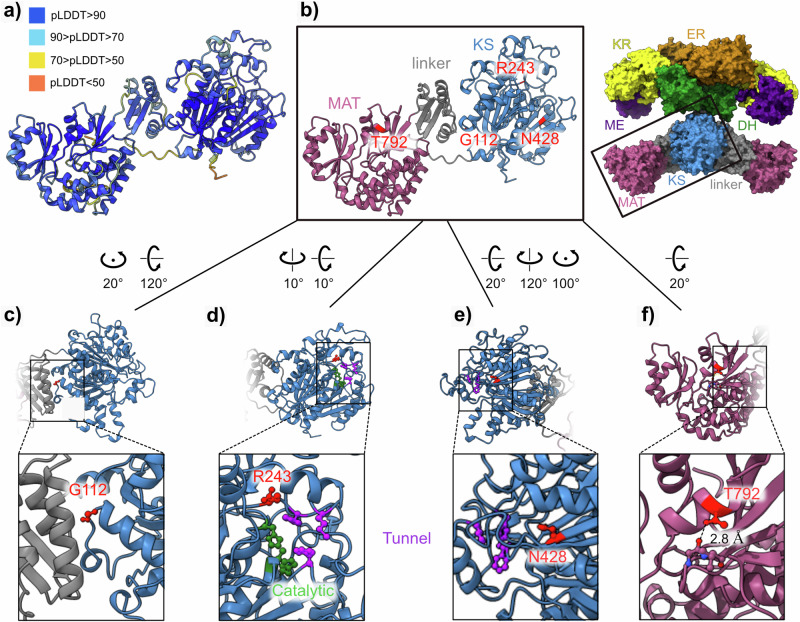
Abstract Polyketide synthases (PKSs) are crucial multidomain enzymes in diverse natural product biosynthesis. Parrots use a type I PKS to produce a unique pigment called psittacofulvin in their feathers. In domesticated budgerigars and lovebirds, the same amino acid substitution (R644W) within malonyl/acetyltransferase (MAT) domain of this enzyme has been shown to cause the blue phenotype with no psittacofulvin pigmentation, proposing a strong evolutionary constraint mechanism. Here, we identified six new causal variants in PKS associated with defective psittacofulvin production in four diverse species, including three nonsense mutations. Remaining three nonsynonymous substitutions reside within the ketoacyl synthase (KS) domain. Heterologous expression of these PKS variants in yeast confirmed complete or reduced loss of psittacofulvin production, additionally suggesting presence of an auxiliary polymorphism at the MAT domain. Our findings establish PKS as a functionally conserved key-enzyme determining psittacofulvin-based hues among diverse parrots, highlighting multiple conserved domains essential for the PKS function.
Overview
The claims and statistics of this paper haven’t been checked yet.
Run the check — free, no account needed. It reads the open-access text and checks the sources it cites.
Integrity signals
Registry
Cites
Statistics
Figures
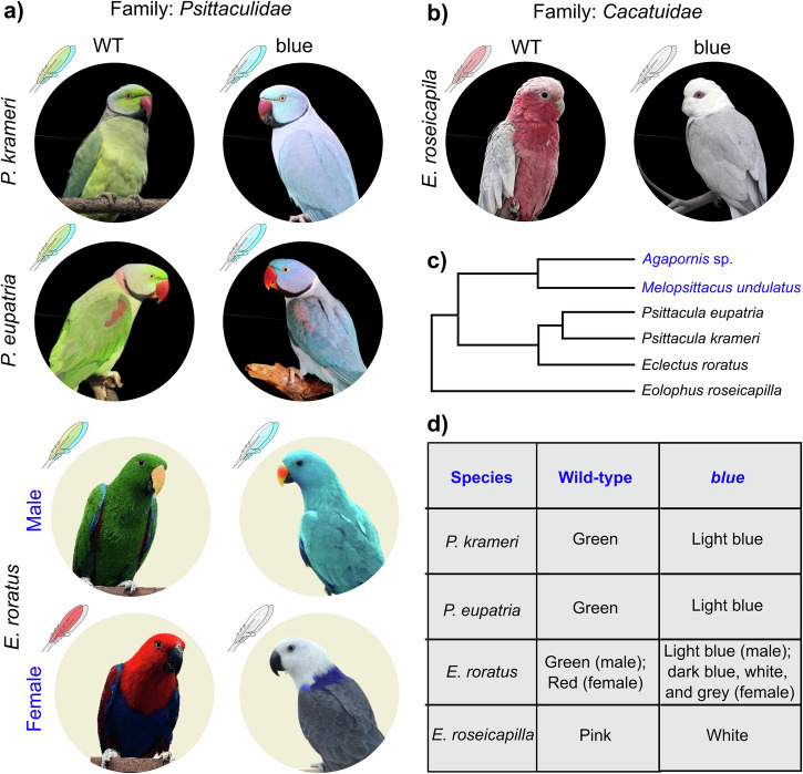
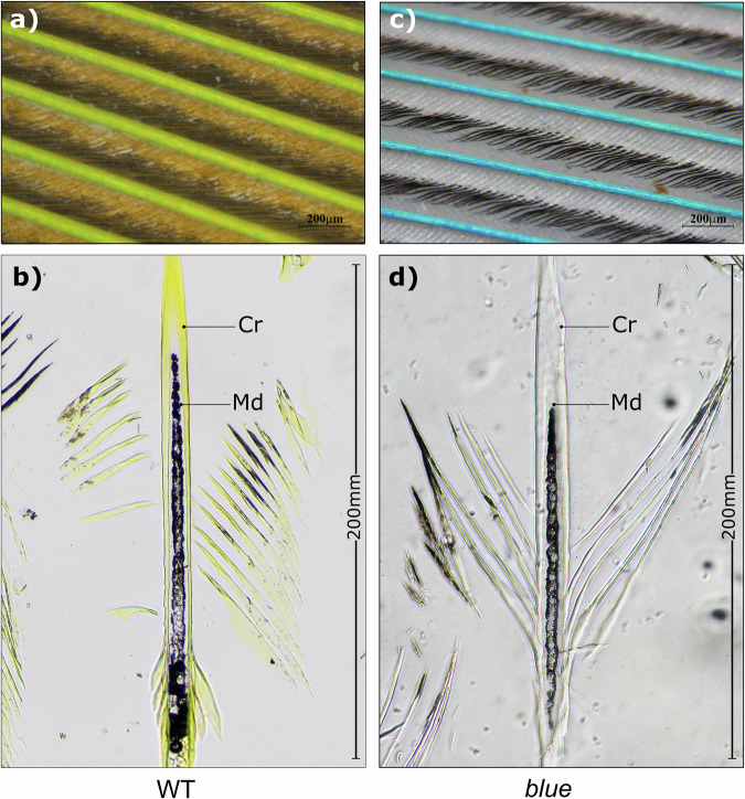
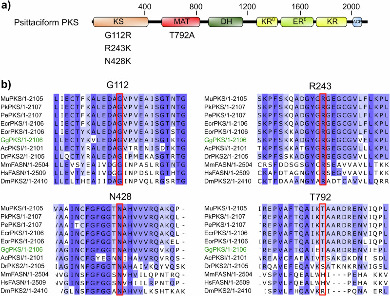
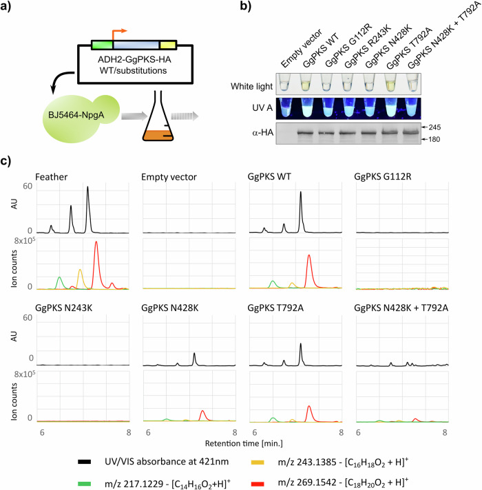
Verification
Claims the paper makes
The claims and statistics of this paper haven’t been checked yet.
Run the check — free, no account needed. It reads the open-access text and checks the sources it cites.
Self-check
Percentages, sums and reported differences re-computed from the paper’s own numbers.
The self-check needs the paper’s full text.
Citations
Cites
Highly accurate protein structure prediction with AlphaFold
Jumper, J., Evans, R., Pritzel, A., Green, T., Figurnov, M., Ronneberger, O., Tunyasuvunakool, K., Bates, R., Žídek, A., Potapenko, A., et al. (2021) · Nature · doi:10.1038/s41586-021-03819-2 · Cited by 43,764
The Evolutionary Origin And Diversification Of Feathers
Prum, R. O., & Brush, A. H. (2002) · The Quarterly Review of Biology · doi:10.1086/341993 · Cited by 331
Research works printed but not linked: 28
- Prum, R. O., Torres, R., Kovach, C., Williamson, S. & Goodman, S. M. Coherent light scattering by nanostructured collagen arrays in the caruncles of the malagasy asities (Eurylaimidae: Aves). J. Exp. Biol. 202, 3507–3522 (1999). · DOI
- Krukenberg, C. F. W. Die federfarbstoffe der psittaciden. Vergleichend-physiologische Studien Reihe 2, 29–36 (1882).
- Stradi, R., Pini, E. & Celentano, G. The chemical structure of the pigments in Ara macao plumage. Compar. Biochem. Physiol. B Biochem. Mol. Biol. 130, 57–63 (2001). · DOI
- Tinbergen, J., Wilts, B. D. & Stavenga, D. G. Spectral tuning of amazon parrot feather coloration by psittacofulvin pigments and spongy structures. J. Exp. Biol. 216, 4358–4364 (2013).
- Masello, J. F. & Quillfeldt, P. Body size, body condition and ornamental feathers of Burrowing Parrots: Variation between years and sexes, assortative mating and influences on breeding success. Emu 103, 149–161 (2003). · DOI
- Burtt, E. H., Schroeder, M. R., Smith, L. A., Sroka, J. E. & McGraw, K. J. Colourful parrot feathers resist bacterial degradation. Biol. Lett. 7, 214–216 (2011). · DOI
- Cooke, T. F. et al. Genetic mapping and biochemical basis of yellow feather pigmentation in budgerigars. Cell 171, 427–439.e21 (2017). · DOI
- Torres, J. P. & Schmidt, E. W. The biosynthetic diversity of the animal world. J. Biol. Chem. 294, 17684 –17692 (2019). · DOI
- Li, F. et al. Sea Urchin Polyketide Synthase SpPks1 Produces the Naphthalene Precursor to Echinoderm Pigments. J. Am. Chem. Soc. 144, 9363–9371 (2022). · DOI
- Ke, F. et al. Convergent evolution of parrot plumage coloration. PNAS Nexus https://doi.org/10.1093/pnasnexus/pgae107 (2024). · DOI
- Adamec, F. et al. Spectroscopic investigation of a brightly colored psittacofulvin pigment from parrot feathers. Chem. Phys. Lett. 648, 195–199 (2016). · DOI
- Arbore, R. et al. A molecular mechanism for bright color variation in parrots. Science 386, eadp7710 (2024). · DOI
- Chan, D. T. C., Poon, E. S. K., Wong, A. T. C. & Sin, S. Y. W. Global trade in parrots – Influential factors of trade and implications for conservation. Glob. Ecol. Conserv. 30, e01784 (2021). · DOI
- Ross, C., Opel, V., Scherlach, K. & Hertweck, C. Biosynthesis of antifungal and antibacterial polyketides by Burkholderia gladioli in coculture with Rhizopus microsporus. Mycoses 57, 48–55 (2014). · DOI
- McGraw, K. J. & Nogare, M. C. Carotenoid pigments and the selectivity of psittacofulvin-based coloration systems in parrots. Compare. Biochem. Physiol. B Biochem. Mol. Biol. 138, 229–233 (2004). · DOI
- D’Alba, L., Kieffer, L. & Shawkey, M. D. Relative contributions of pigments and biophotonic nanostructures to natural color production: a case study in budgerigar (Melopsittacus undulatus) feathers. J. Exp. Biol. 215, 1272–1277 (2012). · DOI
- Leibundgut, M., Maier, T., Jenni, S. & Ban, N. The multienzyme architecture of eukaryotic fatty acid synthases. Curr. Opin. Struct. Biol. 18, 714-725 (2008). · DOI
- Ma, S. M. et al. Complete reconstitution of a highly reducing iterative polyketide synthase. Science 326, 589–592 (2009). · DOI
- Hausmann, F., Arnold, K. E., Marshall, N. J. & Owens, I. P. F. Ultraviolet signals in birds are special. Proc. R. Soc. B Biol. Sci. 270, 61–67 (2003). · DOI
- Maier, T., Leibundgut, M. & Ban, N. The Crystal Structure of a Mammalian Fatty Acid Synthase. https://www.science.org (2008). · DOI
- Ghosh Roy, S. et al. Mutations in SLC45A2 lead to loss of melanin in parrot feathers. G3: Genes, Genomes, Genetics https://doi.org/10.1093/g3journal/jkad254 (2023). · DOI
- Bonin, J. & Homberger, D. Possible convergent evolution of yellow psittacofulvin colors in the feathers of cockatoos (Cacatuidae) and parrots (Psittacidae). FASEB J. 26, 722.23 (2012). · DOI
- De Oliveira Neves, A. C., Galván, I. & Van Den Abeele, D. Impairment of mixed melanin-based pigmentation in parrots. J. Exp. Biol. 223, jeb225912 (2020). · DOI
- Price-Waldman, R. & Stoddard, M. C. Avian coloration genetics: recent advances and emerging questions. J. Heredity 112, 395–416 (2021). · DOI
- Kumar, S. et al. TimeTree 5: an expanded resource for species divergence times. Mol. Biol. Evol. 39, msac174 (2022). · DOI
- Wright, T. F. et al. A multilocus molecular phylogeny of the parrots (Psittaciformes): support for a gondwanan origin during the cretaceous. Mol. Biol. Evol. 25, 2141–2156 (2008). · DOI
- Smith, B. T. et al. Phylogenomic analysis of the parrots of the world distinguishes artifactual from biological sources of gene tree discordance. Syst. Biol. 72, 228–241 (2023). · DOI
- Goddard, T. D. et al. UCSF ChimeraX: meeting modern challenges in visualization and analysis. Protein Sci. 27, 14–25 (2018). · DOI · Paper page
Metadata
- Authors
- Ghosh Roy, S., Brejcha, J., Maršík, P., Bakhrat, A., Abdu, M., Arbore, R., Araújo, P. M., Afonso, S., Carneiro, M., Grossman-Haham, I., & Abdu, U.
- Affiliations
- Charles University (CZ)
- Czech University of Life Sciences Prague (CZ)
- Ben-Gurion University of the Negev (IL)
- Universidade do Porto (PT)
- Corresponding author
- Abdu, Uri
- ORCID
- Venue
- Communications Biology
- Published
- 2025-01-17
- Volume
- 8
- Issue
- 1
- Pages
- 69-69
- ISSN
- 2399-3642 (ISSN-L)
- PMC
- PMC11739604
- OpenAlex
- W4406446488
- Cited by
- 1
- References
- 2
- Citations by year
- 2026: 1
- FWCI
- 0.50
- Type
- journal article
- Language
- en
- Open access
- gold
- doi.org
- Licence
- Locations
- Communications Biology (publishedVersion, cc-by-nc-nd)
- Research Square (acceptedVersion, cc-by)
- PubMed Central (publishedVersion, cc-by-nc-nd)
- PubMed (publishedVersion)
- DOAJ (DOAJ: Directory of Open Access Journals) (submittedVersion, cc-by-sa)
- Funders
- Israel Science Foundation (941/21)
- Topics
- Life Sciences › Biochemistry, Genetics and Molecular Biology › Cell Biology › melanin and skin pigmentation
- Life Sciences › Biochemistry, Genetics and Molecular Biology › Biotechnology › Microbial Metabolism and Applications
- Life Sciences › Immunology and Microbiology › Applied Microbiology and Biotechnology › Microbial Natural Products and Biosynthesis
- Keywords
- polyketide synthase, parrots, feather pigmentation, budgerigars, lovebirds, nonsense mutation, nonsynonymous substitutions, heterologous expression in yeast, natural product biosynthesis, evolutionary constraints
- MeSH terms
- Animals, Feathers, Mutation, Parrots, Pigmentation, Pigments, Biological, Polyketide Synthases
About these checks
Registry, retraction, Cites and statistics results are computed, not judged, with one exception: a printed reference that no index lists is read once by a model before it counts as a citation error. Claims are extracted by a model and kept only when their quote appears verbatim in the text; each cited source is then checked, with a model’s help, for whether it says what the claim relies on. Those verdicts can be wrong — read the claim’s quote and the cited source before relying on one.
Statistics and arithmetic are recomputed, not judged.
Confidence bands are shown only for fields calibrated on our annotated test set. None is calibrated yet, so no band is shown.
What counts as a claim ›How we measure confidence ›How we check this ›
Are you an author, or do you see a problem with this page? Request a review
