Cited by 15DNA Repair MechanismsPlant Molecular Biology ResearchPlant Genetic and Mutation Studies

The DOI matches its Crossref record; 4 of the 4 references it cites were found in a registry or an index.

Identification of plants’ functional counterpart of the metazoan mediator of DNA Damage checkpoint 1

Open access

Published version of a preprint

Abstract

Induction of DNA damage triggers rapid phosphorylation of the histone H2A.X (γH2A.X). In animals, mediator of DNA damage checkpoint 1 (MDC1) binds γH2A.X through a tandem BRCA1 carboxyl-terminal (tBRCT) domain and mediates recruitment of downstream effectors of DNA damage response (DDR). However, readers of this modification in plants have remained elusive. We show that from the Arabidopsis BRCT domain proteome, BCP1-4 proteins with tBRCT domains are involved in DDR. Through its tBRCT domain BCP4 binds γH2A.X in vitro and localizes to DNA damage-induced foci in an H2A.X-dependent manner. BCP4 also contains a domain that interacts directly with NBS1 and thus acts as a functional counterpart of MDC1. We also show that BCP1, that contains two tBRCT domains, co-localizes with γH2A.X but it does not bind γH2A.X suggesting functional similarity with human PAXIP1. A phylogenetic analysis supports that PAXIP1 and MDC1 in metazoa and their plant counterparts evolved independently from common ancestors with tBRCT domains. Collectively, our study reveals missing components and provides mechanistic and evolutionary insights into plant DDR.

Overview

The claims and statistics of this paper haven’t been checked yet.

Run the check — free, no account needed. It reads the open-access text and checks the sources it cites.

A few free checks a day · papers already verified open instantly · results are public

Integrity signals

Registry

Crossref · 10.1038/s44319-024-00107-8

Cites

4 of 4 references verified0 citation errors · 0 not checkable

Statistics

Figures

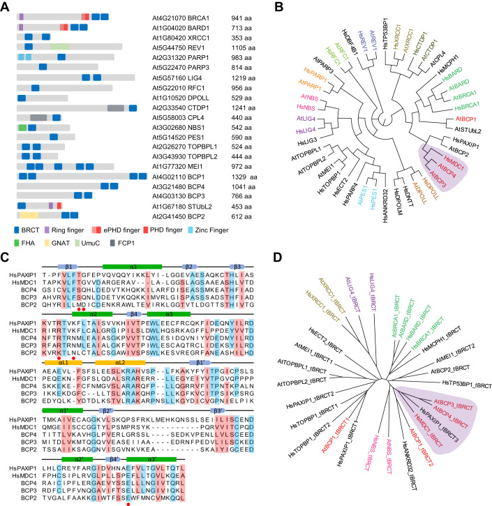
Figure 1 Identification of Arabidopsis proteins with BRCT domains. (A) Schematic representation of 21 Arabidopsis proteins containing BRCT domains. The gene codes, protein names, and length in amino acids (aa) are indicated. (B) Human and Arabidopsis BRCT domain proteins were aligned with MUSCLE and a maximum likelihood circular cladogram was generated with CLC Genomics Workbench ver. 11.0. (C) Sequence alignment of Arabidopsis BCP3 and BCP4 and human MDC1 and PAXIP1 tBRCT domains. For alignment, tBRCT3 of PAXIP1 was used. Residues conserved in at least four proteins are shaded light blue and those similar in at least four proteins are shaded light red. Structural elements of domains are indicated on the top of the alignment according to Stucki et al (2005). Amino acids involved in MDC1 interaction with γH2A.X are indicated with red dots. (D) All human and Arabidopsis tBRCT domains were aligned as in (B) and a maximum likelihood tree illustrating amino acid sequence conservation between tBRCT domains was generated with CLC Genomics Workbench ver. 11.0. (B, D) Human and Arabidopsis proteins with high sequence homology share the same color code. Arabidopsis proteins and tBRCT domains clustering with human MDC1 and PAXIP1 are indicated in red and shaded in purple.
Figure 1 Identification of Arabidopsis proteins with BRCT domains. (A) Schematic representation of 21 Arabidopsis proteins containing BRCT domains. The gene codes, protein names, and length in amino acids (aa) are indicated. (B) Human and Arabidopsis BRCT domain proteins were aligned with MUSCLE and a maximum likelihood circular cladogram was generated with CLC Genomics Workbench ver. 11.0. (C) Sequence alignment of Arabidopsis BCP3 and BCP4 and human MDC1 and PAXIP1 tBRCT domains. For alignment, tBRCT3 of PAXIP1 was used. Residues conserved in at least four proteins are shaded light blue and those similar in at least four proteins are shaded light red. Structural elements of domains are indicated on the top of the alignment according to Stucki et al (2005). Amino acids involved in MDC1 interaction with γH2A.X are indicated with red dots. (D) All human and Arabidopsis tBRCT domains were aligned as in (B) and a maximum likelihood tree illustrating amino acid sequence conservation between tBRCT domains was generated with CLC Genomics Workbench ver. 11.0. (B, D) Human and Arabidopsis proteins with high sequence homology share the same color code. Arabidopsis proteins and tBRCT domains clustering with human MDC1 and PAXIP1 are indicated in red and shaded in purple.CC BY · PubMed Central
Figure EV1 Phylogeny of BCP1, BCP3, and BCP4. (A) Maximum likelihood tree of BCP1 across Viridiplantae. The schematic presentation of BCP1 is shown at the bottom and a PHD finger present in all BCP sequences except in Brassicaceae is indicated. (B) Maximum likelihood tree of BCP3 and BCP4 across Viridiplantae. (A, B) Major clades are indicated by differently colored shading. The non-flowering land plant clade includes sequences from hornworts, mosses, liverworts, lycophytes, and monilophytes. Thuja plicata was the only gymnosperm used in the analysis. (C) Alignment of BCP3 and BCP4 proteins. Identical and conserved amino acids are indicated in red and blue letters, respectively. The positions of introns are indicated by black arrows. The tBRCT domain and three other conserved regions are shown in colored boxes. (D) Schematic representation of Arabidopsis BCP genes with exons indicated by gray boxes and introns by black lines. Exons and introns are drawn to scale according to the lengths of DNA sequences. Positions of T-DNA insertions in bcp mutant lines used for DNA damage sensitivity assays are indicated above each gene. Data information: A list of protein sequences used for the analysis is available from Source data for Fig. 5A,B.
Figure EV1 Phylogeny of BCP1, BCP3, and BCP4. (A) Maximum likelihood tree of BCP1 across Viridiplantae. The schematic presentation of BCP1 is shown at the bottom and a PHD finger present in all BCP sequences except in Brassicaceae is indicated. (B) Maximum likelihood tree of BCP3 and BCP4 across Viridiplantae. (A, B) Major clades are indicated by differently colored shading. The non-flowering land plant clade includes sequences from hornworts, mosses, liverworts, lycophytes, and monilophytes. Thuja plicata was the only gymnosperm used in the analysis. (C) Alignment of BCP3 and BCP4 proteins. Identical and conserved amino acids are indicated in red and blue letters, respectively. The positions of introns are indicated by black arrows. The tBRCT domain and three other conserved regions are shown in colored boxes. (D) Schematic representation of Arabidopsis BCP genes with exons indicated by gray boxes and introns by black lines. Exons and introns are drawn to scale according to the lengths of DNA sequences. Positions of T-DNA insertions in bcp mutant lines used for DNA damage sensitivity assays are indicated above each gene. Data information: A list of protein sequences used for the analysis is available from Source data for Fig. 5A,B.CC BY · PubMed Central
Figure EV2 Primary sequences analysis of BCP proteins. (A) The consensus sequence of the PHD finger from BCP1 derived from the alignment of plant BCP1 proteins. Cysteine and histidine residues characteristic of PHD fingers are underlined. (B) Schematic representation of Arabidopsis BCP1 and human PAXIP1 proteins. Conserved domains and motifs of PAXIP1 and their assigned functions are indicated. A list of plant species and the corresponding protein sequences used for the analysis in (A, B) are available from Source data. (C) Alignment of SQSQ, DWD, and DTQ sequence motifs from BCP4. The signature motifs are shaded in blue and green. (D) Schematic representation of MDC1 proteins from invertebrate, vertebrate (except mammals), and mammalian species. Conserved domains and motifs and their assigned functions are indicated. Source data are available online for this figure.
Figure EV2 Primary sequences analysis of BCP proteins. (A) The consensus sequence of the PHD finger from BCP1 derived from the alignment of plant BCP1 proteins. Cysteine and histidine residues characteristic of PHD fingers are underlined. (B) Schematic representation of Arabidopsis BCP1 and human PAXIP1 proteins. Conserved domains and motifs of PAXIP1 and their assigned functions are indicated. A list of plant species and the corresponding protein sequences used for the analysis in (A, B) are available from Source data. (C) Alignment of SQSQ, DWD, and DTQ sequence motifs from BCP4. The signature motifs are shaded in blue and green. (D) Schematic representation of MDC1 proteins from invertebrate, vertebrate (except mammals), and mammalian species. Conserved domains and motifs and their assigned functions are indicated. Source data are available online for this figure.CC BY · PubMed Central
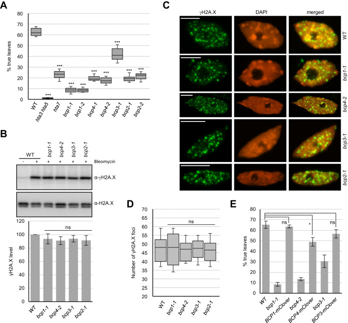
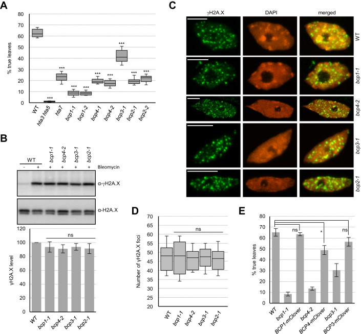
Figure 2 Analysis of bcp mutants. (A) DNA damage sensitivity of BCP mutant lines assessed by true leaf development assay (n = 13 biological replicates, each with 64 seeds). DNA damage-sensitive mutants of H2A.X (hta3 hta5) and H2A.W.7 (hta7) histone variants were used as controls. Data information: Data are represented as a box plot with median and interquartile range (box) and minimal and maximal values (whiskers). ***P ≤ 0.0001 (two-tailed paired Student’s t test). (B) Analysis of γH2A.X levels in bcp mutant seedlings after induction of DNA damage for 2 h. Representative western blots for γH2A.X and H2A.X in BCP mutants. Quantified γH2A.X levels normalized to the total H2A.X (n = 6, three biological replicates with two technical repeats). (C) Analysis of γH2A.X foci in nuclei of WT and bcp mutants. Data information: Maximum intensity projection images from Z-stacks of representative nuclei are shown. Scale bars represent 5 µm. (D) Quantification of γH2A.X foci number in bcp mutants. For each genetic background n = 8 nuclei were used. Data are represented as a box plot with median and interquartile range (box) and minimal and maximal values (whiskers). Data information: P > 0.05, ns (nonsignificant). (two-tailed paired Student’s t test). (E) DNA damage sensitivity of BCP-mClover3 complementing lines (n = 6 biological replicates, each with 64 seeds). Data information: In (B, E), data are presented as mean ± SD. ***P ≤ 0.0001; **P ≤ 0.001; *P ≤ 0.01; P > 0.01, ns (nonsignificant). (two-tailed paired Student’s t test). In (A, E) Seeds were germinated on medium containing 20 μg/ml of zeocin and true leaf development was scored 12 days after germination. Source data are available online for this figure.
Figure 2 Analysis of bcp mutants. (A) DNA damage sensitivity of BCP mutant lines assessed by true leaf development assay (n = 13 biological replicates, each with 64 seeds). DNA damage-sensitive mutants of H2A.X (hta3 hta5) and H2A.W.7 (hta7) histone variants were used as controls. Data information: Data are represented as a box plot with median and interquartile range (box) and minimal and maximal values (whiskers). ***P ≤ 0.0001 (two-tailed paired Student’s t test). (B) Analysis of γH2A.X levels in bcp mutant seedlings after induction of DNA damage for 2 h. Representative western blots for γH2A.X and H2A.X in BCP mutants. Quantified γH2A.X levels normalized to the total H2A.X (n = 6, three biological replicates with two technical repeats). (C) Analysis of γH2A.X foci in nuclei of WT and bcp mutants. Data information: Maximum intensity projection images from Z-stacks of representative nuclei are shown. Scale bars represent 5 µm. (D) Quantification of γH2A.X foci number in bcp mutants. For each genetic background n = 8 nuclei were used. Data are represented as a box plot with median and interquartile range (box) and minimal and maximal values (whiskers). Data information: P > 0.05, ns (nonsignificant). (two-tailed paired Student’s t test). (E) DNA damage sensitivity of BCP-mClover3 complementing lines (n = 6 biological replicates, each with 64 seeds). Data information: In (B, E), data are presented as mean ± SD. ***P ≤ 0.0001; **P ≤ 0.001; *P ≤ 0.01; P > 0.01, ns (nonsignificant). (two-tailed paired Student’s t test). In (A, E) Seeds were germinated on medium containing 20 μg/ml of zeocin and true leaf development was scored 12 days after germination. Source data are available online for this figure.CC BY · PubMed Central
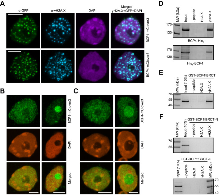
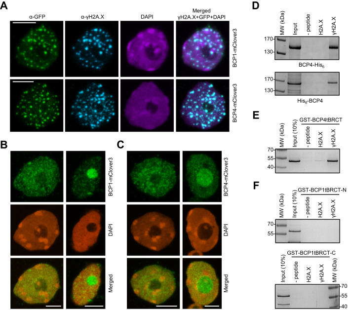
Figure 3 BCP4 and MDC1 share similar properties. (A) BCP1 and BCP4 co-localize with γH2A.X foci. Immunostaining of nuclei from bleomycin-treated seedlings expressing BCP1-mClover3 and BCP4-mClover3 in bcp1 and bcp4 mutants, respectively. (B, C) BCP1 and BCP4 foci formation is dependent on H2A.X Immunostaining of nuclei from bleomycin-treated seedlings expressing BCP1-mClover (B) and BCP4-mClover (C) in bcp1 hta3 hta5 and bcp4 hta3 hta5 genetic backgrounds, respectively. (D) BCP4 binds phosphorylated H2A.X C-terminal peptide. Affinity pull-down with N- and C-terminally His-tagged BCP4 and biotinylated H2A.X peptides. (E) Affinity pull-down with GST-tagged tBRCT domain of BCP4. (F) The tBRCT domains of BCP1 do not bind phosphorylated H2A.X C-terminal peptide. Affinity pull-down with GST-tagged tBRCT domains and biotinylated H2A.X peptides. Data information: In (A–C), maximum intensity projection images from Z-stacks are shown and scale bars represent 5 µm. Proteins were analyzed on 10% (D) or 12% (E, F) SDS-PAGE and gels were stained with Coomassie blue. Source data are available online for this figure.
Figure 3 BCP4 and MDC1 share similar properties. (A) BCP1 and BCP4 co-localize with γH2A.X foci. Immunostaining of nuclei from bleomycin-treated seedlings expressing BCP1-mClover3 and BCP4-mClover3 in bcp1 and bcp4 mutants, respectively. (B, C) BCP1 and BCP4 foci formation is dependent on H2A.X Immunostaining of nuclei from bleomycin-treated seedlings expressing BCP1-mClover (B) and BCP4-mClover (C) in bcp1 hta3 hta5 and bcp4 hta3 hta5 genetic backgrounds, respectively. (D) BCP4 binds phosphorylated H2A.X C-terminal peptide. Affinity pull-down with N- and C-terminally His-tagged BCP4 and biotinylated H2A.X peptides. (E) Affinity pull-down with GST-tagged tBRCT domain of BCP4. (F) The tBRCT domains of BCP1 do not bind phosphorylated H2A.X C-terminal peptide. Affinity pull-down with GST-tagged tBRCT domains and biotinylated H2A.X peptides. Data information: In (A–C), maximum intensity projection images from Z-stacks are shown and scale bars represent 5 µm. Proteins were analyzed on 10% (D) or 12% (E, F) SDS-PAGE and gels were stained with Coomassie blue. Source data are available online for this figure.CC BY · PubMed Central
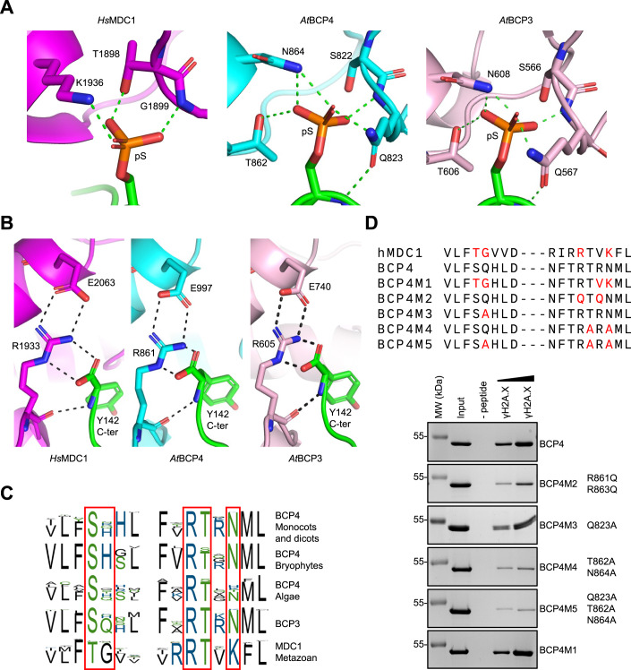
Figure 4 BCP4 binds γH2A.X through a structurally conserved binding pocket. (A) Comparison of interactions of tBRCT domains of MDC1, BCP4, and BCP3 with phosphorylated C-terminal peptide of H2A.X. Published (MDC1; Stucki et al, 2005) and AlphaFold2 predicted (BCP3 and BCP4) contacts of indicated amino acids with pSer of H2A.X are indicated with dotted lines. (B) Contacts of the human Arg1933 with the C-terminal carboxyl group of H2A.X and Glu2063 are conserved in Arabidopsis BCP3 and BCP4. (C) Phosphoserine-binding consensus sequences of BCP3, BCP4, and MDC1. The coloring of amino acids is according to their polarity. (D) Sequence alignment of MDC1 and BCP4 point mutants comprising γH2A.X-binding site. MDC1 and BCP4 amino acids involved and predicted in binding of γH2A.X are indicated in red (top panel). Phosphopeptide pull-down analysis of wild-type and mutant GST-tBRCT domain of BCP4 (bottom panels). GST-tBRCT concentrations used with phosphopeptide were 2.5, and 20 mg/ml, respectively. Input lanes were loaded with 1.5 µg. Proteins were analyzed on 12% SDS-PAGE and gels were stained with Coomassie blue. Source data are available online for this figure.
Figure 4 BCP4 binds γH2A.X through a structurally conserved binding pocket. (A) Comparison of interactions of tBRCT domains of MDC1, BCP4, and BCP3 with phosphorylated C-terminal peptide of H2A.X. Published (MDC1; Stucki et al, 2005) and AlphaFold2 predicted (BCP3 and BCP4) contacts of indicated amino acids with pSer of H2A.X are indicated with dotted lines. (B) Contacts of the human Arg1933 with the C-terminal carboxyl group of H2A.X and Glu2063 are conserved in Arabidopsis BCP3 and BCP4. (C) Phosphoserine-binding consensus sequences of BCP3, BCP4, and MDC1. The coloring of amino acids is according to their polarity. (D) Sequence alignment of MDC1 and BCP4 point mutants comprising γH2A.X-binding site. MDC1 and BCP4 amino acids involved and predicted in binding of γH2A.X are indicated in red (top panel). Phosphopeptide pull-down analysis of wild-type and mutant GST-tBRCT domain of BCP4 (bottom panels). GST-tBRCT concentrations used with phosphopeptide were 2.5, and 20 mg/ml, respectively. Input lanes were loaded with 1.5 µg. Proteins were analyzed on 12% SDS-PAGE and gels were stained with Coomassie blue. Source data are available online for this figure.CC BY · PubMed Central
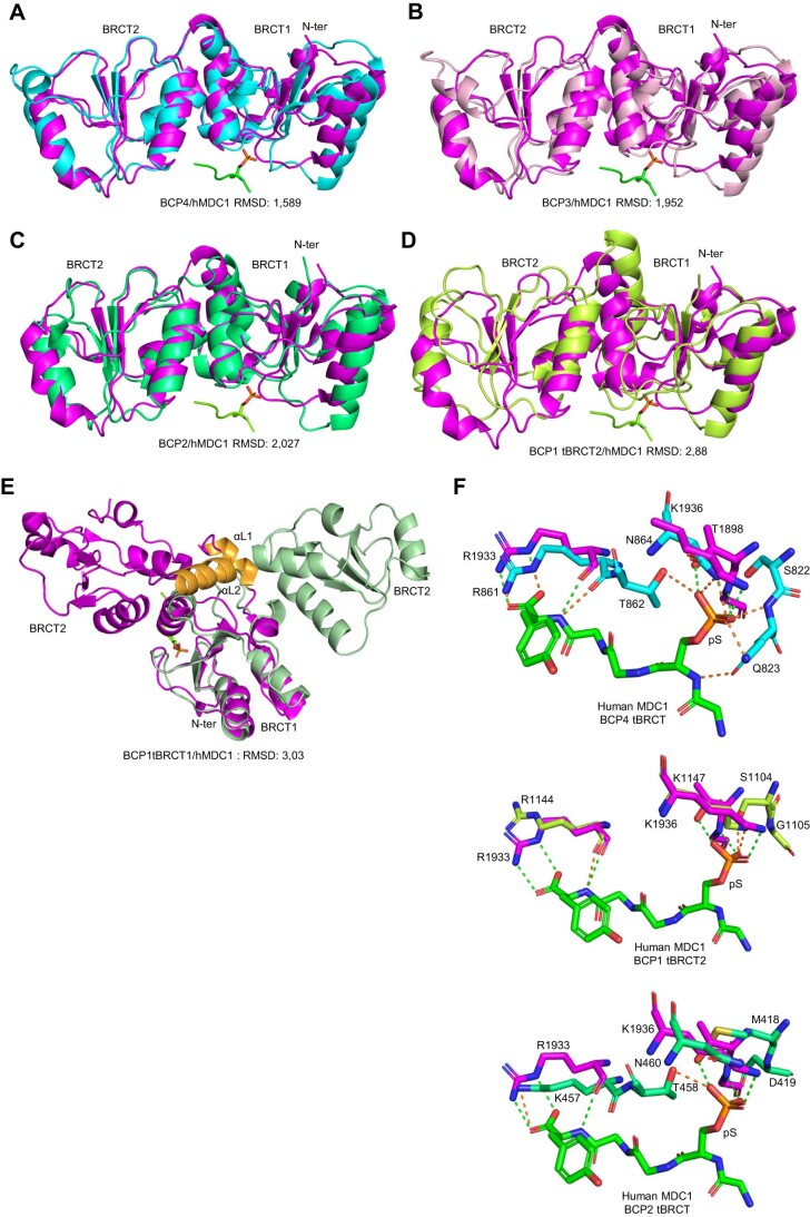
Figure EV3 Predicted structures of BCP proteins. (A–E) AlphaFold2 models of the tBRCT domains of BCP4 (A), BCP3 (B), BCP2 (C), and BCP1 (D, E) superimposed with a structure of human MDC1 in complex with the phosphorylated C-terminal peptide of H2A.X (Stucki et al, 2005). Note that tBRCT1 of BCP1 overlaps only with MDC1 N-terminal BRCT domain. This is presumably due to the absence of alpha helices (αL1 and αL2, in orange) in tBRCT1 of BCP1 connecting two BRCT domains as indicated in orange. In all panels, human MDC1 tBRCT is purple colored. (F) Comparison of interactions of the tBRCT domains of BCP1 and BCP2 with phosphorylated C-terminal peptide of H2A.X. Published (MDC1; Stucki et al, 2005) and AlphaFold2 predicted (BCP1 and BCP2) contacts of amino acids with pSer of H2A.X are indicated respectively with green and orange dotted lines. For a comparison, BCP4 is displayed to indicate reduced abilities of the tBRCT domains of BCP1 and BCP2 to contact pSer of H2A.X. Human MDC1 tBRCT is purple colored.
Figure EV3 Predicted structures of BCP proteins. (A–E) AlphaFold2 models of the tBRCT domains of BCP4 (A), BCP3 (B), BCP2 (C), and BCP1 (D, E) superimposed with a structure of human MDC1 in complex with the phosphorylated C-terminal peptide of H2A.X (Stucki et al, 2005). Note that tBRCT1 of BCP1 overlaps only with MDC1 N-terminal BRCT domain. This is presumably due to the absence of alpha helices (αL1 and αL2, in orange) in tBRCT1 of BCP1 connecting two BRCT domains as indicated in orange. In all panels, human MDC1 tBRCT is purple colored. (F) Comparison of interactions of the tBRCT domains of BCP1 and BCP2 with phosphorylated C-terminal peptide of H2A.X. Published (MDC1; Stucki et al, 2005) and AlphaFold2 predicted (BCP1 and BCP2) contacts of amino acids with pSer of H2A.X are indicated respectively with green and orange dotted lines. For a comparison, BCP4 is displayed to indicate reduced abilities of the tBRCT domains of BCP1 and BCP2 to contact pSer of H2A.X. Human MDC1 tBRCT is purple colored.CC BY · PubMed Central
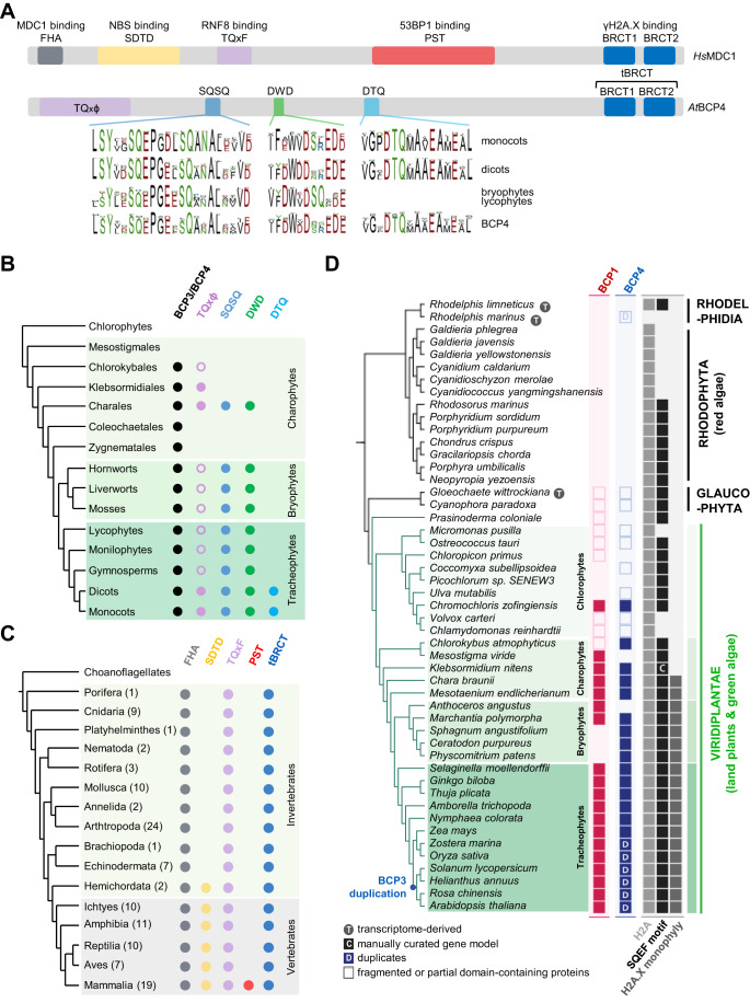
Figure 5 Comparison of BCP4 and MDC1 sequence motifs. (A) Schematic representation of human MDC1 and Arabidopsis BCP4 proteins. Conserved sequence motifs are indicated, and consensus sequences of plant motifs are depicted at the bottom. (B) Summary of the presence/absence of BCP3/BCP4 sequence motifs across Viridiplantae. Open circles denote phylogenetic groups where motif is present in only a subset of analyzed species. (C) In metazoa, functional motifs identified in human MDC1 are unique to mammals. The numbers of MDC1 proteins from each phylogenetic group used for the creation of cladogram are indicated in parentheses. (D) Evolutionary trajectories of BCP1, BCP4, and H2A.X in Archaeplastida. Protein presence is displayed at the tip of each branch, and major groups are denoted next to phylogeny. For H2A.X, (i) the presence of H2A, (ii) SQEF or SQEF-like motifs, and (iii) the presence of a monophyletic clade containing Arabidopsis H2A.X (Fig. EV4) were displayed separately. Data information: Plant and metazoan protein sequences used for the analysis shown in A–C are available in Source data. Source data are available online for this figure.
Figure 5 Comparison of BCP4 and MDC1 sequence motifs. (A) Schematic representation of human MDC1 and Arabidopsis BCP4 proteins. Conserved sequence motifs are indicated, and consensus sequences of plant motifs are depicted at the bottom. (B) Summary of the presence/absence of BCP3/BCP4 sequence motifs across Viridiplantae. Open circles denote phylogenetic groups where motif is present in only a subset of analyzed species. (C) In metazoa, functional motifs identified in human MDC1 are unique to mammals. The numbers of MDC1 proteins from each phylogenetic group used for the creation of cladogram are indicated in parentheses. (D) Evolutionary trajectories of BCP1, BCP4, and H2A.X in Archaeplastida. Protein presence is displayed at the tip of each branch, and major groups are denoted next to phylogeny. For H2A.X, (i) the presence of H2A, (ii) SQEF or SQEF-like motifs, and (iii) the presence of a monophyletic clade containing Arabidopsis H2A.X (Fig. EV4) were displayed separately. Data information: Plant and metazoan protein sequences used for the analysis shown in A–C are available in Source data. Source data are available online for this figure.CC BY · PubMed Central
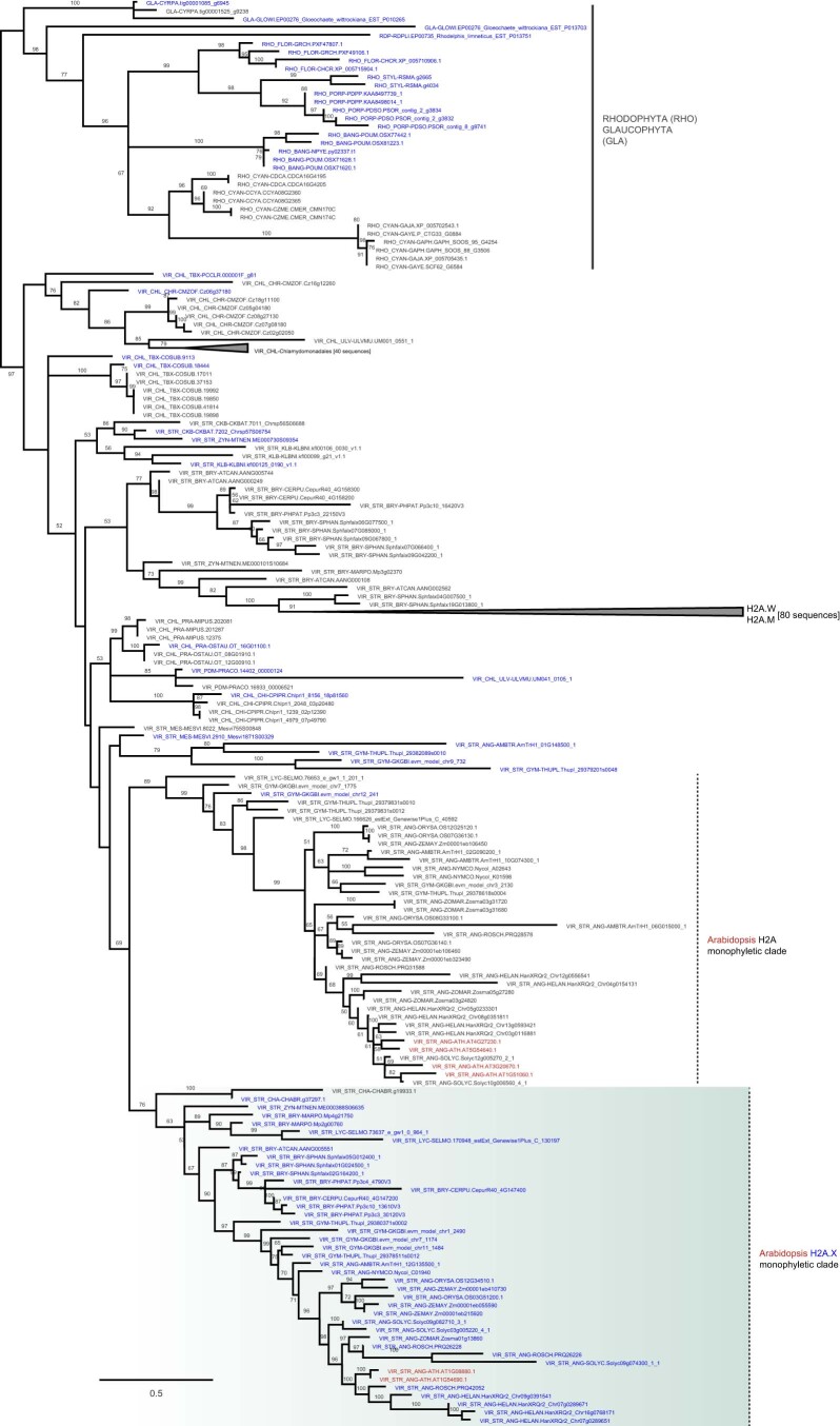
Figure EV4 Phylogeny of H2A variants from Archaeplastida H2A orthogroup. The phylogenetic positions of Arabidopsis H2A variants are marked in red. H2A.X sequences with a SQEF/Y motif or SQEF-like (SQ + E/D + F/I/L/V/Y) motif at the C-terminus are highlighted in blue. Note that in green algae, red algae, and glaucophytes H2A and SQEF/Y motif-containing H2As do not form separate clades.
Figure EV4 Phylogeny of H2A variants from Archaeplastida H2A orthogroup. The phylogenetic positions of Arabidopsis H2A variants are marked in red. H2A.X sequences with a SQEF/Y motif or SQEF-like (SQ + E/D + F/I/L/V/Y) motif at the C-terminus are highlighted in blue. Note that in green algae, red algae, and glaucophytes H2A and SQEF/Y motif-containing H2As do not form separate clades.CC BY · PubMed Central
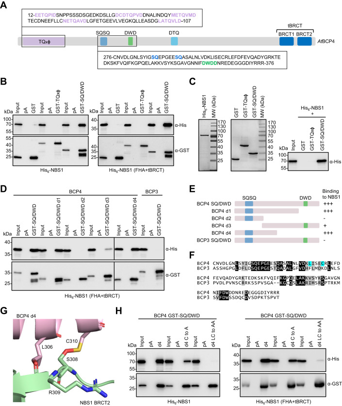
Figure 6 BCP4 interacts with NBS1 of the MRN complex. (A) Schematic presentation of BCP4 with sequences of TQxϕ and SQ/DWD sequences used for protein expression. (B) Co-purification of full-length NBS1 and the FHA+tBRCT domains of NBS1 with GST and GST-tagged TQxϕ or SQ/DWD motifs of BCP4. (C) GST pull-down with independently purified recombinant proteins. Coomassie-stained gels of purified proteins are on two left panels. (D) Co-purification of His6- FHA+tBRCT of NBS1 with the SQ/DWD deletion mutant forms of BCP4. (E) Schematic presentation of BCP4 SQ/DWD deletion mutant forms and summary of interactions with NBS1. (F) Sequence alignment of the SQ/DWD motifs of BCP3 and BCP4. Identical and similar amino acids are highlighted on black and gray background, respectively. Blue background indicates amino acids predicted to interact with NBS1. (G) AlphaFold2 predicted interaction of NBS1 and the SQ/DWD motif of BCP4. Amino acids predicted to interact are indicated with numbers corresponding to full-length proteins. (H) Co-purification of full-length NBS1 or the FHA+tBRCT domains of NBS1 with GST-tagged deletion 4 of BCP4 SQ/DWD motif and its point mutants as indicated. Source data are available online for this figure.
Figure 6 BCP4 interacts with NBS1 of the MRN complex. (A) Schematic presentation of BCP4 with sequences of TQxϕ and SQ/DWD sequences used for protein expression. (B) Co-purification of full-length NBS1 and the FHA+tBRCT domains of NBS1 with GST and GST-tagged TQxϕ or SQ/DWD motifs of BCP4. (C) GST pull-down with independently purified recombinant proteins. Coomassie-stained gels of purified proteins are on two left panels. (D) Co-purification of His6- FHA+tBRCT of NBS1 with the SQ/DWD deletion mutant forms of BCP4. (E) Schematic presentation of BCP4 SQ/DWD deletion mutant forms and summary of interactions with NBS1. (F) Sequence alignment of the SQ/DWD motifs of BCP3 and BCP4. Identical and similar amino acids are highlighted on black and gray background, respectively. Blue background indicates amino acids predicted to interact with NBS1. (G) AlphaFold2 predicted interaction of NBS1 and the SQ/DWD motif of BCP4. Amino acids predicted to interact are indicated with numbers corresponding to full-length proteins. (H) Co-purification of full-length NBS1 or the FHA+tBRCT domains of NBS1 with GST-tagged deletion 4 of BCP4 SQ/DWD motif and its point mutants as indicated. Source data are available online for this figure.CC BY · PubMed Central
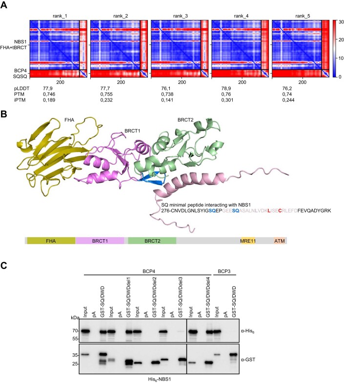
Figure EV5 Predicted structure of NBS1 and its interaction with SQ/DWD region of BCP4. (A) PAE plots of NBS1 (1–324) and BCP4 SQ/DWD (276–325), with calculated predicted local distance difference test (pLDDT), predicted template modeling (PTM), and interface-predicted template modeling (iPTM) scores. (B) AlphaFold2 model of Arabidopsis NBS1 in complex with SQ/DWD region of BCP4 (top panel). A sequence of BCP4 minimal peptide interacting with NBS1 is indicated with Leu and Cys residues involved in interaction with NBS1 highlighted in red. Schematic presentation of NBS1 with indicated conserved domains (bottom panel). (C) Co-purification of NBS1 with GST-tagged deletion 4 of BCP4 SQ/DWD motif and its point mutants. Source data are available online for this figure.
Figure EV5 Predicted structure of NBS1 and its interaction with SQ/DWD region of BCP4. (A) PAE plots of NBS1 (1–324) and BCP4 SQ/DWD (276–325), with calculated predicted local distance difference test (pLDDT), predicted template modeling (PTM), and interface-predicted template modeling (iPTM) scores. (B) AlphaFold2 model of Arabidopsis NBS1 in complex with SQ/DWD region of BCP4 (top panel). A sequence of BCP4 minimal peptide interacting with NBS1 is indicated with Leu and Cys residues involved in interaction with NBS1 highlighted in red. Schematic presentation of NBS1 with indicated conserved domains (bottom panel). (C) Co-purification of NBS1 with GST-tagged deletion 4 of BCP4 SQ/DWD motif and its point mutants. Source data are available online for this figure.CC BY · PubMed Central

Verification

Claims the paper makes

The claims and statistics of this paper haven’t been checked yet.

Run the check — free, no account needed. It reads the open-access text and checks the sources it cites.

A few free checks a day · papers already verified open instantly · results are public

Self-check

Percentages, sums and reported differences re-computed from the paper’s own numbers.

The self-check needs the paper’s full text.

Citations

Cites

Research works printed but not linked: 60

  1. Akutsu N, Iijima K, Hinata T, Tauchi H (2007) Characterization of the plant homolog of Nijmegen breakage syndrome 1: Involvement in DNA repair and recombination. Biochem Biophys Res Commun 353:394–408 · DOI
  2. Almawi AW, Matthews LA, Guarne A (2017) FHA domains: phosphopeptide binding and beyond. Prog Biophys Mol Biol 127:105–110 · DOI
  3. Buchfink B, Xie C, Huson DH (2015) Fast and sensitive protein alignment using DIAMOND. Nat Methods 12:59–60 · DOI · Paper page
  4. Callebaut I, Mornon PJ (1997) From BRCA1 to RAP1: a widespread BRCT module closely associated with DNA repair. FEBS Lett 400:25–30 · DOI
  5. Chapman JR, Jackson PS (2008) Phospho-dependent interactions between NBS1 and MDC1 mediate chromatin retention of the MRN complex at sites of DNA damage. EMBO Rep 9:795–801 · DOI
  6. Cho CH, Park SI, Huang T-Y, Lee Y, Ciniglia C, Yadavalli HC, Yang SW, Bhattacharya D, Yoon HS (2023) Genome-wide signatures of adaptation to extreme environments in red algae. Nat Commun 14:10 · DOI · Paper page
  7. Ciccia A, Elledge JS (2010) The DNA damage response: making it safe to play with knives. Mol Cell 40:179–204 · DOI · Paper page
  8. Coster G, Goldberg M (2010) The cellular response to DNA damage: a focus on MDC1 and its interacting proteins. Nucleus 1:166–178 · DOI
  9. Dronamraju R, Mason JM (2009) Recognition of double strand breaks by a mutator protein (MU2) in Drosophila melanogaster. PLoS Genet 5:e1000473 · DOI
  10. Escribano-Diaz C, Durocher D (2013) DNA repair pathway choice-a PTIP of the hat to 53BP1. EMBO Rep 14:665–666 · DOI
  11. Evans R, O’Neill M, Pritzel A, Antropova N, Senior A, Green T, Žídek A, Bates R, Blackwell S, Yim J et al (2022) Protein complex prediction with AlphaFold-Multimer. Preprint at https://doi.org/10.1101/2021.10.04.463034 · DOI · Paper page
  12. Fan T, Kang H, Wu D, Zhu X, Huang L, Wu J, Zhu Y (2022) Arabidopsis γ-H2A.X-INTERACTING PROTEIN participates in DNA damage response and safeguards chromatin stability. Nat Commun 13:7942 · DOI
  13. Ferrand J, Plessier A, Polo SE (2020) Control of the chromatin response to DNA damage: Histone proteins pull the strings. Semin Cell Dev Biol 113:75–87 · DOI
  14. Gatti M, Pinat S, Maiolica A, Rocchio F, Prato GM, Aebersold R, Penengo L (2015) RNF168 promotes noncanonical K27 ubiquitination to signal DNA damage. Cell Rep 10:226–238 · DOI
  15. Gong Z, Cho Y-W, Kim J-E, Ge K, Chen J (2009) Accumulation of Pax2 transactivation domain interaction protein (PTIP) at sites of DNA breaks via RNF8-dependent pathway is required for cell survival after DNA damage. J Biol Chem 284:7284–7293 · DOI
  16. Hale CJ, Potok ME, Lopez J, Do T, Liu A, Gallego-Bartolome J, Michaels SD, Jacobsen SE (2016) Identification of multiple proteins coupling transcriptional gene silencing to genome stability in Arabidopsis thaliana. PLoS Genet 12:e1006092 · DOI
  17. Hari FJ, Spycher C, Jungmichel S, Pavic L, Stucki M (2010) A divalent FHA/BRCT-binding mechanism couples the MRE11-RAD50-NBS1 complex to damaged chromatin. EMBO Rep 11:387–392 · DOI
  18. Jowsey PA, Doherty AJ, Rouse J (2004) Human PTIP facilitates ATM-mediated activation of p53 and promotes cellular resistance to ionizing radiation. J Biol Chem 279:55562–55569 · DOI
  19. Kasravi A, Walter MF, Brand S, Mason JM, Biessmann H (1999) Molecular cloning and tissue-specific expression of the mutator2 Gene (mu2) in Drosophila melanogaster. Genetics 152:1025–1035 · DOI
  20. Katoh K, Standley DM (2013) MAFFT multiple sequence alignment software version 7: improvements in performance and usability. Mol Biol Evol 30:772–780 · DOI · Paper page
  21. Kolas NK, Chapman JR, Nakada S, Ylanko J, Chahwan R, Sweeney FD, Panier S, Mendez M, Wildenhain J, Thomson TM et al (2007) Orchestration of the DNA-damage response by the RNF8 ubiquitin ligase. Science 318:1637–1640 · DOI
  22. Lafarge S, Montané MH (2003) Characterization of Arabidopsis thaliana ortholog of the human breast cancer susceptibility gene 1: AtBRCA1, strongly induced by gamma rays. Nucleic Acids Res 31:1148–1155 · DOI
  23. Lanz MC, Dibitetto D, Smolka MB (2019) DNA damage kinase signaling: checkpoint and repair at 30 years. EMBO J 38:e101801 · DOI
  24. Leebens-Mack et al (2019) One thousand plant transcriptomes and the phylogenomics of green plants. Nature 574:679–685 · DOI · Paper page
  25. Letunić I, Bork P (2021) Interactive Tree Of Life (iTOL) v5: an online tool for phylogenetic tree display and annotation. Nucleic Acids Res 49(W1):W293–W296 · DOI
  26. Leung CCY, Glover MJN (2011) BRCT domains: easy as one, two, three. Cell Cycle 10:2461–2470 · DOI
  27. Lloyd J, Chapman JR, Clapperton JA, Haire FL, Hartsuiker E, Li J, Carr AM, Jackson SP, Smerdon SJ (2009) A supramodular FHA/BRCT-repeat architecture mediates Nbs1 adaptor function in response to DNA damage. Cell 139:100–111 · DOI · Paper page
  28. Lorković ZJ, Park C, Goiser M, Jiang D, Kurzbauer MT, Schlögelhofer P, Berger F (2017) Compartmentalization of DNA damage response between heterochromatin and euchromatin is mediated by distinct H2A histone variants. Curr Biol 27:1192–1199 · DOI
  29. Lou Z, Minter-Dykhouse K, Franco S, Gostissa M, Rivera MA, Celeste A, Manis JP, van Deursen J, Nussenzweig A, Paull TT et al (2006) MDC1 maintains genomic stability by participating in the amplification of ATM-dependent DNA damage signals. Mol Cell 21:187–200 · DOI
  30. Mailand N, Bekker-Jensen S, Faustrup H, Melander F, Bartek J, Lukas C, Lukas J (2007) RNF8 ubiquitylates histones at DNA double-strand breaks and promotes assembly of repair proteins. Cell 131:887–900 · DOI
  31. Malik HS, Henikoff S (2003) Phylogenomics of the nucleosome. Nat Struct Biol 10:882–891 · DOI
  32. Manke IA, Lowery DM, Nguyen A, Yaffe MB (2003) BRCT repeats as phosphopeptide-binding modules involved in protein targeting. Science 302:636–639 · DOI · Paper page
  33. Mathilde G, Ghislaine G, Daniel V, Georges P (2003) The Arabidopsis MEI1 gene encodes a protein with five BRCT domains that is involved in meiosis-specific DNA repair events independent of SPO11-induced DSBs. Plant J 35:465–475 · DOI
  34. Melander F, Bekker-Jensen S, Falck J, Bartek J, Mailand N, Lukas J (2008) Phosphorylation of SDT repeats in the MDC1 N terminus triggers retention of NBS1 at the DNA damage-modified chromatin. J Cell Biol 181:213–226 · DOI
  35. Minh BQ, Schmidt HA, Chernomor O, Schrempf D, Woodhams MD, von Haeseler A, Lanfear R (2020) IQ-TREE 2: New models and efficient methods for phylogenetic inference in the genomic era. Mol Biol Evol 37:1530–1534 · DOI · Paper page
  36. Mirman Z, de Lange T (2020) 53BP1: a DSB escort. Genes Dev 34:7–23 · DOI
  37. Muñoz IM, Jowsey PA, Toth R, Rouse J (2007) Phospho-epitope binding by the BRCT domains of hPTIP controls multiple aspects of the cellular response to DNA damage. Nucleic Acids Res 35:5312–5322 · DOI
  38. Nisa MU, Huang Y, Benhamed M, Raynaud C (2019) The plant DNA damage response: Signaling pathways leading to growth inhibition and putative role in response to stress conditions. Front Plant Sci 10:653 · DOI
  39. Paull TT, Rogakou EP, Yamazaki V, Kirchgessner CU, Gellert M, Bonner WM (2000) A critical role for histone H2AX in recruitment of repair factors to nuclear foci after DNA damage. Curr Biol 10:886–895 · DOI · Paper page
  40. Qiu H, Price DC, Yang EC, Yoon HS, Bhattacharya D (2015) Evidence of ancient genome reduction in red algae (Rhodophyta). J Phycol 51:624–636 · DOI
  41. Reidt W, Wurz R, Wanieck K, Ch HH, Puchta H (2006) A homologue of the breast cancer-associated gene BARD1 is involved in DNA repair in plants. EMBO J 25:4326–4337 · DOI
  42. Rogakou EP, Boon C, Redon C, Bonner WM (1999) Megabase chromatin domains involved in DNA double-strand breaks in vivo. J Cell Biol 146:905–916 · DOI · Paper page
  43. Rogakou EP, Pilch DR, Orr AH, Ivanova VS, Bonner WM (1998) DNA double-stranded breaks induce histone H2AX phosphorylation on serine 139. J Biol Chem 273:5858–5868 · DOI · Paper page
  44. Rosa M, Mittelsten Scheid O (2014) DNA damage sensitivity assays with Arabidopsis seedlings. Bio Protoc 4:e1093 · DOI
  45. Salguero I, Belotserkovskaya R, Coates J, Sczaniecka-Clift M, Demir M, Jhujh S, Wilson MD, Jackson SP (2019) MDC1 PST-repeat region promotes histone H2AX-independent chromatin association and DNA damage tolerance. Nat Commun 10:5191 · DOI
  46. Spycher C, Miller ES, Townsend K, Pavic L, Morrice NA, Janscak P, Stewart GS, Stucki M (2008) Constitutive phosphorylation of MDC1 physically links the MRE11-RAD50-NBS1 complex to damaged chromatin. J Cell Biol 181:227–240 · DOI
  47. Stucki M, Clapperton JA, Mohammad D, Yaffe MB, Smerdon SJ, Jackson SP (2005) MDC1 directly binds phosphorylated histone H2AX to regulate cellular responses to DNA double-strand breaks. Cell 123:1213–1226 · DOI
  48. Turinetto V, Giachino C (2015) Multiple facets of histone variant H2AX: a DNA double-strand-break marker with several biological functions. Nucleic Acids Res 43:2489–2498 · DOI
  49. Vladejić J, Yang F, Dvořák Tomaštíková E, Doležel J, Palecek JJ, Pecinka A (2022) Analysis of BRCT5 domain-containing proteins reveals a new component of DNA damage repair in Arabidopsis. Front Plant Sci 13:1023358 · DOI
  50. Wang X, Takenaka K, Takeda S (2010) PTIP promotes DNA double-strand break repair through homologous recombination. Genes Cells 15:243–254 · DOI
  51. Waterworth WM, Altun C, Armstrong SJ, Roberts N, Dean PJ, Young K, Weil FC, Bray MC, West EC (2007) NBS1 is involved in DNA repair and plays a synergistic role with ATM in mediating meiotic homologous recombination in plants. Plant J 52:41–52 · DOI
  52. Williams SR, Dodson EG, Limbo O, Yamada Y, Williams SJ, Guenther G, Classen S, Glover JNM, Iwasaki H, Russell P, Tainer AJ (2009) Nbs1 flexibly tethers Ctp1 and Mre11-Rad50 to coordinate DNA double-strand break processing and repair. Cell 139:87–99 · DOI · Paper page
  53. Woods NT, Mesquita RD, Sweet M, Carvalho MA, Li X, Liu Y, Nguyen H, Thomas CE, Iversen Jr ES, Marsillac S et al (2012) Charting the landscape of tandem BRCT domain-mediated protein interactions. Sci Signal 5(242):rs6 · DOI
  54. Wu L, Luo K, Lou Z, Chen J (2008) MDC1 regulates intra-S-phase checkpoint by targeting NBS1 to DNA double-strand breaks. Proc Natl Acad Sci USA 105:11200–11205 · DOI
  55. Xu Y, Xu D (2020) Repair pathway choice for double-strand breaks. Essays Biochem 64:765–777 · DOI
  56. Yelagandula R, Stroud H, Holec S, Zhou K, Feng S, Zhong X, Muthurajan UM, Nie X, Kawashima T, Groth M et al (2014) The histone variant H2A.W defines heterochromatin and promotes chromatin condensation in Arabidopsis. Cell 158:98–109 · DOI
  57. Yoshiyama K, Conklin PA, Huefner ND, Britt AB (2009) Suppressor of gamma response 1 (SOG1) encodes a putative transcription factor governing multiple responses to DNA damage. Proc Natl Acad Sci USA 106:12843–12848 · DOI
  58. Yoshiyama KO, Sakaguchi K, Kimura S (2013) DNA damage response in plants: conserved and variable response compared to animals. Biology 2:1338–1356 · DOI
  59. Yu C, Hou L, Huang Y, Cui X, Xu S, Wang L, Yan S (2023) The multi-BRCT domain protein DDRM2 promotes the recruitment of RAD51 to DNA damage sites to facilitate homologous recombination. New Phytol 238:1073–1084 · DOI
  60. Zhang J, Ma Z, Treszezamsky A, Powell SN (2005) MDC1 interacts with Rad51 and facilitates homologous recombination. Nat Struct Mol Biol 12:902–909 · DOI

Metadata

Authors
Lorković, Z. J., Klingenbrunner, M., Cho, C. H., & Berger, F.
Affiliations
Gregor Mendel Institute of Molecular Plant Biology (AT)
Corresponding author
  • Lorković, Zdravko J
  • Berger, Frédéric
Venue
EMBO Reports
Published
2024-03-04
Volume
25
Issue
4
Pages
1936-1961
ISSN
  • 1469-221X (ISSN-L)
  • 1469-3178
OpenAlex
W4392381579
Cited by
15
References
4
Citations by year
2026: 5 · 2025: 6 · 2024: 4
FWCI
2.04
Type
journal article
Language
en
Open access
Locations
  • EMBO Reports (publishedVersion, cc-by)
  • bioRxiv (Cold Spring Harbor Laboratory) (acceptedVersion)
  • PubMed Central (publishedVersion, cc-by)
  • PubMed (publishedVersion)
  • DOAJ (DOAJ: Directory of Open Access Journals) (submittedVersion, cc-by-sa)
Funders
  • Austrian Science Fund (PAT 1104523)
  • National Research Foundation of Korea (RS-2023-00248097)
  • PlantS and ProTech facilitis at the Vienna BioCenter Core Facilities
  • BioOptics Facility and Molecular Biology Services from IMP/IMBA/GMI
  • Core funding from the Gregor Mendel Institute
Topics
  • Life Sciences › Biochemistry, Genetics and Molecular Biology › Molecular Biology › DNA Repair Mechanisms
  • Life Sciences › Agricultural and Biological Sciences › Plant Science › Plant Molecular Biology Research
  • Life Sciences › Agricultural and Biological Sciences › Plant Science › Plant Genetic and Mutation Studies
Keywords
MDC1, DNA damage response, γH2AX, histone H2A.X phosphorylation, BRCT domain, NBS1, Arabidopsis thaliana, DNA damage foci
MeSH terms
Animals, DNA Damage, DNA Repair, Humans, Nuclear Proteins, Phosphorylation, Phylogeny, Cell Cycle Proteins

About these checks

Registry, retraction, Cites and statistics results are computed, not judged, with one exception: a printed reference that no index lists is read once by a model before it counts as a citation error. Claims are extracted by a model and kept only when their quote appears verbatim in the text; each cited source is then checked, with a model’s help, for whether it says what the claim relies on. Those verdicts can be wrong — read the claim’s quote and the cited source before relying on one.

Statistics and arithmetic are recomputed, not judged.

Confidence bands are shown only for fields calibrated on our annotated test set. None is calibrated yet, so no band is shown.

What counts as a claim ›How we measure confidence ›How we check this ›

Are you an author, or do you see a problem with this page? Request a review