

The DOI matches its Crossref record; 1 of the 1 references it cites were found in a registry or an index.
Origin of flaw-tolerance in nacre
Open accessAbstract
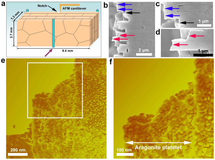
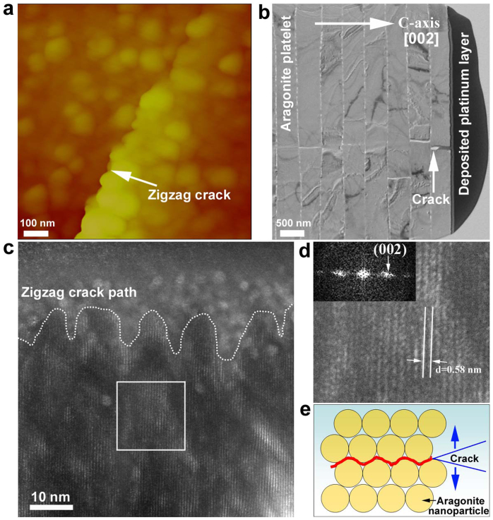
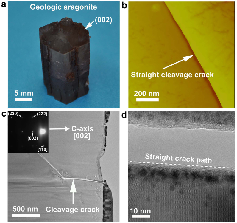
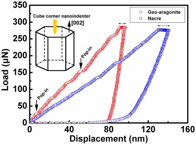
Over the past decades, our understanding of nacre's toughening origin has long stayed at the level of crack deflection along the biopolymer interface between aragonite platelets. It has been widely thought that the ceramic aragonite platelets in nacre invariably remain shielded from the propagating crack. Here we report an unexpected experimental observation that the propagating crack, surprisingly, invades the aragonite platelet following a zigzag crack propagation trajectory. The toughening origin of previously-thought brittle aragonite platelet is ascribed to its unique nanoparticle-architecture, which tunes crack propagation inside the aragonite platelet in an intergranular manner. For comparison, we also investigated the crack behavior in geologic aragonite mineral (pure monocrystal) and found that the crack propagates in a cleavage fashion, in sharp contrast with the intergranular cracking in the aragonite platelet of nacre. These two fundamentally different cracking mechanisms uncover a new toughening strategy in nacre's hierarchical flaw-tolerance design.
Overview
The claims and statistics of this paper haven’t been checked yet.
Run the check — free, no account needed. It reads the open-access text and checks the sources it cites.
Integrity signals
Registry
Cites
Statistics
Figures
Verification
Claims the paper makes
The claims and statistics of this paper haven’t been checked yet.
Run the check — free, no account needed. It reads the open-access text and checks the sources it cites.
Claims other papers make about this one
Each is checked against this paper’s own text: does it say what the citing paper says it does?
The check wasn’t confident either way, so it gives no verdict — never disputed.
In the citing paper
“suggesting that natural design in limpet teeth is optimized towards theoretical strength limits.”
From Extreme strength observed in limpet teeth (2015) · cites this paper as [36]
Experimental — accuracy not yet measured
Self-check
Percentages, sums and reported differences re-computed from the paper’s own numbers.
The self-check needs the paper’s full text.
Citations
Cites
The conflicts between strength and toughness
Ritchie, R. O. (2011) · Nature Materials · doi:10.1038/nmat3115 · Cited by 3,989
Research works printed but not linked: 38
- Kumar, S. & Curtin, W. A. Crack interaction with microstructure. Mater. Today 10, 34–44 (2007). · DOI
- Chakravarthy, S. S. & Curtin, W. A. Origin of Plasticity Length-Scale Effects in Fracture. Phys. Rev. Lett. 105, 115502 (2010). · DOI
- Hasnaoui, A., Van Swygenhoven, H. & Derlet, P. M. Dimples on nanocrystalline fracture surfaces as evidence for shear plane formation. Science 300, 1550–1552 (2003). · DOI
- Lawn, B. R., Padture, N. P., Cai, H. D. & Guiberteau, F. Making ceramics ductile. Science 263, 1114–1116 (1994). · DOI
- Ovid'ko, I. A. & Sheinerman, A. G. Micromechanisms for improved fracture toughness in nanoceramics. Rev. Adv. Mater. Sci. 29, 105–125 (2011).
- Currey, J. D. Mechanical-properties of mother of pearl in tension. Proc. R. Soc. Lond. B 196, 443–463 (1977). · DOI
- Jackson, A. P., Vincent, J. F. V. & Turner, R. M. The mechanical design of nacre. Proc. R. Soc. Lond. B 234, 415–440 (1988). · DOI
- Smith, B. L. et al. Molecular mechanistic origin of the toughness of natural adhesives, fibres and composites. Nature 399, 761–763 (1999). · DOI
- Kamat, S., Su, X., Ballarini, R. & Heuer, A. H. Structural basis for the fracture toughness of the shell of the conch Strombus gigas. Nature 405, 1036–1040 (2000). · DOI
- Ortiz, C. & Boyce, M. C. Materials science - Bioinspired structural materials. Science 319, 1053–1054 (2008). · DOI
- Meyers, M. A., Chen, P.-Y., Lin, A. Y.-M. & Seki, Y. Biological materials: Structure and mechanical properties. Prog. Mater. Sci. 53, 1–206 (2008). · DOI · Paper page
- Espinosa, H. D., Rim, J. E., Barthelat, F. & Buehler, M. J. Merger of structure and material in nacre and bone - Perspectives on de novo biomimetic materials. Prog. Mater. Sci. 54, 1059–1100 (2009). · DOI
- Wang, R. & Gupta, H. S. Deformation and Fracture Mechanisms of Bone and Nacre. Annu. Rev. Mater. Res. 41, 41–73 (2011). · DOI
- Wang, R. Z., Wen, H. B., Cui, F. Z., Zhang, H. B. & Li, H. D. Observations of damage morphologies in nacre during deformation and fracture. J. Mater. Sci. 30, 2299–2304 (1995). · DOI
- Gao, H. J., Ji, B. H., Jager, I. L., Arzt, E. & Fratzl, P. Materials become insensitive to flaws at nanoscale: Lessons from nature. Proc. Natl. Acad. Sci. USA 100, 5597–5600 (2003). · DOI · Paper page
- Li, X. D., Chang, W. C., Chao, Y. J., Wang, R. Z. & Chang, M. Nanoscale structural and mechanical characterization of a natural nanocomposite material: The shell of red abalone. Nano Lett. 4, 613–617 (2004). · DOI
- Rousseau, M. et al. Multiscale structure of sheet nacre. Biomaterials 26, 6254–6262 (2005). · DOI
- Li, X. D. & Huang, Z. W. Unveiling the Formation Mechanism of Pseudo-Single-Crystal Aragonite Platelets in Nacre. Phys. Rev. Lett. 102, 075502 (2009). · DOI
- Huang, Z. & Li, X. Order-disorder transition of aragonite nanoparticles in nacre. Phys. Rev. Lett. 109, 025501 (2012). · DOI
- Li, X. D., Xu, Z.-H. & Wang, R. Z. In situ observation of nanograin rotation and deformation in nacre. Nano Lett. 6, 2301–2304 (2006). · DOI
- Sun, J. Y. & Bhushan, B. Hierarchical structure and mechanical properties of nacre: a review. Rsc Advances 2, 7617–7632 (2012). · DOI
- Bruet, B. J. F. et al. Nanoscale morphology and indentation of individual nacre tablets from the gastropod mollusc Trochus niloticus. J. Mater. Res. 20, 2400–2419 (2005). · DOI
- Barthelat, F., Li, C.-M., Comi, C. & Espinosa, H. D. Mechanical properties of nacre constituents and their impact on mechanical performance. J. Mater. Res. 21, 1977–1986 (2006). · DOI
- Katti, K. S., Mohanty, B. & Katti, D. R. Nanomechanical properties of nacre. J. Mater. Res. 21, 1237–1242 (2006). · DOI
- Huang, Z. W. et al. Uncovering high-strain rate protection mechanism in nacre. Sci. Rep. 1, 148 (2011). · DOI
- Fratzl, P., Gupta, H. S., Fischer, F. D. & Kolednik, O. Hindered crack propagation in materials with periodically varying Young's modulus - Lessons from biological materials. Adv. Mater. 19, 2657–2661 (2007). · DOI
- Kolednik, O., Predan, J., Fischer, F. D. & Fratzl, P. Bioinspired Design Criteria for Damage-Resistant Materials with Periodically Varying Microstructure. Adv. Funct. Mater. 21, 3634–3641 (2011). · DOI
- Bei, H., Lu, Z. P. & George, E. P. Theoretical strength and the onset of plasticity in bulk metallic glasses investigated by nanoindentation with a spherical indenter. Phys. Rev. Lett. 93, 125504 (2004). · DOI
- Bei, H., George, E. P., Hay, J. L. & Pharr, G. M. Influence of indenter tip geometry on elastic deformation during nanoindentation. Phys. Rev. Lett. 95, 045501 (2005). · DOI
- Hofmann, D. C. et al. Designing metallic glass matrix composites with high toughness and tensile ductility. Nature 451, 1085–1089 (2008). · DOI
- Demetriou, M. D. et al. A damage-tolerant glass. Nat. Mater. 10, 123–128 (2011). · DOI
- Kim, Y.-Y. et al. An artificial biomineral formed by incorporation of copolymer micelles in calcite crystals. Nat. Mater. 10, 890–896 (2011). · DOI
- Cranford, S. W., Tarakanova, A., Pugno, N. M. & Buehler, M. J. Nonlinear material behaviour of spider silk yields robust webs. Nature 482, 72–78 (2012). · DOI
- Omenetto, F. G. & Kaplan, D. L. Spider webs: Damage control. Nat. Mater. 11, 273–274 (2012). · DOI
- Imbeni, V., Kruzic, J. J., Marshall, G. W., Marshall, S. J. & Ritchie, R. O. The dentin-enamel junction and the fracture of human teeth. Nat. Mater. 4, 229–232 (2005). · DOI
- Tai, K., Dao, M., Suresh, S., Palazoglu, A. & Ortiz, C. Nanoscale heterogeneity promotes energy dissipation in bone. Nat. Mater. 6, 454–462 (2007). · DOI
- Seto, J. et al. Structure-property relationships of a biological mesocrystal in the adult sea urchin spine. Proc. Natl. Acad. Sci. USA 109, 3699–3704 (2012). · DOI
- Bruet, B. J. F., Song, J., Boyce, M. C. & Ortiz, C. Materials design principles of ancient fish armour. Nat. Mater. 7, 748–756 (2008). · DOI
Cited by
Sorted by citations
Extreme strength observed in limpet teeth
Barber, A. H., Lu, D., & Pugno, N. M. (2015) · Journal of the Royal Society Interface · doi:10.1098/rsif.2014.1326 · Cited by 212
Metadata
- Authors
- Huang, Z., & Li, X.
- Affiliations
- University of South Carolina (US)
- Corresponding author
- Li, Xiaodong
- Venue
- Scientific Reports
- Published
- 2013-04-22
- Volume
- 3
- Issue
- 1
- Pages
- 1693-1693
- ISSN
- 2045-2322 (ISSN-L)
- PMC
- PMC3631768
- OpenAlex
- W2076735776
- Cited by
- 111
- References
- 1
- Citations by year
- 2026: 3 · 2025: 3 · 2024: 8 · 2023: 14 · 2022: 11 · 2021: 5 · 2020: 12 · 2019: 11 · 2018: 2 · 2017: 12 · 2016: 13 · 2015: 12 · 2014: 4 · 2013: 1
- FWCI
- 3.82
- Type
- journal article
- Language
- en
- Open access
- gold
- doi.org
- Licence
- Locations
- Scientific Reports (publishedVersion, cc-by-nc-nd)
- Europe PMC (PubMed Central) (publishedVersion, cc-by-nc-nd)
- PubMed Central (publishedVersion, cc-by-nc-nd)
- PubMed (publishedVersion)
- Funders
- North Carolina State University
- University of South Carolina
- Army Research Office
- Topics
- Physical Sciences › Materials Science › Biomaterials › Calcium Carbonate Crystallization and Inhibition
- Physical Sciences › Materials Science › Metals and Alloys › Microstructure and mechanical properties
- Physical Sciences › Engineering › Mechanical Engineering › Advanced Materials and Mechanics
- Keywords
- nacre, aragonite platelets, crack propagation, toughening mechanisms, flaw tolerance, intergranular fracture, nanoparticle structure, biominerals, hierarchical design, cleavage fracture
- MeSH terms
- Hardness, Materials Testing, Tensile Strength, Compressive Strength, Nacre
About these checks
Registry, retraction, Cites and statistics results are computed, not judged, with one exception: a printed reference that no index lists is read once by a model before it counts as a citation error. Claims are extracted by a model and kept only when their quote appears verbatim in the text; each cited source is then checked, with a model’s help, for whether it says what the claim relies on. Those verdicts can be wrong — read the claim’s quote and the cited source before relying on one.
Statistics and arithmetic are recomputed, not judged.
Confidence bands are shown only for fields calibrated on our annotated test set. None is calibrated yet, so no band is shown.
What counts as a claim ›How we measure confidence ›How we check this ›
Are you an author, or do you see a problem with this page? Request a review
How to cite
Cite CoChat for the checks on this page, and the paper for its own findings.
- This page
- CoChat. (2026). Citation check: Origin of flaw-tolerance in nacre. https://app.cochat.ai/papers/10.1038/srep01693
- The paper
- Huang, Z., & Li, X. (2013). Origin of flaw-tolerance in nacre. Scientific Reports. https://doi.org/10.1038/srep01693
