

The DOI matches its Crossref record; 158 of the 158 references it cites were found in a registry or an index.
Current progress, challenges and future prospects of diagnostic and therapeutic interventions in Alzheimer's disease
Open accessAbstract
The diverse pathological mechanisms and their implications for the development of effective diagnostic and therapeutic interventions in Alzheimer's disease are presented with current progress, challenges and future prospects.
Overview
The claims and statistics of this paper haven’t been checked yet.
Run the check — free, no account needed. It reads the open-access text and checks the sources it cites.
Integrity signals
Registry
Cites
Statistics
Figures
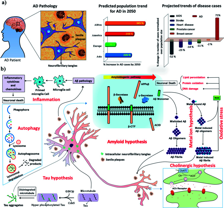
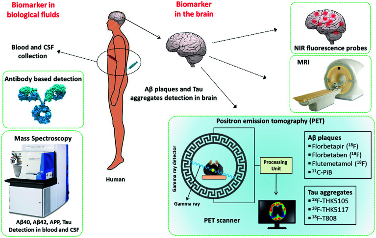
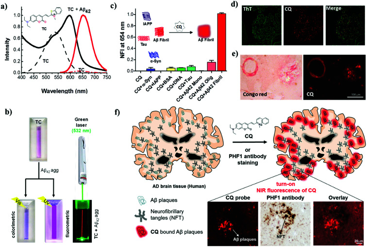
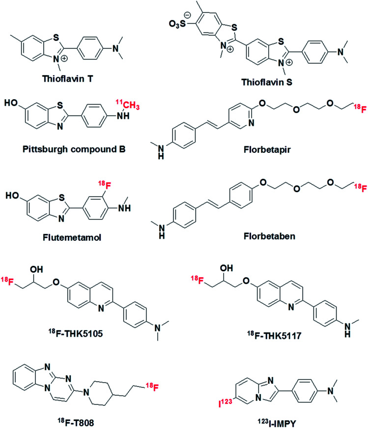
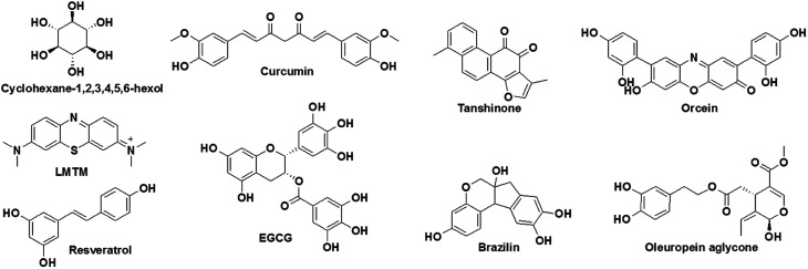
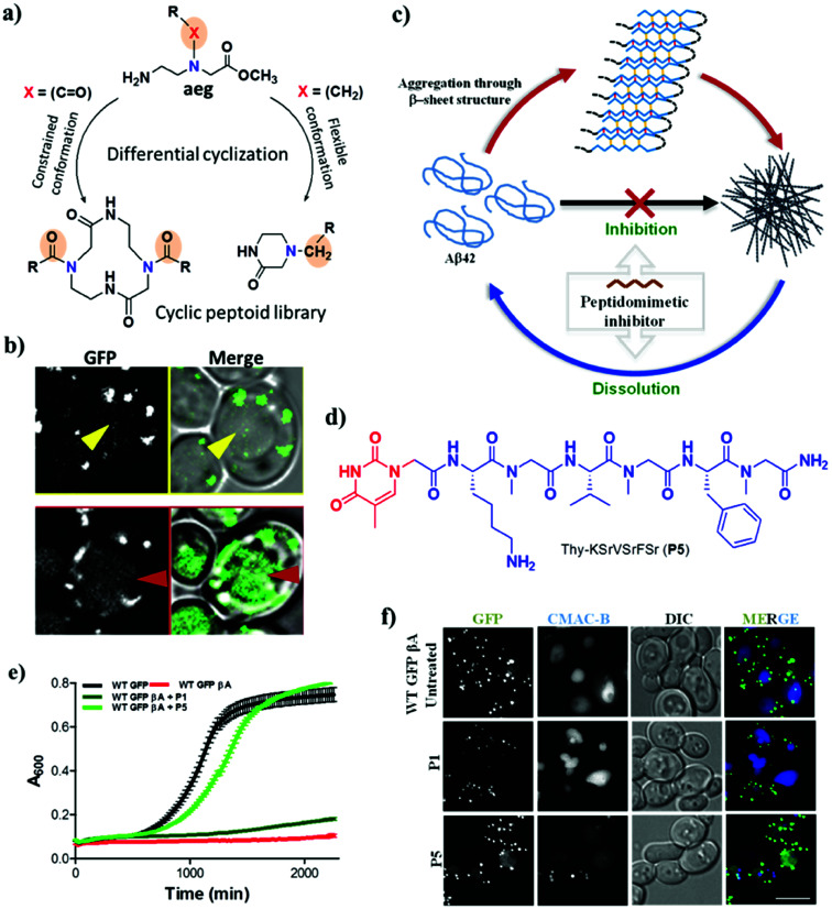
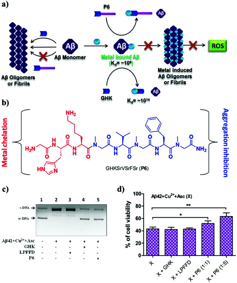
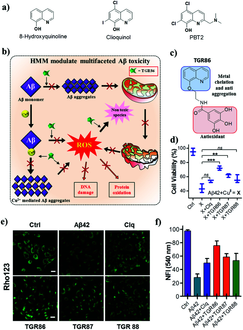
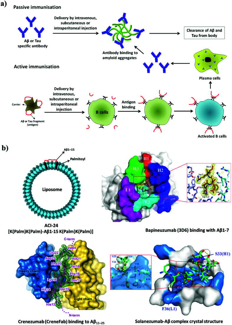
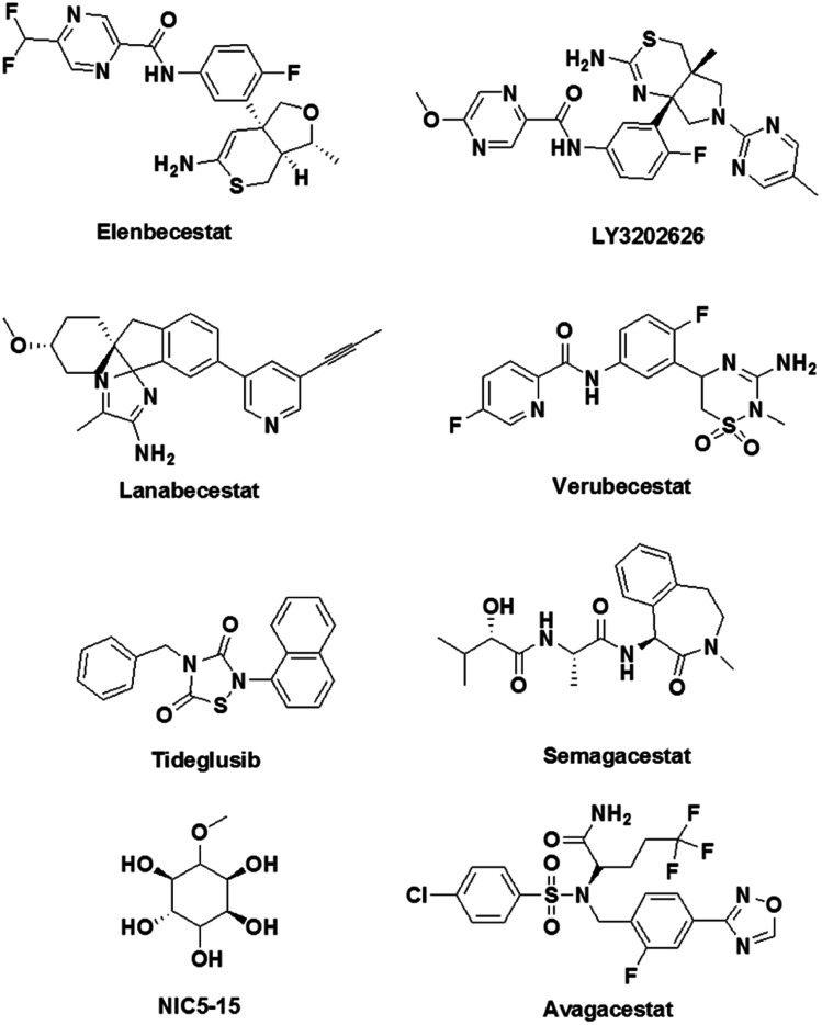
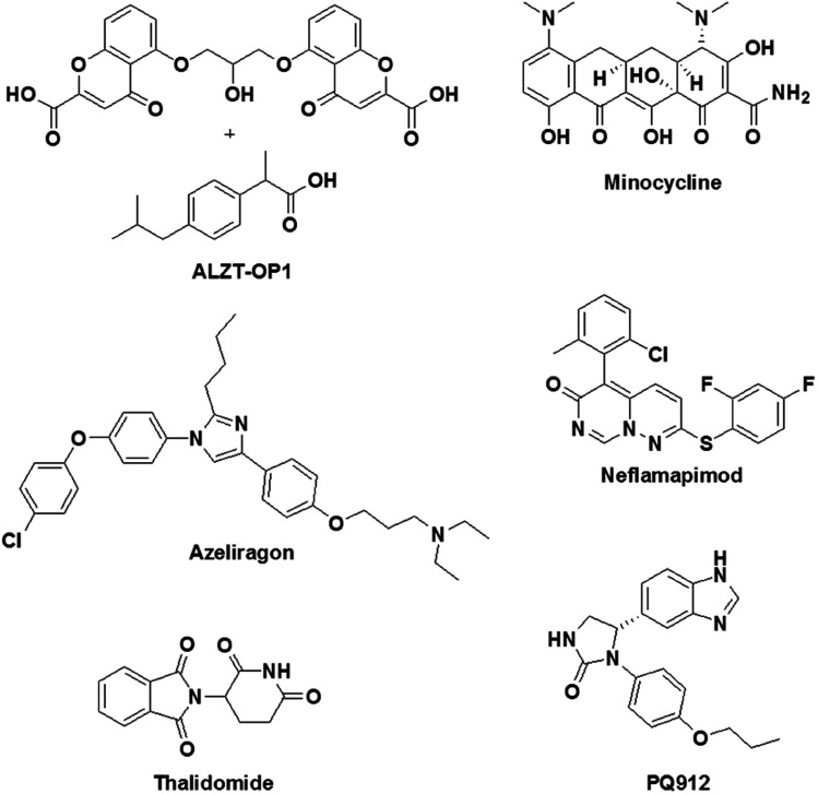
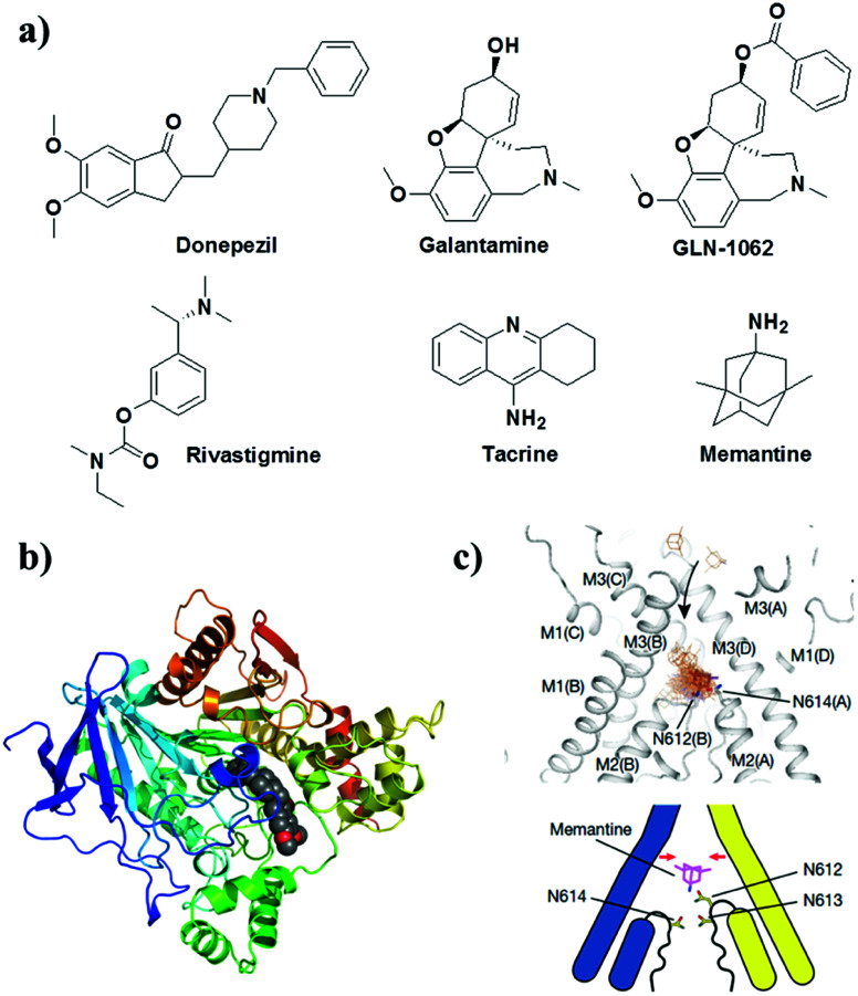
Verification
Claims the paper makes
The claims and statistics of this paper haven’t been checked yet.
Run the check — free, no account needed. It reads the open-access text and checks the sources it cites.
Claims other papers make about this one
Each is checked against this paper’s own text: does it say what the citing paper says it does?
In the citing paper
“Various growing factors for AD like presenelin 1 proteins, oxidative stress, tau neurofibrillary tangles can cause dysfunction of autophagic vacuoles and hence can contribute in the pathogenesis of AD [32, 33].”
From Recent Advancements in Pathogenesis, Diagnostics and Treatment of Alzheimer’s Disease (2020) · cites this paper as [32]
Self-check
Percentages, sums and reported differences re-computed from the paper’s own numbers.
The self-check needs the paper’s full text.
Citations
Cites
The amyloid hypothesis of Alzheimer's disease at 25 years
Selkoe, D. J., & Hardy, J. (2016) · EMBO Molecular Medicine · doi:10.15252/emmm.201606210 · Cited by 6,164
Pushko, P., Pumpens, P., & Grens, E. (2013) · Intervirology · doi:10.1159/000346773 · Cited by 5,975
Cillà, S. D., Farruggio, S., Vujosevic, S., Raina, G., Filippini, D. A., Gatti, V., Clemente, N., Mary, D., Vezzola, D., Casini, G., et al. (2017) · Cellular Physiology and Biochemistry · doi:10.1159/000479441 · Cited by 5,154
2016 Alzheimer's disease facts and figures
Alzheimer's Association (2016) · Alzheimer's & Dementia · doi:10.1016/j.jalz.2016.03.001 · Cited by 5,069
The global prevalence of dementia: a systematic review and metaanalysis.
Prince, M., Bryce, R., Albanese, E., Wimo, A., Ribeiro, W., & Ferri, C. P. (2013) · Alzheimer's & Dementia · doi:10.1016/j.jalz.2012.11.007 · Cited by 4,941
Inflammation and Alzheimer’s disease
Akiyama, H. (2000) · Neurobiology of Aging · doi:10.1016/s0197-4580(00)00124-x · Cited by 4,514
Imaging brain amyloid in Alzheimer's disease with Pittsburgh Compound‐B
Klunk, W. E., Engler, H., Nordberg, A., Wang, Y., Blomqvist, G., Holt, D. P., Bergström, M., Savitcheva, I., Huang, G., Estrada, S., et al. (2004) · Annals of Neurology · doi:10.1002/ana.20009 · Cited by 4,496
The Alzheimer’s disease neuroimaging initiative (ADNI): MRI methods
Jack, C. R., Bernstein, M. A., Fox, N. C., Thompson, P., Alexander, G., Harvey, D., Borowski, B., Britson, P. J., L. Whitwell, J., Ward, C., et al. (2008) · Journal of Magnetic Resonance Imaging · doi:10.1002/jmri.21049 · Cited by 4,347
Autophagy: cellular and molecular mechanisms
Glick, D. R., Barth, S., & Macleod, K. F. (2010) · The Journal of Pathology · doi:10.1002/path.2697 · Cited by 3,995
Protein aggregation and neurodegenerative disease
Ross, C. A., & Poirier, M. A. (2004) · Nature Medicine · doi:10.1038/nm1066 · Cited by 3,454
Research works printed but not linked: 7
- Steele, 2013, Postdoc J., 1, 21
- Newberg, 2006, J. Nucl. Med., 47, 748
- Hung, 2017, J. Biomed. Sci., 24, 47, 10.1186/s12929-017-0355-7 · DOI · Paper page
- Mark, 2002, Curr. Opin. Drug Discovery Dev., 2, 417
- Guo, 2013, Neural Regener. Res., 8, 2003
- Zhang, 2014, Front. Cell. Neurosci., 8, 427
- Dolan, 2010, Curr. Opin. Drug Discovery Dev., 13, 595
Cited by
Sorted by citations
Recent Advancements in Pathogenesis, Diagnostics and Treatment of Alzheimer’s Disease
Khan, S., Barve, K. H., & Kumar, M. S. (2020) · Current Neuropharmacology · doi:10.2174/1570159x18666200528142429 · Cited by 820
“Various growing factors for AD like presenelin 1 proteins, oxidative stress, tau neurofibrillary tangles can cause dysfunction of autophagic vacuoles and hence can contribute in the pathogenesis of AD [32, 33].”
BackgroundBiomolecules-derived biomaterials
Datta, L. P., Manchineella, S., & Govindaraju, T. (2019) · Biomaterials · doi:10.1016/j.biomaterials.2019.119633 · Cited by 187
Self-citationFasae, K. D., Abolaji, A. O., Faloye, T. R., Odunsi, A., Oyetayo, B. O., Enya, J. I., Rotimi, J. A., Akinyemi, R., Whitworth, A. J., & Aschner, M. (2021) · Journal of Trace Elements in Medicine and Biology · doi:10.1016/j.jtemb.2021.126779 · Cited by 161
Rehman, M. U., Sehar, N., Dar, N. J., Khan, A., Arafah, A., Rashid, S., Rashid, S. M., & Ganaie, M. A. (2023) · Neuroscience & Biobehavioral Reviews · doi:10.1016/j.neubiorev.2022.104961 · Cited by 130
Antioxidant Berberine-Derivative Inhibits Multifaceted Amyloid Toxicity
Rajasekhar, K., Samanta, S., Bagoband, V., Murugan, N. A., & Govindaraju, T. (2020) · iScience · doi:10.1016/j.isci.2020.101005 · Cited by 100
Self-citation
Metadata
- Authors
- Rajasekhar, K., & Govindaraju, T.
- Affiliations
- Jawaharlal Nehru Centre for Advanced Scientific Research (IN)
- Corresponding author
- Govindaraju, Thimmaiah
- Venue
- RSC Advances
- Publisher
- Royal Society of Chemistry (RSC)
- Published
- 2018
- Volume
- 8
- Issue
- 42
- Pages
- 23780-23804
- ISSN
- 2046-2069 (ISSN-L)
- PMC
- PMC9081849
- OpenAlex
- W2811135471
- Cited by
- 150
- References
- 158
- Citations by year
- 2026: 4 · 2025: 16 · 2024: 27 · 2023: 20 · 2022: 17 · 2021: 24 · 2020: 25 · 2019: 13 · 2018: 4
- FWCI
- 2.17
- Type
- journal article
- Language
- en
- Open access
- gold
- doi.org
- Licence
- Locations
- RSC Advances (publishedVersion, cc-by-nc)
- PubMed Central (publishedVersion, cc-by-nc)
- PubMed (publishedVersion)
- Funders
- Department of Science and Technology, Ministry of Science and Technology (DST/SJF/CSA-02/2015-2016)
- Sheikh Saqr Laboratory
- Jawaharlal Nehru Centre for Advanced Scientific Research
- Topics
- Life Sciences › Neuroscience › Cellular and Molecular Neuroscience › Alzheimer's disease research and treatments
- Health Sciences › Medicine › Psychiatry and Mental health › Dementia and Cognitive Impairment Research
- Health Sciences › Medicine › Pharmacology (medical) › Cholinesterase and Neurodegenerative Diseases
- Keywords
- Alzheimer's disease, Alzheimer's disease pathogenesis, early diagnosis, therapeutic interventions, drug development, neurotoxicity, clinical trials
About these checks
Registry, retraction, Cites and statistics results are computed, not judged, with one exception: a printed reference that no index lists is read once by a model before it counts as a citation error. Claims are extracted by a model and kept only when their quote appears verbatim in the text; each cited source is then checked, with a model’s help, for whether it says what the claim relies on. Those verdicts can be wrong — read the claim’s quote and the cited source before relying on one.
Statistics and arithmetic are recomputed, not judged.
Confidence bands are shown only for fields calibrated on our annotated test set. None is calibrated yet, so no band is shown.
What counts as a claim ›How we measure confidence ›How we check this ›
Are you an author, or do you see a problem with this page? Request a review
