

The DOI matches its Crossref record; the references it cites are not checked yet.
Oleic acid differentially affects lipid droplet storage of de novo synthesized lipids in hepatocytes and adipocytes
Open accessPublished version of a preprint
Abstract
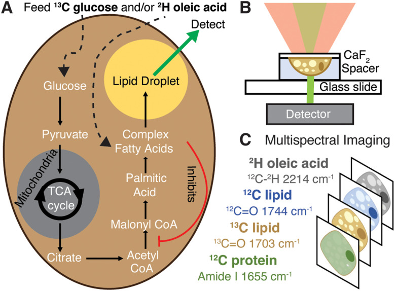
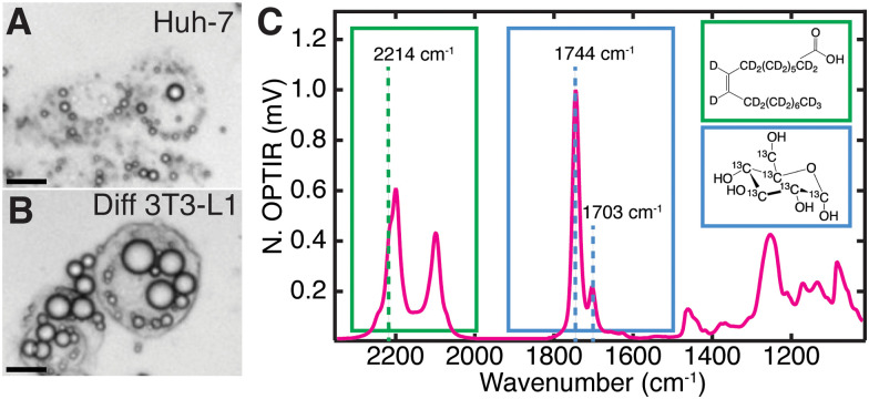
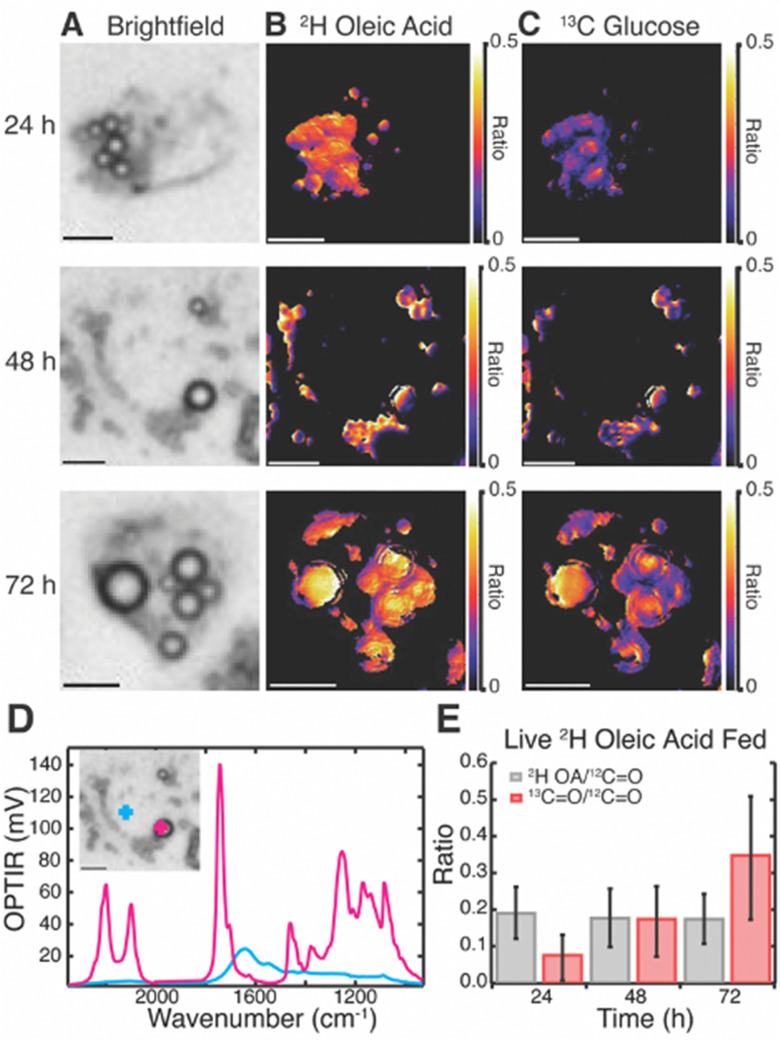
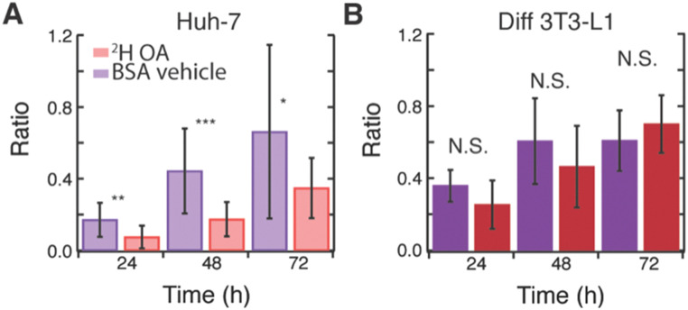
This work introduces a multiplexed sub-micron vibrational imaging technique to monitor differential effects of oleic acid in adipocytes and hepatocytes.
Overview
The claims and statistics of this paper haven’t been checked yet.
Run the check — free, no account needed. It reads the open-access text and checks the sources it cites.
Integrity signals
Registry
Cites
Statistics
Figures
Verification
Claims the paper makes
The claims and statistics of this paper haven’t been checked yet.
Run the check — free, no account needed. It reads the open-access text and checks the sources it cites.
Claims other papers make about this one
Each is checked against this paper’s own text: does it say what the citing paper says it does?
The source backs part of the claim, not all of it — or backs it for a different population, condition or comparison.
In the citing paper
“Oleic acid primarily promotes the formation of larger lipid droplets and facilitates the safe esterification of fatty acids into triglycerides [148,149,150,151], whereas palmitic acid is more strongly associated with mitochondrial fragmentation, increased oxidative metabolism, and ROS formation in both the cytosol and mitochondria.”
From The HepG2 Cell Line as a Model for Studying Metabolic Dysfunction-Associated Steatotic Liver Disease (2026) · cites this paper as [148]
Experimental — accuracy not yet measured
Self-check
Percentages, sums and reported differences re-computed from the paper’s own numbers.
The self-check needs the paper’s full text.
Citations
The citation lists haven’t been fetched yet
References 42 · Cited by 32
Research works printed but not linked: 40
- Cheemerla, 2021, Clin. Liver Dis., 17, 365, 10.1002/cld.1061 · DOI · Paper page
- Riazi, 2022, Lancet Gastroenterol. Hepatol., 7, 851, 10.1016/S2468-1253(22)00165-0 · DOI · Paper page
- Ameer, 2014, Metabolism, 63, 895, 10.1016/j.metabol.2014.04.003 · DOI
- Eynaudi, 2021, Front. Nutr., 8, 775382, 10.3389/fnut.2021.775382 · DOI · Paper page
- Natali, 2007, J. Lipid Res., 48, 1966, 10.1194/jlr.M700051-JLR200 · DOI
- Ducheix, 2017, PLoS One, 12, e0181393, 10.1371/journal.pone.0181393 · DOI
- Fujimoto, 2006, Biol. Pharm. Bull., 29, 2174, 10.1248/bpb.29.2174 · DOI
- Rohwedder, 2014, J. Cell Sci., 127, 3104
- Priore, 2017, Oxid. Med. Cell. Longev., 2017, 9076052, 10.1155/2017/9076052 · DOI
- Hannah, 2001, J. Biol. Chem., 276, 4365, 10.1074/jbc.M007273200 · DOI · Paper page
- Ou, 2001, Proc. Natl. Acad. Sci. U. S. A., 98, 6027, 10.1073/pnas.111138698 · DOI
- Shao, 2014, Lipids, 49, 403, 10.1007/s11745-014-3883-x · DOI
- Li, 2014, Sci. Rep., 4, 6807, 10.1038/srep06807 · DOI
- Shi, 2020, Nat. Methods, 17, 844, 10.1038/s41592-020-0883-z · DOI
- Shuster, 2023, J. Phys. Chem. B, 127, 2918, 10.1021/acs.jpcb.2c08812 · DOI
- Qi, 2012, J. Lipid Res., 53, 1106, 10.1194/jlr.M020156 · DOI
- Prouteau, 2018, Biomolecules, 8, 160, 10.3390/biom8040160 · DOI
- Samuel, 2020, Commun. Biol., 3, 1, 10.1038/s42003-020-1100-4 · DOI
- Schultz, 2011, Annu. Rev. Anal. Chem. Palo Alto Calif, 4, 343, 10.1146/annurev-anchem-061010-114048 · DOI
- Wei, 2016, Acc. Chem. Res., 49, 1494, 10.1021/acs.accounts.6b00210 · DOI
- Chen, 2021, Nat. Commun., 12, 3405, 10.1038/s41467-021-23700-0 · DOI
- Palonpon, 2013, Curr. Opin. Chem. Biol., 17, 708, 10.1016/j.cbpa.2013.05.021 · DOI
- Centrone, 2015, Annu. Rev. Anal. Chem. Palo Alto Calif, 8, 101, 10.1146/annurev-anchem-071114-040435 · DOI
- Spadea, 2021, Anal. Chem., 93, 3938, 10.1021/acs.analchem.0c04846 · DOI
- Shams, 2023, Front. Microbiol., 14, 1077106, 10.3389/fmicb.2023.1077106 · DOI
- Bouzy, 2023, Anal. Methods, 15, 1620, 10.1039/D2AY01197B · DOI
- Zhang, 2016, Sci. Adv., 2, e1600521, 10.1126/sciadv.1600521 · DOI
- Wei, 2016, Acc. Chem. Res., 49, 1494, 10.1021/acs.accounts.6b00210 · DOI
- Yamawaki, 2022, Spectrochim. Acta, Part A, 265, 120290, 10.1016/j.saa.2021.120290 · DOI
- A.Derenne , et al. , in Encyclopedia of Biophysics , ed. G. C. K. Roberts , Springer , Berlin, Heidelberg , 2013 , pp. 1074–1081 · DOI
- Olzmann, 2019, Nat. Rev. Mol. Cell Biol., 20, 137, 10.1038/s41580-018-0085-z · DOI · Paper page
- Muhamadali, 2015, Anal. Chem., 87, 4578, 10.1021/acs.analchem.5b00892 · DOI
- Ricchi, 2009, J. Gastroenterol. Hepatol., 24, 830, 10.1111/j.1440-1746.2008.05733.x · DOI
- Ducheix, 2017, PLoS One, 12, e0181393, 10.1371/journal.pone.0181393 · DOI
- Palomer, 2018, Trends Endocrinol. Metab., 29, 178, 10.1016/j.tem.2017.11.009 · DOI
- Alsabeeh, 2018, Biochim. Biophys. Acta, 1863, 143, 10.1016/j.bbalip.2017.11.006 · DOI · Paper page
- Kohjima, 2008, Int. J. Mol. Med., 21, 507
- Song, 2018, Nutrients, 10, 1383, 10.3390/nu10101383 · DOI
- Sekiya, 2007, J. Lipid Res., 48, 1581, 10.1194/jlr.M700033-JLR200 · DOI
- Shimano, 1997, J. Clin. Invest., 100, 2115, 10.1172/JCI119746 · DOI · Paper page
Metadata
- Authors
- Castillo, H. B., Shuster, S. O., Tarekegn, L. H., & Davis, C. M.
- Affiliations
- Yale University (US)
- Corresponding author
- Davis, Caitlin M.
- Venue
- Chemical Communications
- Publisher
- Royal Society of Chemistry (RSC)
- Published
- 2024
- Volume
- 60
- Issue
- 23
- Pages
- 3138-3141
- ISSN
- 1359-7345 (ISSN-L)
- 1364-548X
- PMC
- PMC10939124
- OpenAlex
- W4391347979
- Cited by
- 32
- References
- 42
- Citations by year
- 2026: 16 · 2025: 8 · 2024: 8
- FWCI
- 5.14
- Type
- journal article
- Language
- en
- Open access
- hybrid
- doi.org
- Licence
- Locations
- Chemical Communications (publishedVersion, cc-by)
- bioRxiv (Cold Spring Harbor Laboratory) (acceptedVersion, cc-by-nc-nd)
- PubMed (publishedVersion)
- PubMed Central (publishedVersion, cc-by)
- PubMed Central (submittedVersion, cc-by-nc-nd)
- Funders
- National Science Foundation (DGE-2139841)
- Research Corporation for Science Advancement
- Gordon and Betty Moore Foundation
- National Institute of General Medical Sciences (R35 GM151146, T32 GM008283)
- Topics
- Life Sciences › Biochemistry, Genetics and Molecular Biology › Biochemistry › Lipid metabolism and biosynthesis
- Life Sciences › Biochemistry, Genetics and Molecular Biology › Cancer Research › Cancer, Lipids, and Metabolism
- Health Sciences › Medicine › Cardiology and Cardiovascular Medicine › Lipid metabolism and disorders
- Keywords
- oleic acid, de novo lipogenesis, hepatocytes, adipocytes, lipid metabolism
About these checks
Registry, retraction, Cites and statistics results are computed, not judged, with one exception: a printed reference that no index lists is read once by a model before it counts as a citation error. Claims are extracted by a model and kept only when their quote appears verbatim in the text; each cited source is then checked, with a model’s help, for whether it says what the claim relies on. Those verdicts can be wrong — read the claim’s quote and the cited source before relying on one.
Statistics and arithmetic are recomputed, not judged.
Confidence bands are shown only for fields calibrated on our annotated test set. None is calibrated yet, so no band is shown.
What counts as a claim ›How we measure confidence ›How we check this ›
Are you an author, or do you see a problem with this page? Request a review
How to cite
Cite CoChat for the checks on this page, and the paper for its own findings.
- This page
- CoChat. (2026). Citation check: Oleic acid differentially affects lipid droplet storage of de novo synthesized lipids in hepatocytes and adipocytes. https://app.cochat.ai/papers/10.1039/d3cc04829b
- The paper
- Castillo, H. B., Shuster, S. O., Tarekegn, L. H., & Davis, C. M. (2024). Oleic acid differentially affects lipid droplet storage of de novo synthesized lipids in hepatocytes and adipocytes. Chemical Communications. https://doi.org/10.1039/d3cc04829b
