Cited by 137

The DOI matches its Crossref record.

Differential Intrinsic Enzymatic Activity of Syk and Zap-70 Protein-tyrosine Kinases

Latour, Sylvain, Chow, Lionel M.L., Veillette, Andrá·1996·Journal of Biological Chemistry

Overview

The claims and statistics of this paper haven’t been checked yet.

Run the check — free, no account needed. It reads the open-access text and checks the sources it cites.

A few free checks a day · papers already verified open instantly · results are public

Integrity signals

Registry

Crossref · 10.1074/jbc.271.37.22782

Cites

Statistics

Figures

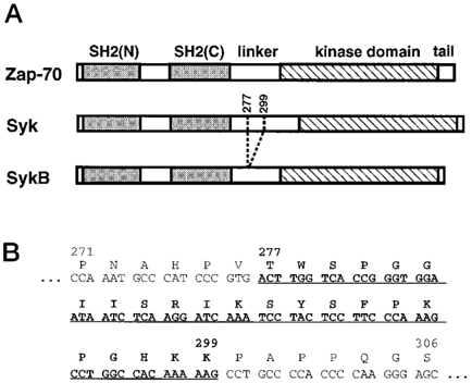
FIG. 1 Structure of Syk family kinases. A , schematic representation of the primary structure of Zap-70, Syk, and SykB. The position of the sequence deleted in the linker region of SykB (amino acids 277-299) is indicated. B , partial cDNA sequence of the mouse Syk linker region. Corresponding amino acids are shown above. The exon sequence missing in the sykB cDNA is shown in bold and underlined .
FIG. 1 Structure of Syk family kinases. A , schematic representation of the primary structure of Zap-70, Syk, and SykB. The position of the sequence deleted in the linker region of SykB (amino acids 277-299) is indicated. B , partial cDNA sequence of the mouse Syk linker region. Corresponding amino acids are shown above. The exon sequence missing in the sykB cDNA is shown in bold and underlined .CC-BY · From the PDF
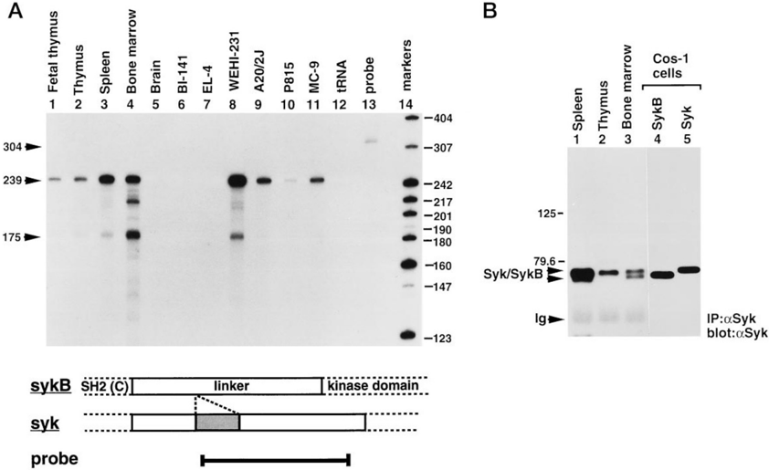
FIG. 2 Expression of sykB transcripts and polypeptides in mouse hemopoietic cells. A , ribonuclease protection assays. The expression of syk and sykB RNAs in various mouse tissues and cell lines was determined by ribonuclease protection assays. The probe used was 304 nucleotides in length and covered the most 3 9 portion of the linker region of syk (see bottom for schema). Protection of this riboprobe by syk transcripts was expected to yield a 239-nucleotide fragment, while shielding by sykB RNAs was predicted to result in a fragment of 175 nucleotides. The migrations of the full-length syk probe, and of the two types of protected fragments, are indicated on the left , while those of radiolabeled size markers are shown on the right . Exposure: 3 days. B , expression of Syk proteins in normal mouse hemopoietic cells. Hemopoietic cells were isolated from BALB/c mice. After depletion of erythrocytes, they were lysed, and Syk proteins were immunoprecipitated from 1 mg of cellular proteins, using a rabbit anti-Syk serum. Syk polypeptides were subsequently detected by anti-Syk immunoblotting. As controls, Syk proteins were immunoprecipitated from lysates (20 m g) of COS-1 cells transfected with a mouse syk or sykB cDNA. The positions of Syk, SykB, heavy chain of Ig, and prestained molecular mass markers are shown on the left . Exposure: 1 h.
FIG. 2 Expression of sykB transcripts and polypeptides in mouse hemopoietic cells. A , ribonuclease protection assays. The expression of syk and sykB RNAs in various mouse tissues and cell lines was determined by ribonuclease protection assays. The probe used was 304 nucleotides in length and covered the most 3 9 portion of the linker region of syk (see bottom for schema). Protection of this riboprobe by syk transcripts was expected to yield a 239-nucleotide fragment, while shielding by sykB RNAs was predicted to result in a fragment of 175 nucleotides. The migrations of the full-length syk probe, and of the two types of protected fragments, are indicated on the left , while those of radiolabeled size markers are shown on the right . Exposure: 3 days. B , expression of Syk proteins in normal mouse hemopoietic cells. Hemopoietic cells were isolated from BALB/c mice. After depletion of erythrocytes, they were lysed, and Syk proteins were immunoprecipitated from 1 mg of cellular proteins, using a rabbit anti-Syk serum. Syk polypeptides were subsequently detected by anti-Syk immunoblotting. As controls, Syk proteins were immunoprecipitated from lysates (20 m g) of COS-1 cells transfected with a mouse syk or sykB cDNA. The positions of Syk, SykB, heavy chain of Ig, and prestained molecular mass markers are shown on the left . Exposure: 1 h.CC-BY · From the PDF
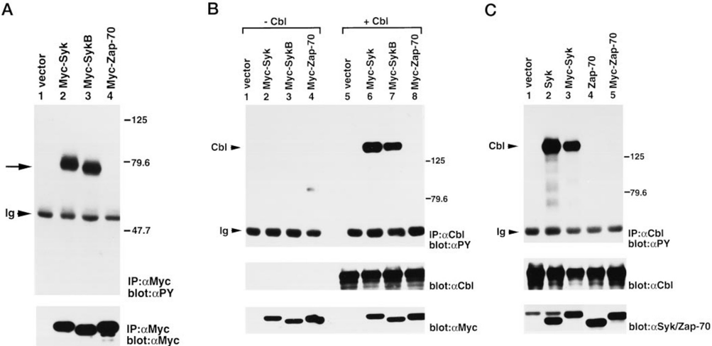
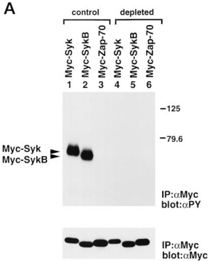
FIG. 3 Impact of Syk family kinases on intracellular protein tyrosine phosphorylation in COS-1 cells. COS-1 cells were transiently transfected with the indicated DNAs. Equivalent amounts of lysates (250 m g) were subjected to immunoprecipitation and immunoblotting with the indicated antibodies. A , tyrosine phosphorylation of Myc-tagged proteins. Myc-tagged proteins were immunoprecipitated with mAb 9E10 and immunoblotted with rabbit anti-phosphotyrosine antibodies ( top panel ) or anti-Myc antibodies ( bottom panel ). The migrations of Myc-Syk and Myc-SykB, as well as of heavy chain of Ig, are indicated on the left , whereas those of prestained molecular weight markers are shown on the right . Exposures: top panel , 12 h; bottom panel , 4 h. B and C , COS-1 cells were also transfected with the indicated DNAs, in the absence or presence of a mouse ccbl cDNA. c-Cbl was recovered by immunoprecipitation from 250 m g of cellular proteins and probed by immunoblotting with anti-phosphotyrosine antibodies. The abundance of c-Cbl, and of the various Syk-related enzymes, was determined by immunoblotting of equivalent amounts (30 m g) of cell lysates with the indicated antibodies. B , comparison of Myc-Syk, Myc-SykB, and Myc-Zap-70. The migrations of c-Cbl and heavy chain of Ig are indicated on the left ; those of prestained molecular mass markers are shown on the right . Exposures: top panel , 24 h; middle panel , 8 h; bottom panel , 2 h. C , comparison of Myc-tagged and nontagged versions of Syk and Zap-70. All cells were transfected with the ccbl cDNA. For the bottom panel, lanes 1-3 were immunoblotted with anti-Syk antibodies, whereas lanes 4 and 5 were reacted with anti-Zap-70 antibodies. The migrations of c-Cbl and heavy chain of Ig are indicated on the left ; those of prestained molecular mass markers are shown on the right . Exposures: top panel , 8 h; middle panel , 8 h; bottom panel , 8 h.
FIG. 3 Impact of Syk family kinases on intracellular protein tyrosine phosphorylation in COS-1 cells. COS-1 cells were transiently transfected with the indicated DNAs. Equivalent amounts of lysates (250 m g) were subjected to immunoprecipitation and immunoblotting with the indicated antibodies. A , tyrosine phosphorylation of Myc-tagged proteins. Myc-tagged proteins were immunoprecipitated with mAb 9E10 and immunoblotted with rabbit anti-phosphotyrosine antibodies ( top panel ) or anti-Myc antibodies ( bottom panel ). The migrations of Myc-Syk and Myc-SykB, as well as of heavy chain of Ig, are indicated on the left , whereas those of prestained molecular weight markers are shown on the right . Exposures: top panel , 12 h; bottom panel , 4 h. B and C , COS-1 cells were also transfected with the indicated DNAs, in the absence or presence of a mouse ccbl cDNA. c-Cbl was recovered by immunoprecipitation from 250 m g of cellular proteins and probed by immunoblotting with anti-phosphotyrosine antibodies. The abundance of c-Cbl, and of the various Syk-related enzymes, was determined by immunoblotting of equivalent amounts (30 m g) of cell lysates with the indicated antibodies. B , comparison of Myc-Syk, Myc-SykB, and Myc-Zap-70. The migrations of c-Cbl and heavy chain of Ig are indicated on the left ; those of prestained molecular mass markers are shown on the right . Exposures: top panel , 24 h; middle panel , 8 h; bottom panel , 2 h. C , comparison of Myc-tagged and nontagged versions of Syk and Zap-70. All cells were transfected with the ccbl cDNA. For the bottom panel, lanes 1-3 were immunoblotted with anti-Syk antibodies, whereas lanes 4 and 5 were reacted with anti-Zap-70 antibodies. The migrations of c-Cbl and heavy chain of Ig are indicated on the left ; those of prestained molecular mass markers are shown on the right . Exposures: top panel , 8 h; middle panel , 8 h; bottom panel , 8 h.CC-BY · From the PDF
Image 4
CC-BY · From the PDF
Image 5
CC-BY · From the PDF
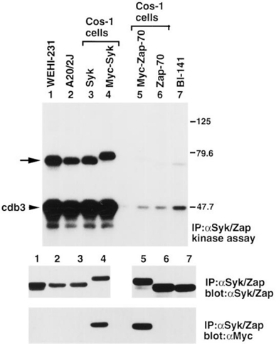
FIG. 6 Activity of Syk family kinases expressed in hemopoietic cells. Syk ( lanes 1-4 ) or Zap-70 ( lanes 5-7 ) was immunoprecipitated from cell lysates using the appropriate polyclonal rabbit antisera. The following amounts of cellular proteins were utilized: WEHI-231, 0.5 mg; A20/2J, 2 mg; BI-141, 2 mg; and COS-1 cells, 100 m g. The activity of the enzymes was measured in immune complex kinase reactions ( top panel ). Reactions were conducted for 5 min, in the presence of the cytoplasmic domain of erythrocyte band 3 (cdb3). Prior to autoradiography, the gel was treated with 1 M KOH, to remove phosphoserine and phosphothreonine. The abundance of Syk or Zap-70 was determined by immunoblotting of parallel immunoprecipitates with anti-Syk ( middle panel, lanes 1-4 ), anti-Zap-70 ( middle panel, lanes 5-7 ), or anti-Myc antibodies ( bottom panel ). The position of the Syk family kinases is indicated by an arrow on the left . The location of cdb3 is also shown on the left , whereas those of prestained molecular mass markers are indicated on the right . Exposures: top panel , 15 min; middle panel, lanes 1-4 , 3 h; lanes 5-7 , 1 h; bottom panel , 1 h.
FIG. 6 Activity of Syk family kinases expressed in hemopoietic cells. Syk ( lanes 1-4 ) or Zap-70 ( lanes 5-7 ) was immunoprecipitated from cell lysates using the appropriate polyclonal rabbit antisera. The following amounts of cellular proteins were utilized: WEHI-231, 0.5 mg; A20/2J, 2 mg; BI-141, 2 mg; and COS-1 cells, 100 m g. The activity of the enzymes was measured in immune complex kinase reactions ( top panel ). Reactions were conducted for 5 min, in the presence of the cytoplasmic domain of erythrocyte band 3 (cdb3). Prior to autoradiography, the gel was treated with 1 M KOH, to remove phosphoserine and phosphothreonine. The abundance of Syk or Zap-70 was determined by immunoblotting of parallel immunoprecipitates with anti-Syk ( middle panel, lanes 1-4 ), anti-Zap-70 ( middle panel, lanes 5-7 ), or anti-Myc antibodies ( bottom panel ). The position of the Syk family kinases is indicated by an arrow on the left . The location of cdb3 is also shown on the left , whereas those of prestained molecular mass markers are indicated on the right . Exposures: top panel , 15 min; middle panel, lanes 1-4 , 3 h; lanes 5-7 , 1 h; bottom panel , 1 h.CC-BY · From the PDF
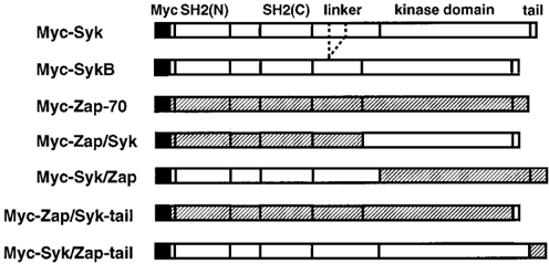
FIG. 7 Chimeras between Syk and Zap-70. Schematic representations of the various chimeras created between Syk and Zap-70. Note that all polypeptides carried the amino-terminal Myc-derived epitope.
FIG. 7 Chimeras between Syk and Zap-70. Schematic representations of the various chimeras created between Syk and Zap-70. Note that all polypeptides carried the amino-terminal Myc-derived epitope.CC-BY · From the PDF
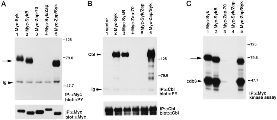
FIG. 8 Enzymatic function of Syk/Zap and Zap/Syk. The enzymatic function of Myc-Syk/Zap and Myc-Zap/Syk was compared with that of Myc-Syk, Myc-SykB, and Myc-Zap-70. A , tyrosine phosphorylation of Myc-tagged proteins. The extent of tyrosine phosphorylation of the chimeras was determined by immunoblotting of anti-Myc immunoprecipitates with anti-phosphotyrosine antibodies ( top panel ), as outlined in the legend of Fig. 3 A . The abundance of the polypeptides was ascertained by anti-Myc immunoblotting of parallel immunoprecipitates ( bottom panel ). The migration of the various Myc-tagged polypeptides is highlighted by an arrow on the left . The position of heavy chain of Ig is shown on the left , while those of prestained molecular weight markers are indicated on the right . Exposures: top panel , 12 h; bottom panel , 2 h. B , tyrosine phosphorylation of c-Cbl. COS-1 cells were transfected with the indicated DNAs, in the presence of a mouse ccbl cDNA. c-Cbl was recovered by immunoprecipitation from 250 m g of cellular proteins and probed by immunoblotting with anti-phosphotyrosine antibodies ( top panel ). The abundance of c-Cbl was determined by stripping the immunoblot and re-hybridizing with anti-Cbl antibodies ( bottom panel ). The migrations of c-Cbl and heavy chain of Ig are indicated on the left ; those of prestained molecular mass markers are shown on the right . Exposures: top panel , 6 h; bottom panel , 4 h. C , immune complex kinase assays. Myc-tagged proteins were immunoprecipitated with mAb 9E10 from 100 m g of total cellular proteins. Kinase reactions were conducted for 5 min, in the presence of the cytoplasmic domain of erythrocyte band 3 (cdb3). The position of the Syk family kinases is indicated by an arrow on the left . The location of the cytoplasmic domain of erythrocyte band 3 (cdb3) is also shown on the left , whereas those of prestained molecular mass markers are indicated on the right . Exposure, 5 min.
FIG. 8 Enzymatic function of Syk/Zap and Zap/Syk. The enzymatic function of Myc-Syk/Zap and Myc-Zap/Syk was compared with that of Myc-Syk, Myc-SykB, and Myc-Zap-70. A , tyrosine phosphorylation of Myc-tagged proteins. The extent of tyrosine phosphorylation of the chimeras was determined by immunoblotting of anti-Myc immunoprecipitates with anti-phosphotyrosine antibodies ( top panel ), as outlined in the legend of Fig. 3 A . The abundance of the polypeptides was ascertained by anti-Myc immunoblotting of parallel immunoprecipitates ( bottom panel ). The migration of the various Myc-tagged polypeptides is highlighted by an arrow on the left . The position of heavy chain of Ig is shown on the left , while those of prestained molecular weight markers are indicated on the right . Exposures: top panel , 12 h; bottom panel , 2 h. B , tyrosine phosphorylation of c-Cbl. COS-1 cells were transfected with the indicated DNAs, in the presence of a mouse ccbl cDNA. c-Cbl was recovered by immunoprecipitation from 250 m g of cellular proteins and probed by immunoblotting with anti-phosphotyrosine antibodies ( top panel ). The abundance of c-Cbl was determined by stripping the immunoblot and re-hybridizing with anti-Cbl antibodies ( bottom panel ). The migrations of c-Cbl and heavy chain of Ig are indicated on the left ; those of prestained molecular mass markers are shown on the right . Exposures: top panel , 6 h; bottom panel , 4 h. C , immune complex kinase assays. Myc-tagged proteins were immunoprecipitated with mAb 9E10 from 100 m g of total cellular proteins. Kinase reactions were conducted for 5 min, in the presence of the cytoplasmic domain of erythrocyte band 3 (cdb3). The position of the Syk family kinases is indicated by an arrow on the left . The location of the cytoplasmic domain of erythrocyte band 3 (cdb3) is also shown on the left , whereas those of prestained molecular mass markers are indicated on the right . Exposure, 5 min.CC-BY · From the PDF
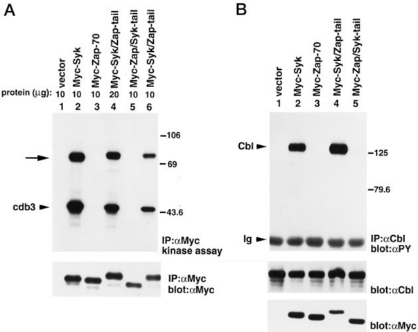
FIG. 9 Enzymatic function of Syk/Zap-tail and Zap/Syk-tail. As in the legend of Fig. 8, except that Myc-Syk/Zap-tail and Myc-Zap/Syk-tail were evaluated. A , immune complex kinase reactions. Myc-tagged proteins were immunoprecipitated from 10 or 20 m g of total cellular proteins, as indicated. Kinase reactions were conducted for 2 min, in the presence of the cytoplasmic domain of erythrocyte band 3 (cdb3). The position of the Syk family kinases is indicated by an arrow on the left . The location of the cytoplasmic domain of erythrocyte band 3 (cdb3) is also shown on the left , whereas those of prestained molecular mass markers are indicated on the right . Exposure: top panel , 7 min; bottom panel , 1.5 h. B , tyrosine phosphorylation of c-Cbl. The migrations of c-Cbl and heavy chain of Ig are indicated on the left ; those of prestained molecular mass markers are shown on the right . Exposures: top panel , 2 h; middle panel , 1.5 h; bottom panel , 6 h.
FIG. 9 Enzymatic function of Syk/Zap-tail and Zap/Syk-tail. As in the legend of Fig. 8, except that Myc-Syk/Zap-tail and Myc-Zap/Syk-tail were evaluated. A , immune complex kinase reactions. Myc-tagged proteins were immunoprecipitated from 10 or 20 m g of total cellular proteins, as indicated. Kinase reactions were conducted for 2 min, in the presence of the cytoplasmic domain of erythrocyte band 3 (cdb3). The position of the Syk family kinases is indicated by an arrow on the left . The location of the cytoplasmic domain of erythrocyte band 3 (cdb3) is also shown on the left , whereas those of prestained molecular mass markers are indicated on the right . Exposure: top panel , 7 min; bottom panel , 1.5 h. B , tyrosine phosphorylation of c-Cbl. The migrations of c-Cbl and heavy chain of Ig are indicated on the left ; those of prestained molecular mass markers are shown on the right . Exposures: top panel , 2 h; middle panel , 1.5 h; bottom panel , 6 h.CC-BY · From the PDF

Verification

Claims the paper makes

The claims and statistics of this paper haven’t been checked yet.

Run the check — free, no account needed. It reads the open-access text and checks the sources it cites.

A few free checks a day · papers already verified open instantly · results are public

Claims other papers make about this one

Each is checked against this paper’s own text: does it say what the citing paper says it does?

Self-check

Percentages, sums and reported differences re-computed from the paper’s own numbers.

The self-check needs the paper’s full text.

Citations

Cites

No references could be resolved yet.

Research works printed but not linked: 42

  1. van Oers, 1995, Semin. Immunol., 7, 227, 10.1006/smim.1995.0027 · DOI
  2. Law, 1996, Mol. Cell. Biol., 16, 1305, 10.1128/MCB.16.4.1305 · DOI
  3. Iwashima, 1994, Science, 263, 1136, 10.1126/science.7509083 · DOI
  4. Couture, 1994, Mol. Cell. Biol., 14, 5249
  5. Chan, 1994, J. Immunol., 152, 4758, 10.4049/jimmunol.152.10.4758 · DOI · Paper page
  6. Kurosaki, 1994, J. Exp. Med., 179, 1725, 10.1084/jem.179.5.1725 · DOI
  7. Rowley, 1995, J. Biol. Chem., 270, 11590, 10.1074/jbc.270.19.11590 · DOI
  8. Shiue, 1995, J. Biol. Chem., 270, 10498, 10.1074/jbc.270.18.10498 · DOI
  9. Kimura, 1996, Mol. Cell. Biol., 16, 1471, 10.1128/MCB.16.4.1471 · DOI
  10. Neumeister, 1995, Mol. Cell. Biol., 15, 3171, 10.1128/MCB.15.6.3171 · DOI
  11. Takata, 1994, EMBO J., 13, 1341, 10.1002/j.1460-2075.1994.tb06387.x · DOI · Paper page
  12. Taylor, 1995, Mol. Cell. Biol., 15, 4149, 10.1128/MCB.15.8.4149 · DOI
  13. Qian, 1996, J. Exp. Med., 183, 611, 10.1084/jem.183.2.611 · DOI
  14. Arpaia, 1994, Cell, 76, 947, 10.1016/0092-8674(94)90368-9 · DOI
  15. Chan, 1994, Science, 264, 1599, 10.1126/science.8202713 · DOI
  16. Elder, 1994, Science, 264, 1596, 10.1126/science.8202712 · DOI
  17. Negishi, 1995, Nature, 376, 435, 10.1038/376435a0 · DOI
  18. Cheng, 1995, Nature, 378, 303, 10.1038/378303a0 · DOI · Paper page
  19. Turner, 1995, Nature, 378, 298, 10.1038/378298a0 · DOI · Paper page
  20. Kolanus, 1993, Cell, 74, 171, 10.1016/0092-8674(93)90304-9 · DOI
  21. Rivera, 1995, Mol. Cell. Biol., 15, 1582, 10.1128/MCB.15.3.1582 · DOI
  22. Kim, 1979, J. Immunol., 122, 549, 10.4049/jimmunol.122.2.549 · DOI
  23. Reske-Kunz, 1985, Eur. J. Immunol., 15, 1048, 10.1002/eji.1830151017 · DOI
  24. Flück, 1995, Biochem. Biophys. Res. Commun., 213, 273, 10.1006/bbrc.1995.2126 · DOI
  25. Richards, 1996, J. Biol. Chem., 271, 6458, 10.1074/jbc.271.11.6458 · DOI
  26. Weil, 1995, J. Biol. Chem., 270, 2791, 10.1074/jbc.270.6.2791 · DOI
  27. Fournel, 1996, J. Exp. Med., 183, 301, 10.1084/jem.183.1.301 · DOI
  28. Evan, 1985, Mol. Cell. Biol., 5, 3610
  29. Abraham, 1991, Nature, 350, 62, 10.1038/350062a0 · DOI
  30. Timson-Gauen, 1994, Mol. Cell. Biol., 14, 3729, 10.1128/mcb.14.6.3729-3741.1994 · DOI
  31. Chow, 1992, Mol. Cell. Biol., 12, 1226
  32. Veillette, 1988, Cell, 55, 301, 10.1016/0092-8674(88)90053-0 · DOI
  33. Veillette, 1988, Oncogene Res., 2, 385
  34. Yagi, 1994, Biochem. Biophys. Res. Commun., 200, 28, 10.1006/bbrc.1994.1409 · DOI
  35. Rowley, 1995, J. Biol. Chem., 270, 12659, 10.1074/jbc.270.21.12659 · DOI
  36. Donovan, 1994, J. Biol. Chem., 269, 22921, 10.1016/S0021-9258(17)31595-8 · DOI
  37. Panchamoorthy, 1996, J. Biol. Chem., 271, 3187, 10.1074/jbc.271.6.3187 · DOI
  38. Low, 1987, J. Biol. Chem., 262, 4592, 10.1016/S0021-9258(18)61234-7 · DOI
  39. Greenberg, 1996, Proc. Natl. Acad. Sci. U. S. A., 93, 1103, 10.1073/pnas.93.3.1103 · DOI
  40. Chan, 1995, EMBO J., 14, 2499, 10.1002/j.1460-2075.1995.tb07247.x · DOI
  41. Wange, 1995, J. Biol. Chem., 270, 18730, 10.1074/jbc.270.32.18730 · DOI
  42. Kong, 1995, Immunity, 2, 485, 10.1016/1074-7613(95)90029-2 · DOI

Cited by

See all 137

Metadata

Authors
Latour, S., Chow, L. M. L., & Veillette, A.
Venue
Journal of Biological Chemistry
Publisher
Elsevier BV
Published
1996-09
Volume
271
Issue
37
Pages
22782-22790
ISSN
0021-9258
Cited by
137
References
0
Language
en

About these checks

Registry, retraction, Cites and statistics results are computed, not judged, with one exception: a printed reference that no index lists is read once by a model before it counts as a citation error. Claims are extracted by a model and kept only when their quote appears verbatim in the text; each cited source is then checked, with a model’s help, for whether it says what the claim relies on. Those verdicts can be wrong — read the claim’s quote and the cited source before relying on one.

Statistics and arithmetic are recomputed, not judged.

Confidence bands are shown only for fields calibrated on our annotated test set. None is calibrated yet, so no band is shown.

What counts as a claim ›How we measure confidence ›How we check this ›

Are you an author, or do you see a problem with this page? Request a review

How to cite

Cite CoChat for the checks on this page, and the paper for its own findings.

This page
CoChat. (2026). Citation check: Differential Intrinsic Enzymatic Activity of Syk and Zap-70 Protein-tyrosine Kinases. https://app.cochat.ai/papers/10.1074/jbc.271.37.22782
The paper
Latour, S., Chow, L. M. L., & Veillette, A. (1996). Differential Intrinsic Enzymatic Activity of Syk and Zap-70 Protein-tyrosine Kinases. Journal of Biological Chemistry. https://doi.org/10.1074/jbc.271.37.22782