

The DOI matches its Crossref record.
Structural Requirements for PAK Activation by Rac GTPases
Overview
The claims and statistics of this paper haven’t been checked yet.
Run the check — free, no account needed. It reads the open-access text and checks the sources it cites.
Integrity signals
Registry
Cites
Statistics
Figures
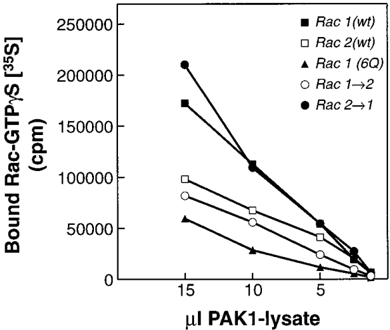
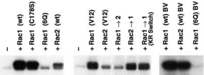
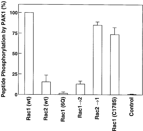
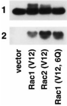
Verification
Claims the paper makes
The claims and statistics of this paper haven’t been checked yet.
Run the check — free, no account needed. It reads the open-access text and checks the sources it cites.
Claims other papers make about this one
Each is checked against this paper’s own text: does it say what the citing paper says it does?
In the citing paper
“RAC2-GTP drives diverse cellular functions through association and activation of downstream effector proteins including p67 phox11 and PAK1.12”
From Dominant activating RAC2 mutation with lymphopenia, immunodeficiency, and cytoskeletal defects (2019) · cites this paper as [12]
In the citing paper
“The ability of RAC2 to cycle between GDP-and GTP-bound forms allows it to act as a molecular switch because only active RAC2-GTP can bind to p67 phox35 or PAK1.12”
From Dominant activating RAC2 mutation with lymphopenia, immunodeficiency, and cytoskeletal defects (2019) · cites this paper as [12]
Self-check
Percentages, sums and reported differences re-computed from the paper’s own numbers.
The self-check needs the paper’s full text.
Citations
Cites
No references could be resolved yet.
Research works printed but not linked: 44
- Hall, 1994, Annu. Rev. Cell Biol., 10, 31, 10.1146/annurev.cb.10.110194.000335 · DOI
- Ridley, 1992, Cell, 70, 389, 10.1016/0092-8674(92)90163-7 · DOI
- Ridley, 1992, Cell, 70, 401, 10.1016/0092-8674(92)90164-8 · DOI
- Kozma, 1995, Mol. Cell. Biol., 16, 5069, 10.1128/MCB.16.9.5069 · DOI
- Nobes, 1995, Cell, 81, 53, 10.1016/0092-8674(95)90370-4 · DOI
- Coso, 1995, Cell, 81, 1137, 10.1016/S0092-8674(05)80018-2 · DOI · Paper page
- Minden, 1995, Cell, 81, 1147, 10.1016/S0092-8674(05)80019-4 · DOI
- Olson, 1995, Science, 269, 1270, 10.1126/science.7652575 · DOI
- Zhang, 1995, J. Biol. Chem., 270, 23934, 10.1074/jbc.270.41.23934 · DOI · Paper page
- Bagrodia, 1995, J. Biol. Chem., 270, 27995, 10.1074/jbc.270.47.27995 · DOI
- Hill, 1995, Cell, 81, 1159, 10.1016/S0092-8674(05)80020-0 · DOI
- Qiu, 1995, Nature, 374, 457, 10.1038/374457a0 · DOI
- Khosravi-Far, 1995, Mol. Cell. Biol., 15, 6443, 10.1128/MCB.15.11.6443 · DOI · Paper page
- Abo, 1991, Nature, 353, 668, 10.1038/353668a0 · DOI
- Knaus, 1991, Science, 254, 1512, 10.1126/science.1660188 · DOI · Paper page
- Heyworth, 1993, Mol. Biol. Cell, 4, 261, 10.1091/mbc.4.3.261 · DOI
- Dorseuil, 1996, J. Biol. Chem., 271, 83, 10.1074/jbc.271.1.83 · DOI
- Manser, 1994, Nature, 367, 40, 10.1038/367040a0 · DOI
- Polverino, 1995, J. Biol. Chem., 270, 26067, 10.1074/jbc.270.44.26067 · DOI
- Dharmawardhane, 1997, J. Cell Biol., 138, 1265, 10.1083/jcb.138.6.1265 · DOI
- Sells, 1997, Curr. Biol., 7, 202, 10.1016/S0960-9822(97)70091-5 · DOI · Paper page
- Brzeska, 1997, Proc. Natl. Acad. Sci. U. S. A., 94, 1092, 10.1073/pnas.94.4.1092 · DOI
- Wu, 1996, J. Biol. Chem., 271, 31787, 10.1074/jbc.271.50.31787 · DOI
- Tauzon, 1984, J. Biol. Chem., 259, 541, 10.1016/S0021-9258(17)43695-7 · DOI
- Knaus, 1995, Science, 269, 221, 10.1126/science.7618083 · DOI
- Jackson, 1990, Proc. Natl. Acad. Sci. U. S. A., 87, 3042, 10.1073/pnas.87.8.3042 · DOI
- Knaus, 1992, J. Biol. Chem., 267, 23575, 10.1016/S0021-9258(18)35877-0 · DOI
- Burbelo, 1995, J. Biol. Chem., 270, 29071, 10.1074/jbc.270.49.29071 · DOI
- Symons, 1996, Cell, 84, 723, 10.1016/S0092-8674(00)81050-8 · DOI
- Sells, 1997, Trends Cell Biol., 7, 162, 10.1016/S0962-8924(97)01003-9 · DOI
- Bagrodia, 1995, J. Biol. Chem., 270, 22731, 10.1074/jbc.270.39.22731 · DOI
- Bokoch, 1996, J. Biol. Chem., 271, 25746, 10.1074/jbc.271.42.25746 · DOI
- Lamarche, 1996, Cell, 87, 519, 10.1016/S0092-8674(00)81371-9 · DOI
- Westwick, 1997, Mol. Cell. Biol., 17, 1324, 10.1128/MCB.17.3.1324 · DOI
- Diekmann, 1995, EMBO J., 14, 5297, 10.1002/j.1460-2075.1995.tb00214.x · DOI · Paper page
- Hancock, 1990, Cell, 63, 133, 10.1016/0092-8674(90)90294-O · DOI
- Kreck, 1994, J. Biol. Chem., 269, 4161, 10.1016/S0021-9258(17)41757-1 · DOI · Paper page
- Kreck, 1996, Biochemistry, 35, 15683, 10.1021/bi962064l · DOI
- Joseph, 1995, J. Biol. Chem., 270, 29079, 10.1074/jbc.270.49.29079 · DOI
- Jackson, 1994, Proc. Natl. Acad. Sci. U. S. A., 91, 12730, 10.1073/pnas.91.26.12730 · DOI
- Shirataki, 1991, J. Biol. Chem., 266, 20672, 10.1016/S0021-9258(18)54761-X · DOI
- Manser, 1994, Nature, 363, 364, 10.1038/363364a0 · DOI
- Manser, 1995, J. Biol. Chem., 270, 25070, 10.1074/jbc.270.42.25070 · DOI
- Manser, 1998, Mol. Cell, 1, 183, 10.1016/S1097-2765(00)80019-2 · DOI
Cited by
Doherty, G. J., & McMahon, H. T. (2009) · Annual Review of Biochemistry · doi:10.1146/annurev.biochem.78.081307.110540 · Cited by 3,075
Dominant activating RAC2 mutation with lymphopenia, immunodeficiency, and cytoskeletal defects
Hsu, A. P., Donkó, A., Arrington, M. E., Swamydas, M., Fink, D., Das, A., Escobedo, O., Bonagura, V., Szabolcs, P., Steinberg, H. N., et al. (2019) · Blood · doi:10.1182/blood-2018-11-886028 · Cited by 92
Metadata
- Authors
- Knaus, U. G., Wang, Y., Reilly, A. M., Warnock, D., & Jackson, J. H.
- Venue
- Journal of Biological Chemistry
- Publisher
- Elsevier BV
- Published
- 1998-08
- Volume
- 273
- Issue
- 34
- Pages
- 21512-21518
- ISSN
- 0021-9258
- Cited by
- 111
- References
- 0
- Language
- en
About these checks
Registry, retraction, Cites and statistics results are computed, not judged, with one exception: a printed reference that no index lists is read once by a model before it counts as a citation error. Claims are extracted by a model and kept only when their quote appears verbatim in the text; each cited source is then checked, with a model’s help, for whether it says what the claim relies on. Those verdicts can be wrong — read the claim’s quote and the cited source before relying on one.
Statistics and arithmetic are recomputed, not judged.
Confidence bands are shown only for fields calibrated on our annotated test set. None is calibrated yet, so no band is shown.
What counts as a claim ›How we measure confidence ›How we check this ›
Are you an author, or do you see a problem with this page? Request a review
