Cited by 198

The DOI matches its Crossref record.

Characterization of a Two-component Alkanesulfonate Monooxygenase from Escherichia coli

Eichhorn, Eric, van der Ploeg, Jan R., Leisinger, Thomas·1999·Journal of Biological Chemistry

Overview

The claims and statistics of this paper haven’t been checked yet.

Run the check — free, no account needed. It reads the open-access text and checks the sources it cites.

A few free checks a day · papers already verified open instantly · results are public

Integrity signals

Registry

Crossref · 10.1074/jbc.274.38.26639

Cites

Statistics

Figures

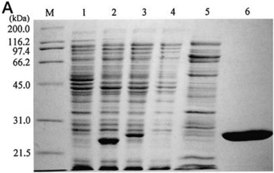
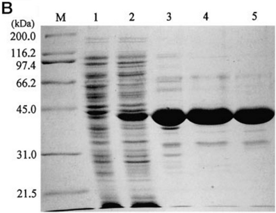
FIG. 1 Purification of the two components of the alkanesulfonate monooxygenase system from E. coli . Protein samples (30 m g) were analyzed at different stages of purification on 12% SDS-PAGE gels under reducing and denaturing conditions, and stained with Coomassie Brilliant Blue. A , purification of NAD(P)H:FMN oxidoreductase SsuE. M, molecular weight markers (with molecular masses indicated on the left in kDa); lane 1, cell extract of uninduced E. coli BL21(DE3)(pME4287); lane 2, extract of cells producing wild type SsuE from pME4146; lane 3, extract of induced E. coli BL21(DE3)(pME4287) cells producing histidine-tagged SsuE; lane 4, wash with binding buffer; lane 5, wash with 60 mM imidazole; lane 6, purified SsuE. B , purification of alkanesulfonate monooxygenase SsuD. M, molecular weight markers (with molecular masses indicated on the left in kDa): lane 1, cell extract of uninduced E. coli BL21(DE3)(pME4282); lane 2, extract of induced cells; lane 3, fraction after Fractogel EMD SO 3 2 (S) chromatography; lane 4, fraction after Fractogel EMD TMAE (S) chromatography; lane 5, purified SsuD obtained after phenyl-Sepharose Fast Flow 66 chromatography.
FIG. 1 Purification of the two components of the alkanesulfonate monooxygenase system from E. coli . Protein samples (30 m g) were analyzed at different stages of purification on 12% SDS-PAGE gels under reducing and denaturing conditions, and stained with Coomassie Brilliant Blue. A , purification of NAD(P)H:FMN oxidoreductase SsuE. M, molecular weight markers (with molecular masses indicated on the left in kDa); lane 1, cell extract of uninduced E. coli BL21(DE3)(pME4287); lane 2, extract of cells producing wild type SsuE from pME4146; lane 3, extract of induced E. coli BL21(DE3)(pME4287) cells producing histidine-tagged SsuE; lane 4, wash with binding buffer; lane 5, wash with 60 mM imidazole; lane 6, purified SsuE. B , purification of alkanesulfonate monooxygenase SsuD. M, molecular weight markers (with molecular masses indicated on the left in kDa): lane 1, cell extract of uninduced E. coli BL21(DE3)(pME4282); lane 2, extract of induced cells; lane 3, fraction after Fractogel EMD SO 3 2 (S) chromatography; lane 4, fraction after Fractogel EMD TMAE (S) chromatography; lane 5, purified SsuD obtained after phenyl-Sepharose Fast Flow 66 chromatography.CC-BY · From the PDF
Image 2
CC-BY · From the PDF
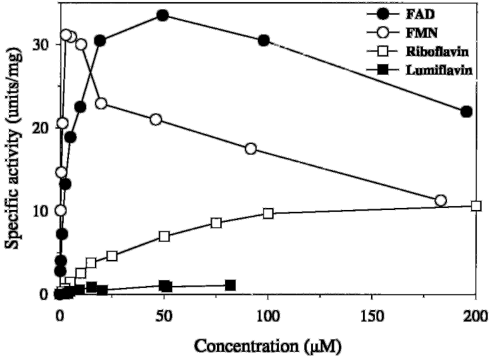
FIG. 2 Activity of NAD(P)H:FMN oxidoreductase SsuE in the presence of various flavin substrates. Activity measurements were performed under aerobic conditions as described under 'Experimental Procedures' in reaction mixtures containing 47 pmol of SsuE, 200 m M NADPH and varying concentrations of FMN, FAD, riboflavin, or lumiflavin in 20 mM imidazole, 50 mM NaCl, 20 mM Tris-HCl, pH 7.9. Initial rates were calculated from the slope of the absorbance decrease at 340 nm by regression analysis. Each point represents the mean of two independent experiments.
FIG. 2 Activity of NAD(P)H:FMN oxidoreductase SsuE in the presence of various flavin substrates. Activity measurements were performed under aerobic conditions as described under 'Experimental Procedures' in reaction mixtures containing 47 pmol of SsuE, 200 m M NADPH and varying concentrations of FMN, FAD, riboflavin, or lumiflavin in 20 mM imidazole, 50 mM NaCl, 20 mM Tris-HCl, pH 7.9. Initial rates were calculated from the slope of the absorbance decrease at 340 nm by regression analysis. Each point represents the mean of two independent experiments.CC-BY · From the PDF
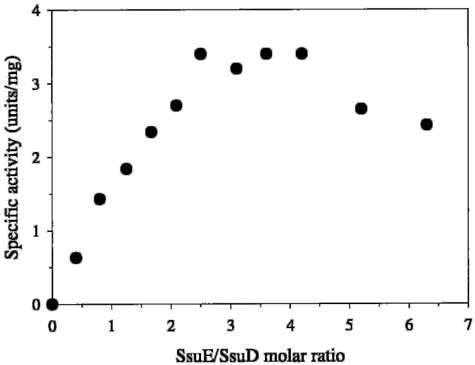
FIG. 3 Influence of the molar ratio of the two components on alkanesulfonate desulfonation. The effect of the SsuE/SsuD molar ratio on alkanesulfonate desulfonation by SsuD was investigated by keeping the SsuD concentration constant at 55 nM and varying the concentration of SsuE in the reaction mixture described under 'Experimental Procedures.' Sulfite release was followed over 3 min and initial rates calculated from the slope of absorbance increase at 430 nm by regression analysis. Each point corresponds to the mean value of two independent experiments.
FIG. 3 Influence of the molar ratio of the two components on alkanesulfonate desulfonation. The effect of the SsuE/SsuD molar ratio on alkanesulfonate desulfonation by SsuD was investigated by keeping the SsuD concentration constant at 55 nM and varying the concentration of SsuE in the reaction mixture described under 'Experimental Procedures.' Sulfite release was followed over 3 min and initial rates calculated from the slope of absorbance increase at 430 nm by regression analysis. Each point corresponds to the mean value of two independent experiments.CC-BY · From the PDF
Image 5
CC-BY · From the PDF
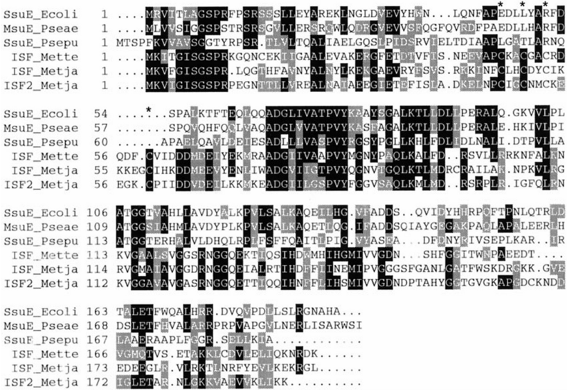
FIG. 5 Alignment of SsuE with flavoprotein sequences. SsuE_ E. coli is NAD(P)H:FMN oxidoreductase from E. coli (Swiss-Prot accession number P80644), MsuE_Pseae is NADH:FMN oxidoreductase from Pseudomonas aeruginosa (TrEMBL accession number O31038), SsuE_Psepu is NADH:FMN oxidoreductase from Pseudomonas putida (TrEMBL accession number O85762) , ISF_Mette is iron-sulfur flavoprotein from M. thermophila (TrEMBL accession number Q50562), ISF_Metja and ISF2_Metja are putative iron-sulfur flavoproteins from Methanococcus janaschii (TrEMBL accession numbers Q58141 and Q58483). Sequences were aligned with the ClustalW program (30). Amino acid residues identical in at least three proteins are indicated in white on a black background. Amino acid residues conserved in at least three sequences are indicated in white on a gray background. Cysteine residues involved in the iron-sulfur cluster of flavoproteins are marked with an asterisk .
FIG. 5 Alignment of SsuE with flavoprotein sequences. SsuE_ E. coli is NAD(P)H:FMN oxidoreductase from E. coli (Swiss-Prot accession number P80644), MsuE_Pseae is NADH:FMN oxidoreductase from Pseudomonas aeruginosa (TrEMBL accession number O31038), SsuE_Psepu is NADH:FMN oxidoreductase from Pseudomonas putida (TrEMBL accession number O85762) , ISF_Mette is iron-sulfur flavoprotein from M. thermophila (TrEMBL accession number Q50562), ISF_Metja and ISF2_Metja are putative iron-sulfur flavoproteins from Methanococcus janaschii (TrEMBL accession numbers Q58141 and Q58483). Sequences were aligned with the ClustalW program (30). Amino acid residues identical in at least three proteins are indicated in white on a black background. Amino acid residues conserved in at least three sequences are indicated in white on a gray background. Cysteine residues involved in the iron-sulfur cluster of flavoproteins are marked with an asterisk .CC-BY · From the PDF
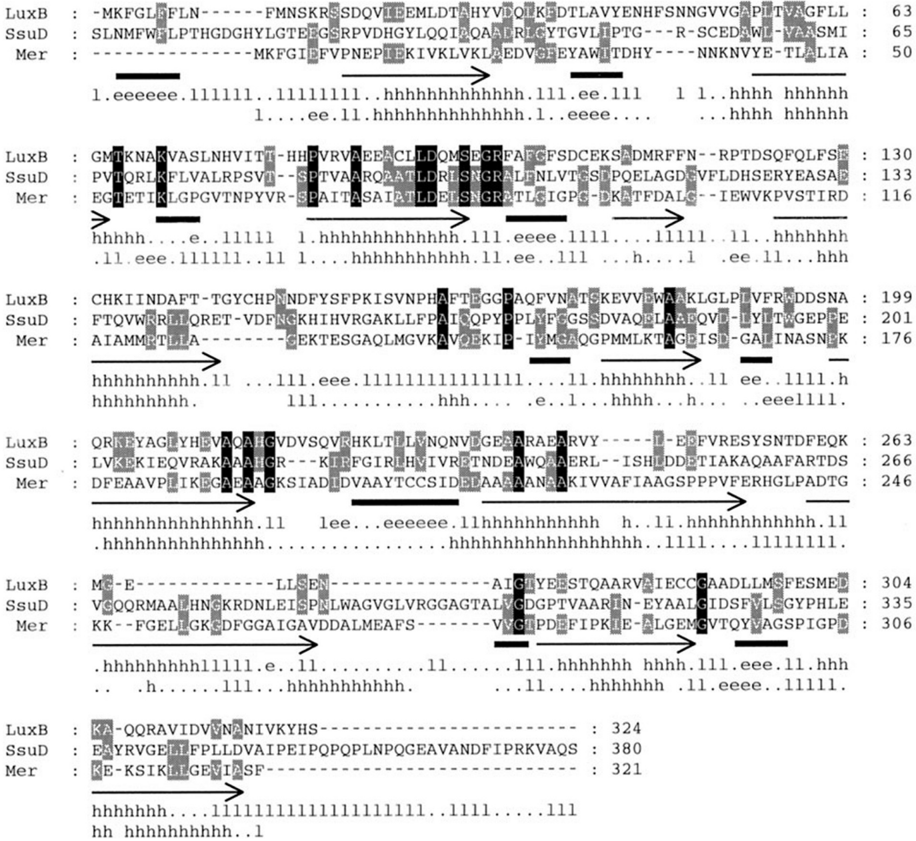
FIG. 6 Alignment of SsuD with the flavin-dependent enzymes Mer and LuxB. The following sequences were aligned with the program ClustalW (30): SsuD from E. coli (Swiss-Prot accession number P80645), NtaA from C. heintzii (Swiss-Prot accession number P54989), SnaA from S. pristinaespiralis (Swiss-Prot accession number P54991), SoxA from Rhodococcus sp. strain IGTS8 (Swiss-Prot accession number P54995), Mer from M. thermoautotrophicum (TrEMBL accession number Q50744), and LuxB and LuxA from Vibrio harveyi (Swiss-Prot accession numbers P07739 and P07740). Only the alignment between SsuD, Mer, and LuxB is shown. Identical residues in all three sequences are in white on a black background, identical residues occurring in two sequences are in white on a gray background. The secondary structure elements of LuxB (27) are shown below the sequence as arrows ( a -helices) or thick lines ( b -sheets). The predicted secondary structure elements of SsuD and Mer were calculated with the program PredictProtein (31) and are indicated by h for a -helices, e for b -sheets, and l for loops. Dots represent lack of defined secondary structure.
FIG. 6 Alignment of SsuD with the flavin-dependent enzymes Mer and LuxB. The following sequences were aligned with the program ClustalW (30): SsuD from E. coli (Swiss-Prot accession number P80645), NtaA from C. heintzii (Swiss-Prot accession number P54989), SnaA from S. pristinaespiralis (Swiss-Prot accession number P54991), SoxA from Rhodococcus sp. strain IGTS8 (Swiss-Prot accession number P54995), Mer from M. thermoautotrophicum (TrEMBL accession number Q50744), and LuxB and LuxA from Vibrio harveyi (Swiss-Prot accession numbers P07739 and P07740). Only the alignment between SsuD, Mer, and LuxB is shown. Identical residues in all three sequences are in white on a black background, identical residues occurring in two sequences are in white on a gray background. The secondary structure elements of LuxB (27) are shown below the sequence as arrows ( a -helices) or thick lines ( b -sheets). The predicted secondary structure elements of SsuD and Mer were calculated with the program PredictProtein (31) and are indicated by h for a -helices, e for b -sheets, and l for loops. Dots represent lack of defined secondary structure.CC-BY · From the PDF

Verification

Claims the paper makes

The claims and statistics of this paper haven’t been checked yet.

Run the check — free, no account needed. It reads the open-access text and checks the sources it cites.

A few free checks a day · papers already verified open instantly · results are public

Claims other papers make about this one

Each is checked against this paper’s own text: does it say what the citing paper says it does?

Self-check

Percentages, sums and reported differences re-computed from the paper’s own numbers.

The self-check needs the paper’s full text.

Citations

Cites

No references could be resolved yet.

Research works printed but not linked: 28

  1. Quadroni, 1996, Eur. J. Biochem., 239, 773, 10.1111/j.1432-1033.1996.0773u.x · DOI
  2. van der Ploeg, 1996, J. Bacteriol., 178, 5438, 10.1128/jb.178.18.5438-5446.1996 · DOI
  3. Eichhorn, 1997, J. Biol. Chem., 272, 23031, 10.1074/jbc.272.37.23031 · DOI · Paper page
  4. van der Ploeg, 1999, J. Biol. Chem., 274, 10.1074/jbc.274.41.29358 · DOI
  5. Uetz, 1992, J. Bacteriol., 174, 1179, 10.1128/jb.174.4.1179-1188.1992 · DOI
  6. Thibaut, 1995, J. Bacteriol., 177, 5199, 10.1128/jb.177.18.5199-5205.1995 · DOI
  7. Studier, 1990, Methods Enzymol., 185, 60, 10.1016/0076-6879(90)85008-C · DOI
  8. Yanisch-Perron, 1985, Gene (Amst.), 33, 103, 10.1016/0378-1119(85)90120-9 · DOI
  9. Jagura-Burdzy, 1981, J. Bacteriol., 147, 744, 10.1128/jb.147.3.744-751.1981 · DOI
  10. Johnston, 1975, Antonie Leeuwenhoek, 41, 493, 10.1007/BF02565092 · DOI
  11. Riddles, 1983, Methods Enzymol., 91, 49, 10.1016/S0076-6879(83)91010-8 · DOI
  12. Bradford, 1976, Anal. Biochem., 72, 248, 10.1016/0003-2697(76)90527-3 · DOI · Paper page
  13. Stoll, 1990, Methods Enzymol., 182, 24, 10.1016/0076-6879(90)82006-N · DOI
  14. Uria-Nickelsen, 1993, J. Gen. Microbiol., 139, 203, 10.1099/00221287-139-2-203 · DOI
  15. Thysse, 1974, Antonie Leeuwenhoek, 40, 25, 10.1007/BF00394550 · DOI
  16. Higgins, 1996, Microbiology, 142, 251, 10.1099/13500872-142-2-251 · DOI
  17. van der Ploeg, 1998, Microbiology, 144, 2555, 10.1099/00221287-144-9-2555 · DOI
  18. Kertesz, 1999, J. Bacteriol., 181, 1464, 10.1128/JB.181.5.1464-1473.1999 · DOI
  19. Witschel, 1997, J. Bacteriol., 179, 6937, 10.1128/jb.179.22.6937-6943.1997 · DOI
  20. Xi, 1997, Biochem. Biophys. Res. Commun., 230, 73, 10.1006/bbrc.1996.5878 · DOI
  21. Fieschi, 1995, J. Biol. Chem., 270, 30392, 10.1074/jbc.270.51.30392 · DOI
  22. Becker, 1998, J. Biol. Chem., 273, 26462, 10.1074/jbc.273.41.26462 · DOI
  23. Denome, 1994, J. Bacteriol., 176, 6707, 10.1128/jb.176.21.6707-6716.1994 · DOI
  24. Fisher, 1996, J. Biol. Chem., 271, 21956, 10.1074/jbc.271.36.21956 · DOI
  25. Vaupel, 1995, Eur. J. Biochem., 231, 773
  26. Uria-Nickelsen, 1993, FEMS Microbiol. Lett., 123, 43, 10.1111/j.1574-6968.1994.tb07199.x · DOI
  27. Thompson, 1994, Nucleic Acids Res., 22, 4673, 10.1093/nar/22.22.4673 · DOI · Paper page
  28. Rost, 1996, Methods Enzymol., 266, 525, 10.1016/S0076-6879(96)66033-9 · DOI

Cited by

Metadata

Authors
Eichhorn, E., van der Ploeg, J. R., & Leisinger, T.
Venue
Journal of Biological Chemistry
Publisher
Elsevier BV
Published
1999-09
Volume
274
Issue
38
Pages
26639-26646
ISSN
0021-9258
Cited by
198
References
0
Language
en

About these checks

Registry, retraction, Cites and statistics results are computed, not judged, with one exception: a printed reference that no index lists is read once by a model before it counts as a citation error. Claims are extracted by a model and kept only when their quote appears verbatim in the text; each cited source is then checked, with a model’s help, for whether it says what the claim relies on. Those verdicts can be wrong — read the claim’s quote and the cited source before relying on one.

Statistics and arithmetic are recomputed, not judged.

Confidence bands are shown only for fields calibrated on our annotated test set. None is calibrated yet, so no band is shown.

What counts as a claim ›How we measure confidence ›How we check this ›

Are you an author, or do you see a problem with this page? Request a review

How to cite

Cite CoChat for the checks on this page, and the paper for its own findings.

This page
CoChat. (2026). Citation check: Characterization of a Two-component Alkanesulfonate Monooxygenase from Escherichia coli. https://app.cochat.ai/papers/10.1074/jbc.274.38.26639
The paper
Eichhorn, E., van der Ploeg, J. R., & Leisinger, T. (1999). Characterization of a Two-component Alkanesulfonate Monooxygenase from Escherichia coli. Journal of Biological Chemistry. https://doi.org/10.1074/jbc.274.38.26639