

The DOI matches its Crossref record.
Mechanism and Cellular Applications of a Green Fluorescent Protein-based Halide Sensor
Overview
The claims and statistics of this paper haven’t been checked yet.
Run the check — free, no account needed. It reads the open-access text and checks the sources it cites.
Integrity signals
Registry
Cites
Statistics
Figures
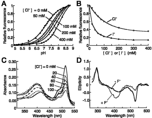
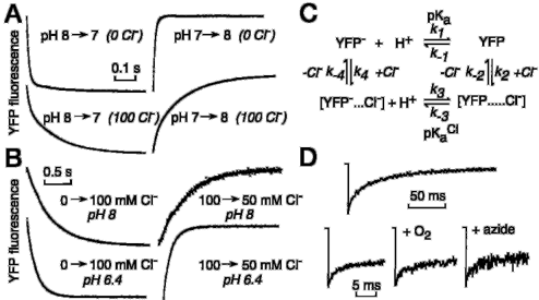
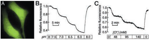
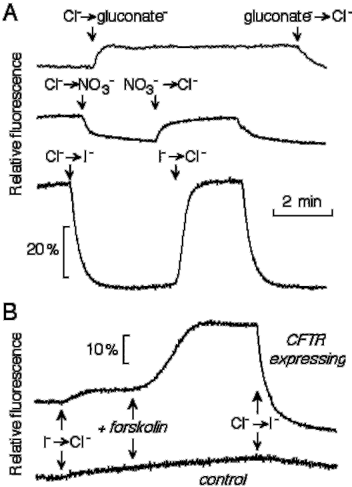
Verification
Claims the paper makes
The claims and statistics of this paper haven’t been checked yet.
Run the check — free, no account needed. It reads the open-access text and checks the sources it cites.
Claims other papers make about this one
Each is checked against this paper’s own text: does it say what the citing paper says it does?
The check wasn’t confident either way, so it gives no verdict — never disputed.
In the citing paper
“However, none of these methods are sufficiently selective or sensitive to rapidly and easily detect intracellular fluoride concentrations in bacteria.”
From Small Molecule Fluoride Toxicity Agonists (2015) · cites this paper as Jayaraman et al., 2000
Experimental — accuracy not yet measured
Self-check
Percentages, sums and reported differences re-computed from the paper’s own numbers.
The self-check needs the paper’s full text.
Citations
Cites
No references could be resolved yet.
Research works printed but not linked: 29
- Mansoura, 1999, Hum. Gene Ther., 10, 861, 10.1089/10430349950018274 · DOI
- Verkman, 1990, Am. J. Physiol., 259, C375, 10.1152/ajpcell.1990.259.3.C375 · DOI
- Biwersi, 1991, Biochemistry, 30, 7879, 10.1021/bi00246a001 · DOI
- Jayaraman, 1999, Am. J. Physiol., 276, C747, 10.1152/ajpcell.1999.276.3.C747 · DOI
- Biwersi, 1994, Anal. Biochem., 219, 139, 10.1006/abio.1994.1242 · DOI
- Jayaraman, 1999, Am. J. Physiol., 277, C1008, 10.1152/ajpcell.1999.277.5.C1008 · DOI
- De Giorgi, 1996, Gene (Amst.), 173, 113, 10.1016/0378-1119(95)00687-7 · DOI
- De Giorgi, 1999, Methods Cell Biol., 58, 75, 10.1016/S0091-679X(08)61949-4 · DOI
- Rizzuto, 1996, Curr. Biol., 6, 183, 10.1016/S0960-9822(02)00451-7 · DOI
- Rizzuto, 1995, Curr. Biol., 5, 635, 10.1016/S0960-9822(95)00128-X · DOI
- Cole, 1996, Science, 273, 797, 10.1126/science.273.5276.797 · DOI
- Kneen, 1998, Biophys. J., 74, 1591, 10.1016/S0006-3495(98)77870-1 · DOI
- Llopis, 1998, Proc. Natl. Acad. Sci. U. S. A., 95, 6803, 10.1073/pnas.95.12.6803 · DOI
- Miesenbock, 1998, Nature, 394, 192, 10.1038/28190 · DOI
- Miyawaki, 1997, Nature, 388, 882, 10.1038/42264 · DOI
- Romoser, 1997, J. Biol. Chem., 272, 13270, 10.1074/jbc.272.20.13270 · DOI
- Siegel, 1997, Neuron, 19, 735, 10.1016/S0896-6273(00)80955-1 · DOI
- Heim, 1995, Nature, 373, 663, 10.1038/373663b0 · DOI
- Heim, 1996, Curr. Biol., 6, 1178, 10.1016/S0960-9822(02)00450-5 · DOI
- Ormo, 1996, Science, 273, 1392, 10.1126/science.273.5280.1392 · DOI
- Elsliger, 1999, Biochemistry, 38, 5296, 10.1021/bi9902182 · DOI
- Wachter, 1998, Structure (Lond.), 6, 1267, 10.1016/S0969-2126(98)00127-0 · DOI
- Wachter, 1999, Curr. Biol., 9, R628, 10.1016/S0960-9822(99)80408-4 · DOI
- Fushimi, 1991, J. Cell Biol., 112, 719, 10.1083/jcb.112.4.719 · DOI
- Partikian, 1998, J. Cell Biol., 140, 821, 10.1083/jcb.140.4.821 · DOI
- Halm, 1992, J. Gen. Physiol., 99, 339, 10.1085/jgp.99.3.339 · DOI
- Swaminathan, 1997, Biophys. J., 72, 1900, 10.1016/S0006-3495(97)78835-0 · DOI
- Periasamy, 1996, Photochem. Photobiol., 63, 265, 10.1111/j.1751-1097.1996.tb03023.x · DOI
- Dawson, 1999, Physiol. Rev., 79, S47, 10.1152/physrev.1999.79.1.S47 · DOI
Cited by
Sorted by citations
Hanson, G. T., Aggeler, R., Oglesbee, D., Cannon, M., Capaldi, R. A., Tsien, R. Y., & Remington, S. J. (2004) · Journal of Biological Chemistry · doi:10.1074/jbc.m312846200 · Cited by 988
Self-citation(2006) · Principles of Fluorescence Spectroscopy · doi:10.1007/978-0-387-46312-4_8 · Cited by 278
Mongeon, R., Venkatachalam, V., & Yellen, G. (2016) · Antioxidants & Redox Signaling · doi:10.1089/ars.2015.6593 · Cited by 100
Small Molecule Fluoride Toxicity Agonists
Nelson, J. W., Plummer, M. S., Blount, K. F., Ames, T. D., & Breaker, R. R. (2015) · Chemistry & Biology · doi:10.1016/j.chembiol.2015.03.016 · Cited by 30
Metadata
- Authors
- Jayaraman, S., Haggie, P., Wachter, R. M., Remington, S. J., & Verkman, A. S.
- Venue
- Journal of Biological Chemistry
- Publisher
- Elsevier BV
- Published
- 2000-03
- Volume
- 275
- Issue
- 9
- Pages
- 6047-6050
- ISSN
- 0021-9258
- Cited by
- 264
- References
- 0
- Language
- en
About these checks
Registry, retraction, Cites and statistics results are computed, not judged, with one exception: a printed reference that no index lists is read once by a model before it counts as a citation error. Claims are extracted by a model and kept only when their quote appears verbatim in the text; each cited source is then checked, with a model’s help, for whether it says what the claim relies on. Those verdicts can be wrong — read the claim’s quote and the cited source before relying on one.
Statistics and arithmetic are recomputed, not judged.
Confidence bands are shown only for fields calibrated on our annotated test set. None is calibrated yet, so no band is shown.
What counts as a claim ›How we measure confidence ›How we check this ›
Are you an author, or do you see a problem with this page? Request a review
How to cite
Cite CoChat for the checks on this page, and the paper for its own findings.
- This page
- CoChat. (2026). Citation check: Mechanism and Cellular Applications of a Green Fluorescent Protein-based Halide Sensor. https://app.cochat.ai/papers/10.1074/jbc.275.9.6047
- The paper
- Jayaraman, S., Haggie, P., Wachter, R. M., Remington, S. J., & Verkman, A. S. (2000). Mechanism and Cellular Applications of a Green Fluorescent Protein-based Halide Sensor. Journal of Biological Chemistry. https://doi.org/10.1074/jbc.275.9.6047
